Raven P.H., Johnson G.B., Mason K.A. Biology (Ninth Edition)
Подождите немного. Документ загружается.

Apago PDF Enhancer
5 9
3 9
P
P
P
P
OH
5-carbon sugar
Nitrogenous base
Phosphate g roup
Phosphodiester bonds
O
O
Adenine
Guanine
NH
2
C
C
N
N
N
C
H
N
C
C H
O
H
H
Cytosine
(both DNA and RNA)
Thymine
(DNA only)
Uracil
(RNA only)
O C
N C
H
N
C
NH
2
H
C
H
Purines Pyrimidines
a.
b.
O
O
O
C
N
C
H
N
C
H
3
C
C
H
H
O
O
C
N
C
H
N
C
H
C
H
NH
2
C
C
N
N
N
C
H
N
C
C H
H
O
4„
5„
1„
3„
2„
4„
5„
1„
3„
2„
4„
5„
1„
3„
2„
4„
5„
1„
3„
2„
4„
5„
1„
3„ 2„
2
8
7 6
3 9
4
5
1
NH
2
O
J
J
P
J
O
-
-
O
J
O
J
CH
2
J
Phosphate group
Sugar
Nitrogenous base
OH
OH in RNA
H in DNA
N N
N
O
N
during the reproduction of organisms. DNA, found primarily
in the nuclear region of cells, contains the genetic information
necessary to build specific organisms.
Cells use a type of RNA called messenger RNA (mRNA)
to direct the synthesis of proteins . mRNA consists of transcribed
single-stranded copies of portions of the DNA. These transcripts
serve as blueprints specifying the amino acid sequences of pro-
teins. This process will be described in detail in chapter 15.
Nucleic acids are nucleotide polymers
Nucleic acids are long polymers of repeating subunits called
nucleotides. Each nucleotide consists of three components: a
pentose, or five-carbon sugar (ribose in RNA and deoxyribose
in DNA); a phosphate (—PO
4
) group; and an organic nitroge-
nous (nitrogen-containing) base (figure 3.14). When a nucleic
acid polymer forms, the phosphate group of one nucleotide
binds to the hydroxyl group from the pentose sugar of another,
releasing water and forming a phosphodiester bond by a dehydra-
tion reaction. A nucleic acid, then, is simply a chain of five-
carbon sugars linked together by phosphodiester bonds with a
nitrogenous base protruding from each sugar (see figure 3.15a).
These chains of nucleotides, polynucleotides, have different ends:
a phosphate on one end and an —OH from a sugar on the
other end. We conventionally refer to these ends as 5´ (“five-
prime,” —PO
4
) and 3´ (“three-prime,” —OH) taken from the
carbon numbering of the sugar (figure 3.15a).
Two types of nitrogenous bases occur in nucleotides
(3.15b). The first type, purines, are large, double-ring molecules
found in both DNA and RNA; the two types of purines are ad-
enine (A) and guanine (G). The second type, pyrimidines, are
smaller, single-ring molecules; they include cytosine (C, in both
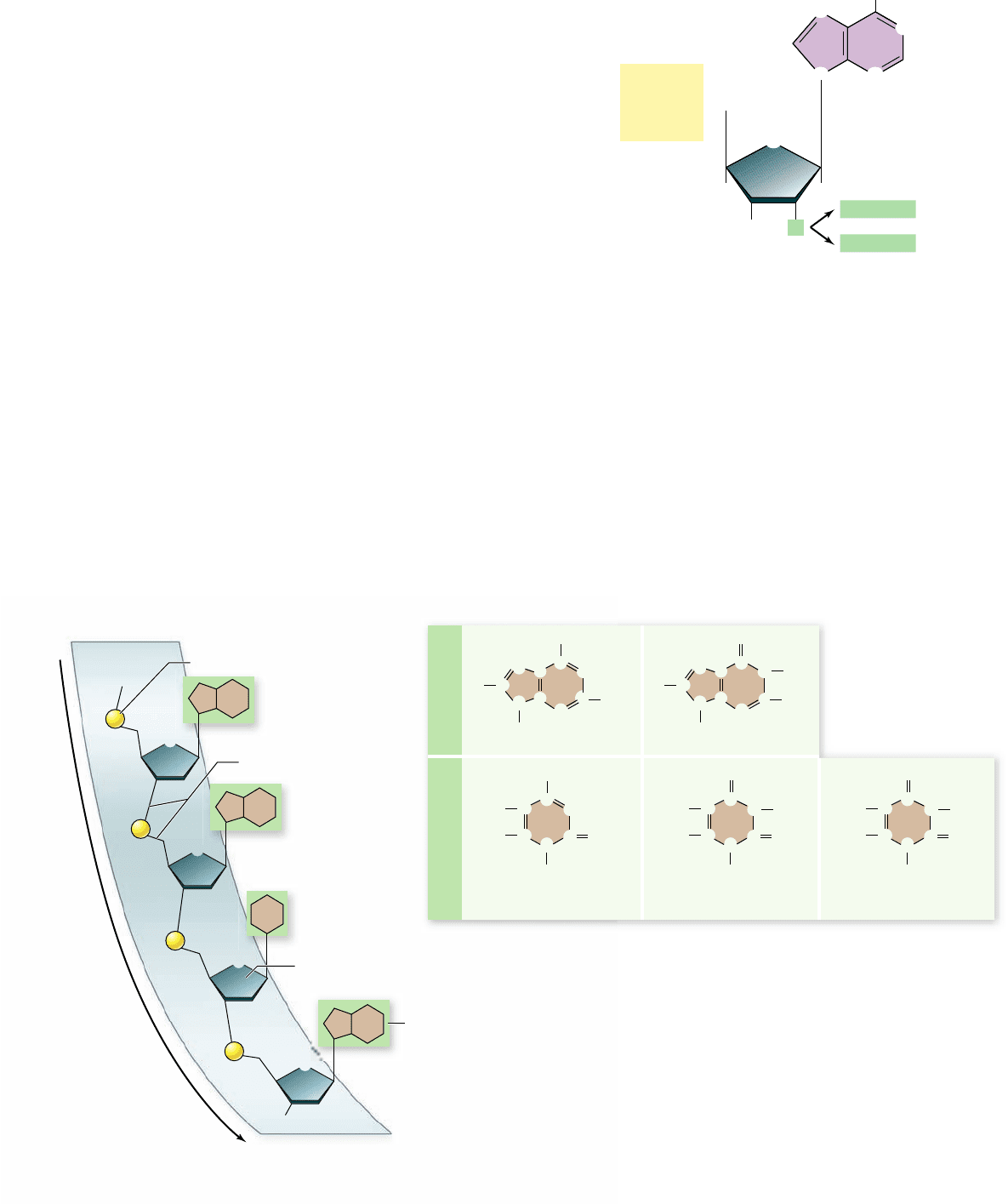
Figure 3.15
The structure of a nucleic acid and the
organic nitrogenous bases. a. In a nucleic acid, nucleotides are
linked to one another via phosphodiester bonds formed between the
phosphate of one nucleotide and the sugar of the next nucleotide. We call
this the sugar-phosphate backbone, and the organic bases protrude from
this chain. The backbone also has different ends: a 5´ phosphate end and
a 3´ hydroxyl end (the numbers come from the numbers in the sugars).
b. The organic nitrogenous bases can be either purines or pyrimidines.
The base thymine is found in DNA. The base uracil is found in RNA.
Figure 3.14
Structure of a nucleotide. The nucleotide
subunits of DNA and RNA are made up of three elements: a ve-
carbon sugar (ribose or deoxyribose), an organic nitrogenous base
(adenine is shown here), and a phosphate group. Notice that all the
numbers on the sugar are given as “primes” (1´, 2´, etc.) to distinguish
them from the numbering on the rings of the bases.
DNA and RNA), thymine (T, in DNA only), and uracil (U, in
RNA only).
DNA carries the genetic code
Organisms use sequences of nucleotides in DNA to encode the
information specifying the amino acid sequences of their proteins.
This method of encoding information is very similar to the way
in which sequences of letters encode information in a sentence.
42
part
I
The Molecular Basis of Life
rav32223_ch03_033-058.indd 42rav32223_ch03_033-058.indd 42 11/6/09 12:43:37 PM11/6/09 12:43:37 PM
Apago PDF Enhancer
Sugar–phosphate
“backbone”
Hydrogen bonds
between
nitrogenous bases
3
„
end
Phosphodiester
bonds
5„ end
P
P
P
P
P
C
G
A
A
OH
T
T
P
P
P
P
P
P
P
P
P
P
P
P
P
P
P
P
P
P
P
P
P
P
DNA
Deoxyribose-
phosphate
backbone
Bases
Hydrogen bonding
occurs between base-pairs
RNA
Ribose-phosphate
backbone
Bases
G
C
G
G
G
C
C
T
A
A
A
A
A
A
T
T
G
U
U
A sentence written in English consists of a combination of the 26
different letters of the alphabet in a certain order; the code of a
DNA molecule consists of different combinations of the four types
of nucleotides in specific sequences, such as CGCTTACG. The
information encoded in DNA is used in the everyday functioning
of the organism and is passed on to the organism’s descendants.
DNA molecules in organisms exist not as single chains
folded into complex shapes, like proteins, but rather as two chains
wrapped about each other in a long linear molecule in eukaryotes,
and a circular molecule in most prokaryotes. The two strands of
a DNA polymer wind around each other like the outside and
inside rails of a spiral staircase. Such a spiral shape is called a
helix, and a helix composed of two chains is called a double
helix. Each step of DNA’s helical staircase is composed of a base-
pair. The pair consists of a base in one chain attracted by hydrogen
bonds to a base opposite it on the other chain (figure 3.16).
The base-pairing rules are rigid: Adenine can pair only
with thymine (in DNA) or with uracil (in RNA), and cytosine
can pair only with guanine. The bases that participate in base-
pairing are said to be complementary to each other. Additional
details of the structure of DNA and how it interacts with RNA in
the production of proteins are presented in chapters 14 and 15 .
RNA is a transcript of a DNA strand
RNA is similar to DNA, but with two major chemical differ-
ences. First, RNA molecules contain ribose sugars, in which the
C-2 is bonded to a hydroxyl group. (In DNA, this hydroxyl
group is replaced by a hydrogen atom.) Second, RNA mole-
cules use uracil in place of thymine. Uracil has the same struc-
ture as thymine, except that one of its carbons lacks a methyl
(—CH
3
) group.
Transcribing the DNA message into a chemically differ-
ent molecule such as RNA allows the cell to distinguish be-
tween the original information-storage molecule and the
transcript. DNA molecules are always double-stranded (except
for a few single-stranded
DNA viruses), whereas the RNA mol-
ecules transcribed from DNA are typically single-stranded
(figure 3.17). These differences allow DNA to store hereditary
information and RNA to use this information to specify the
sequence of amino acids in proteins .
Other nucleotides are vital components
of energy reactions
In addition to serving as subunits of DNA and RNA, nucle-
otide bases play other critical roles in the life of a cell. For ex-
ample, adenine is a key component of the molecule adenosine
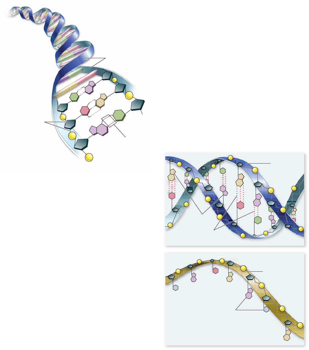
Figure 3.16
The structure of DNA. DNA consists of two
polynucleotide chains running in opposite directions wrapped
about a single helical axis. Hydrogen bond formation (dashed lines)
between the nitrogenous bases, called base-pairing, causes the two
chains of DNA to bind to each other and form a double helix.
Figure 3.17
DNA versus RNA. DNA forms a double helix,
uses deoxyribose as the sugar in its sugar–phosphate backbone, and
uses thymine among its nitrogenous bases. RNA is usually single-
stranded, uses ribose as the sugar in its sugar–phosphate backbone,
and uses uracil in place of thymine.
chapter
3
The Chemical Building Blocks of Life
43www.ravenbiology.com
rav32223_ch03_033-058.indd 43rav32223_ch03_033-058.indd 43 11/6/09 12:43:37 PM11/6/09 12:43:37 PM
Apago PDF Enhancer
4„
5
6
2
3
9
4
8
7
1„
3„ 2„
5„
1
Triphosphate group
5-carbon sugar
Nitrogenous base
(adenine)
CH
2
O
O
:
O
O
:
:
OJPJOJPJOJPJOJ
O
O
:
OH OH
NH
2
N
N
N
N
O
J
J
J
K
K
K
3.4
Proteins: Molecules with Diverse
Structures and Functions
Learning Outcomes
Describe the possible levels of protein structure.1.
Explain how motifs and domains contribute to protein 2.
structure.
Understand the relationship between amino acid 3.
sequence and their three-dimensional structure.
Proteins are the most diverse group of biological macromole-
cules, both chemically and functionally. Because proteins have
so many different functions in cells we could not begin to list
them all. We can, however, group these functions into the fol-
lowing seven categories. This list is a summary only, however;
details are covered in later chapters.
1. Enzyme catalysis. Enzymes are biological catalysts that
facilitate speci c chemical reactions. Because of this
property, the appearance of enzymes was one of the most
important events in the evolution of life. Enzymes are
three-dimensional globular proteins that t snugly around
the molecules they act on. This t facilitates chemical
reactions by stressing particular chemical bonds.
Defense.2. Other globular proteins use their shapes to
“recognize” foreign microbes and cancer cells. These
cell-surface receptors form the core of the body’s
endocrine and immune systems.
Transport.3. A variety of globular proteins transport
small molecules and ions. The transport protein
hemoglobin, for example, transports oxygen in the
blood. Membrane transport proteins help move ions and
molecules across the membrane.
Support.4. Protein bers play structural roles. These
bers include keratin in hair, brin in blood clots, and
collagen. The last one, collagen, forms the matrix of
skin, ligaments, tendons, and bones and is the most
abundant protein in a vertebrate body.
Motion.5. Muscles contract through the sliding motion of
two kinds of protein laments: actin and myosin.
Contractile proteins also play key roles in the cell’s
cytoskeleton and in moving materials within cells.
Regulation.6. Small proteins called hormones serve as
intercellular messengers in animals. Proteins also play
many regulatory roles within the cell—turning on and
shutting off genes during development, for example. In
addition, proteins receive information, acting as
cell-surface receptors.
Storage.7. Calcium and iron are stored in the body by
binding as ions to storage proteins.
Table 3.2 summarizes these functions and includes examples
of the proteins that carry them out in the human body.
Proteins are polymers of amino acids
Proteins are linear polymers made with 20 different amino ac-
ids. Amino acids, as their name suggests, contain an amino
group (—NH
2
) and an acidic carboxyl group (—COOH). The
specific order of amino acids determines the protein’s structure
and function. Many scientists believe amino acids were among
the first molecules formed on the early Earth. It seems highly
likely that the oceans that existed early in the history of the
Earth contained a wide variety of amino acids.
Amino acid structure
The generalized structure of an amino acid is shown here as
amino and carboxyl groups bonded to a central carbon atom,
with an additional hydrogen and a functional side group
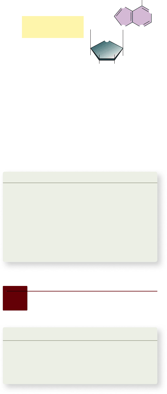
Figure 3.18
ATP. Adenosine
triphosphate (ATP) contains
adenine, a ve-carbon sugar, and
three phosphate groups.
triphosphate (ATP; figure 3.18) —the energy currency of the
cell. Cells use ATP as energy in a variety of transactions, the
way we use money in society. ATP is used to drive energetically
unfavorable chemical reactions, to power transport across
membranes, and to power the movement of cells .
Two other important nucleotide-containing molecules
are nicotinamide adenine dinucleotide (NAD
+
) and flavin
adenine dinucleotide (FAD). These molecules function as
electron carriers in a variety of cellular processes. You will see
the action of these molecules in detail when we discuss photo-
synthesis and respiration (chapters 7–8 ).
Learning Outcomes Review 3.3
A nucleic acid is a polymer composed of alternating phosphate and fi ve-
carbon sugar groups with a nitrogenous base protruding from each sugar.
In DNA, this sugar is deoxyribose. In RNA, the sugar is ribose. RNA also
contains the base uracil instead of thymine. DNA is a double-stranded helix
that stores hereditary information as a specifi c sequence of nucleotide
bases. RNA is a single-stranded molecule consisting of a transcript of a DNA
sequence that directs protein synthesis.
■ If an RNA molecule is copied from a DNA strand, what is
the relationship between the sequence of bases in RNA
and each DNA strand?
44
part
I
The Molecular Basis of Life
rav32223_ch03_033-058.indd 44rav32223_ch03_033-058.indd 44 11/6/09 12:43:39 PM11/6/09 12:43:39 PM

Apago PDF Enhancer
TABLE 3.2
The Many Functions of Protein
Function Class of Protein Examples Examples of Use
Enzyme catalysis Enzymes Glycosidases Cleave polysaccharides
Proteases Break down proteins
Polymerases Synthesize nucleic acids
Kinases Phosphorylate sugars and proteins
Defense Immunoglobulins Antibodies Mark foreign proteins for elimination
Toxins Snake venom Blocks nerve function
Cell-surface antigens MHC* proteins “Self” recognition
Transport Circulating transporters Hemoglobin Carries O
2
and CO
2
in blood
Myoglobin Carries O
2
and CO
2
in muscle
Cytochromes Electron transport
Membrane transporters Sodium–potassium pump Excitable membranes
Proton pump Chemiosmosis
Glucose transporter Transports glucose into cells
Support Fibers Collagen Forms cartilage
Keratin Forms hair, nails
Fibrin Forms blood clots
Motion Muscle Actin Contraction of muscle bers
Myosin Contraction of muscle bers
Regulation Osmotic proteins Serum albumin Maintains osmotic concentration
of blood
Gene regulators lac Repressor Regulates transcription
Hormones Insulin Controls blood glucose levels
Vasopressin Increases water retention by kidneys
Oxytocin Regulates uterine contractions and milk
production
Storage Ion-binding Ferritin Stores iron, especially in spleen
Casein Stores ions in milk
Calmodulin Binds calcium ions
*MHC, major histocompatibility complex.
indicated by R. These components completely fill the bonds of
the central carbon:
R
|
H
2
N—C—COOH
|
H
The unique character of each amino acid is determined
by the nature of the R group. Notice that unless the R group
is an H atom, as in glycine, amino acids are chiral and can
exist as two enantiomeric forms: d or l. In living systems,
only the l-amino acids are found in proteins, and d-amino
acids are rare.
The R group also determines the chemistry of amino ac-
ids. Serine, in which the R group is —CH
2
OH, is a polar mol-
ecule. Alanine, which has —CH
3
as its R group, is nonpolar.
The 20 common amino acids are grouped into five chemical
classes, based on their R group:
Nonpolar amino acids, such as leucine, often have 1.
R groups that contain —CH
2
or —CH
3
.
Polar uncharged amino acids, such as threonine, have 2.
R groups that contain oxygen (or —OH).
Charged amino acids, such as glutamic acid, have 3.
R groups that contain acids or bases that can ionize.
Aromatic amino acids, such as phenylalanine, have R groups 4.
that contain an organic (carbon) ring with alternating single
and double bonds. These are also nonpolar.
Amino acids that have special functions have unique 5.
properties. Some examples are methionine, which is often
the rst amino acid in a chain of amino acids; proline,
which causes kinks in chains; and cysteine, which links
chains together.
chapter
3
The Chemical Building Blocks of Life
45www.ravenbiology.com
rav32223_ch03_033-058.indd 45rav32223_ch03_033-058.indd 45 11/9/09 4:22:59 PM11/9/09 4:22:59 PM
Apago PDF Enhancer
Amino acid Amino acid
Dipeptide
H
J
N
J
C
J
C
J
N
J
C
J
C
J
OH
J J
R
J
H
H
J J
R
H O
J
J
J J
R
O H
J
J
H
J
N
J
C
J
C
J
OH
J J
R
J
H
O H
H
2
O
J
H
O
J
J
H
J
N
J
C
J
C
J
OH
H
J
J
J
Each amino acid affects the shape of a protein differently,
depending on the chemical nature of its side group. For exam-
ple, portions of a protein chain with numerous nonpolar amino
acids tend to fold into the interior of the protein by hydropho-
bic exclusion.
Peptide bonds
In addition to its R group, each amino acid, when ionized, has a
positive amino (NH
3
+
) group at one end and a negative carboxyl
(COO
–
) group at the other. The amino and carboxyl groups on
a pair of amino acids can undergo a dehydration reaction to
form a covalent bond. The covalent bond that links two amino
acids is called a peptide bond (figure 3.19). The two amino
acids linked by such a bond are not free to rotate around the
N—C linkage because the peptide bond has a partial double-
bond character. This is different from the N—C and C—C
bonds to the central carbon of the amino acid. This lack of rota-
tion about the peptide bond is one factor that determines the
structural character of the coils and other regular shapes formed
by chains of amino acids.
A protein is composed of one or more long unbranched
chains. Each chain is called a polypeptide and is composed of
amino acids linked by peptide bonds. The terms protein and
polypeptide tend to be used loosely and may be confusing. For
proteins that include only a single polypeptide chain, the two
terms are synonymous.
The pioneering work of Frederick Sanger in the early
1950s provided the evidence that each kind of protein has a
specific amino acid sequence. Using chemical methods to re-
move successive amino acids and then identify them, Sanger
succeeded in determining the amino acid sequence of insulin.
In so doing he demonstrated clearly that this protein had a de-
fined sequence, which was the same for all insulin molecules in
the solution. Although many different amino acids occur in na-
ture, only 20 commonly occur in proteins. Figure 3.20 illus-
trates these 20 amino acids and their side groups.
Proteins have levels of structure
The shape of a protein determines its function. One way to
study the shape of something as small as a protein is to look at
it with very short wavelength energy—in other words, with X-
rays. X-rays can be passed through a crystal of protein to pro-
duce a diffraction pattern. This pattern can then be analyzed by
a painstaking procedure that allows the investigator to build up
a three-dimensional picture of the position of each atom. The
first protein to be analyzed in this way was myoglobin, and the
related protein hemoglobin was analyzed soon thereafter.
As more and more proteins were studied, a general
principle became evident: In every protein studied, essen-
tially all the internal amino acids are nonpolar ones—amino
acids such as leucine, valine, and phenylalanine. Water’s ten-
dency to hydrophobically exclude nonpolar molecules liter-
ally shoves the nonpolar portions of the amino acid chain
into the protein’s interior (figure 3.21). This tendency forces
the nonpolar amino acids into close contact with one an-
other, leaving little empty space inside. Polar and charged
amino acids are restricted to the surface of the protein, ex-
cept for the few that play key functional roles.
The structure of proteins is usually discussed in terms of a
hierarchy of four levels: primary, secondary, tertiary, and quater-
nary (figure 3.22) . We will examine this view and then integrate
it with a more modern approach arising from our increasing
knowledge of protein structure.
Primary structure: amino acid sequence
The primary structure of a protein is its amino acid sequence.
Because the R groups that distinguish the amino acids play no
role in the peptide backbone of proteins, a protein can consist of
any sequence of amino acids. Thus, because any of 20 different
amino acids might appear at any position, a protein containing
100 amino acids could form any of 20
100
different amino acid
sequences (that’s the same as 10
130
, or 1 followed by 130 zeros—
more than the number of atoms known in the universe). This
important property of proteins permits great diversity.
Consider the protein hemoglobin, the protein your
blood uses to transport oxygen. Hemoglobin is composed of
two α-globin peptide chains and two β-globin peptide chains.
The α-globin chains differ from the β-globin ones in the se-
quence of amino acids. Furthermore, any alteration in the
normal sequence of either of the types of globin proteins,
even by a single amino acid, can have drastic effects on how
the protein functions.
Secondary structure: Hydrogen bonding patterns
The amino acid side groups are not the only portions of pro-
teins that form hydrogen bonds. The peptide groups of the
main chain can also do so. These hydrogen bonds can be with
water or with other peptide groups. If the peptide groups
formed too many hydrogen bonds with water, the proteins
would tend to behave like a random coil and wouldn’t produce
Figure 3.19
The peptide bond. A peptide bond forms when
the amino end of one amino acid joins to the carboxyl end of
another. Reacting amino and carboxyl groups are shown in red and
nonreacting groups are highlighted in green. Notice that the
resulting dipeptide still has an amino end and a carboxyl end.
Because of the partial double-bond nature of peptide bonds, the
resulting peptide chain cannot rotate freely around these bonds.
46
part
I
The Molecular Basis of Life
rav32223_ch03_033-058.indd 46rav32223_ch03_033-058.indd 46 11/6/09 12:43:40 PM11/6/09 12:43:40 PM
Apago PDF Enhancer
J
J
NonaromaticAromatic
Nonpolar Polar uncharged Charged
Special function
CH
2
CH
2
H
3
N
;
J
C
J
C
J
O
:
JJJ
CH
3
JJ
CH
2
J
OH
J
H
J
J
O
J
J
S
J
H
J
S
NH
3
;
J
C
J
C
J
O
:
CH
2
CH
2
CH
J
C
J
O
:
O
J
CH
2
J
J
J
J
J
J
NH
2
;
Proline
(Pro)
Methionine
(Met)
Cysteine
(Cys)
Alanine
(Ala)
H
3
N
;
J
C
J
C
J
O
:
JJ
CH
3
OH
J
J
Serine
(Ser)
H
3
N
;
J
C
J
C
J
O
:
JJ
CH
2
OH
J
J
J
OH
Glycine
(Gly)
H
3
N
;
J
C
J
C
J
O
:
JJ
H
OH
J
J
Valine
(Val)
H
3
N
;
J
C
J
C
J
O
:
JJ
OH
J
J
CH
3
CH
3
CH
J
J
Leucine
(Leu)
CH
2
H
3
N
;
J
C
J
C
J
O
:
JJJ
OH
J
J
CH
3
CH
3
CH
J
J
Asparagine
(Asn)
CH
2
H
3
N
;
J
C
J
C
J
O
:
JJJ
OH
J
J
NH
2
O
C
J
J
J
Aspartic acid
(Asp)
CH
2
H
3
N
;
J
C
J
C
J
O
:
JJJ
OH
J
J
O
:
O
C
J
J
J
Lysine
(Lys)
CH
2
CH
2
H
3
N
;
J
C
J
C
J
O
:
JJJ
CH
2
J
NH
3
;
JJ
OH
J
J
CH
2
Glutamic acid
(Glu)
CH
2
CH
2
H
3
N
;
J
C
J
C
J
O
:
JJJJ
OH
J
J
O
:
O
C
J
J
J
Glutamine
(Gln)
CH
2
CH
2
H
3
N
;
J
C
J
C
J
O
:
JJJJ
OH
J
J
NH
2
O
C
J
J
J
Isoleucine
(Ile)
H
J
C
J
CH
3
CH
2
H
3
N
;
J
C
J
C
J
O
:
JJ
CH
3
JJ
OH
J
J
Threonine
(Thr)
H
J
C
J
OH
CH
3
H
3
N
;
J
C
J
C
J
O
:
JJJ
OH
J
J
Histidine
(His)
CH
2
H
3
N
;
J
C
J
C
J
O
:
CH
C
J
N
HC
J
NH
;
H O
H
JJJ
J
J
J
J
K
J
Tryptophan
(Trp)
CH
2
H
3
N
;
J
C
J
C
J
O
:
—
NH
—
C
—
——
—
H O
J
J
J
Phenylalanine
(Phe)
CH
2
H
3
N
;
J
C
J
C
J
O
:
HO
JJJ
J
J
Tyrosine
(Tyr)
OH
J
CH
2
H
3
N
;
J
C
J
C
J
O
:
H O
JJJ
J
J
Arginine
(Arg)
CH
2
CH
2
H
3
N
;
J
C
J
C
J
O
:
JJJ
NH
J
NH
2
J
C
JJ
OH
J
J
CH
2
NH
2
;
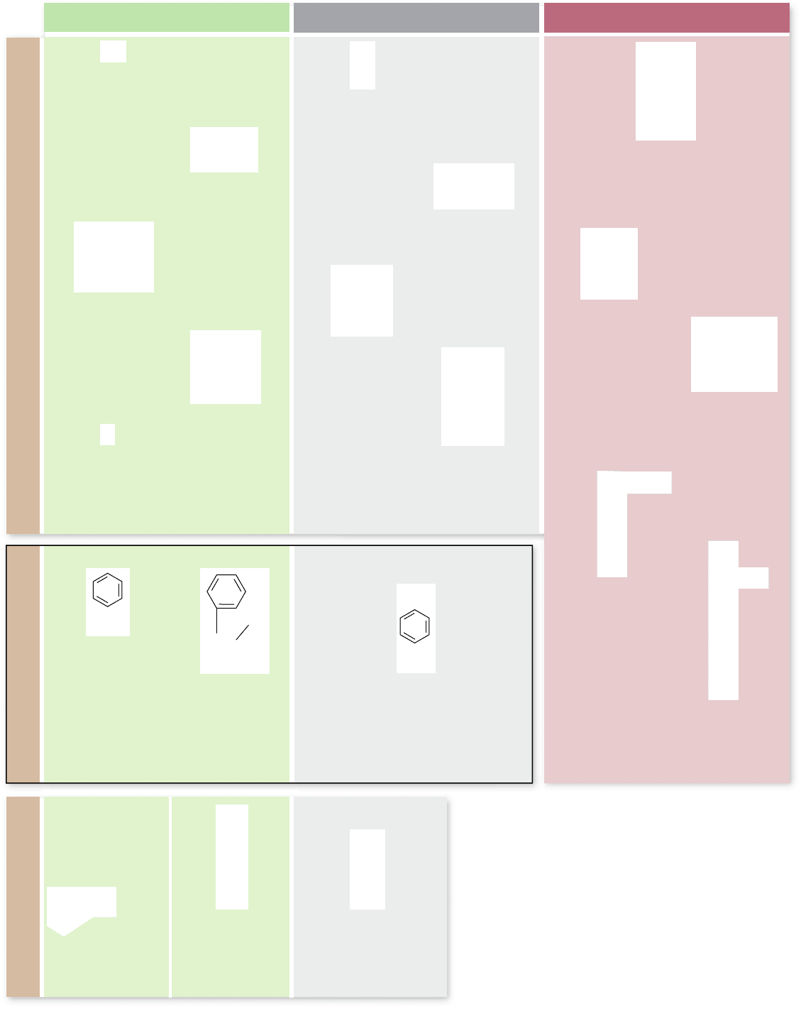
Figure 3.20
The 20 common amino acids. Each
amino acid has the same chemical backbone, but differs in
the side, or R, group. Seven of the amino acids are
nonpolar because they have —CH
2
or —CH
3
in their
R groups. Two of the seven contain ring structures with
alternating double and single bonds, which classi es them
also as aromatic. Another ve are polar because they have
oxygen or a hydroxyl group in their R groups. Five others
are capable of ionizing to a charged form. The remaining
three special-function amino acids have chemical
properties that allow them to help form links between
protein chains or kinks in proteins.
chapter
3
The Chemical Building Blocks of Life
47www.ravenbiology.com
rav32223_ch03_033-058.indd 47rav32223_ch03_033-058.indd 47 11/6/09 12:43:41 PM11/6/09 12:43:41 PM
Apago PDF Enhancer
N
N
Disulfide bridge
J
J
J
J
J
J
J
J
J
J
J
J
J
J
J
J
J
J
J
J
J
J J
J
J
J J
J
J
J J
J
J
J J J
J
J
J
J
J
J
J J
J
J
J
J
J
J
J
J
J
J
O
O
C C
C
H
H
C C
C
H
H
S S
C
C
C
O
N
R
C
N
C
C
H
H
R
R
Hydrogen bond
O
N
O
:
Ionic bond
C
C
C
C
C
O
(CH
2
)
4
CH
2
O
O
N
NH
3
;
CH
3
CH
2
CH
3
CH
3
CH
3
CH
3
CH
3
CH
3
C
H
C
Hydrophobic
exclusion
a. b.
J
J
J
J
J
J
J
J
J
J
C
C
O
O
N
N
C
C
CH
3
CH
3
van der Waals
attraction
d.
e.
c.
the kinds of globular structures that are common in proteins.
Linus Pauling suggested that the peptide groups could interact
with one another if the peptide was coiled into a spiral that he
called the α helix. We now call this sort of regular interaction
of groups in the peptide backbone secondary structure. An-
other form of secondary structure can occur between regions of
peptide aligned next to each other to form a planar structure
called a β sheet. These can be either parallel or antiparallel
depending on whether the adjacent sections of peptide are ori-
ented in the same direction, or opposite direction.
These two kinds of secondary structure create regions of
the protein that are cylindrical (α helices) and planar (β sheets).
A protein’s final structure can include regions of each type of
secondary structure. For example, DNA-binding proteins usu-
ally have regions of α helix that can lay across DNA and inter-
act directly with the bases of DNA. Porin proteins that form
holes in membranes are composed of β sheets arranged to
form a pore in the membrane. Finally in hemoglobin, the α-
and β-globin peptide chains that make up the final molecule
each have characteristic regions of secondary structure.
Tertiary structure: Folds and links
The final folded shape of a globular protein is called its tertiary
structure. This tertiary structure contains regions that have
secondary structure and determines how these are further ar-
ranged in space to produce the overall structure. A protein is
initially driven into its tertiary structure by hydrophobic exclu-
sion from water. Ionic bonds between oppositely charged
R groups bring regions into close proximity, and disulfide bonds
(covalent links between two cysteine R groups) lock particular
regions together. The final folding of a protein is determined
by its primary structure—the chemical nature of its side groups
(see figure 3.21 and 3.22). Many small proteins can be fully
unfolded (“denatured”) and will spontaneously refold into their
characteristic shape. Other larger proteins tend to associate to-
gether and form insoluble clumps when denatured, such as the
film that can form when you heat milk for hot chocolate.
The tertiary structure is stabilized by a number of forces
including hydrogen bonding between R groups of different ami-
no acids, electrostatic attraction between R groups with opposite
charge (also called salt bridges), hydrophobic exclusion of non-
polar R groups, and covalent bonds in the form of disulfides. The
stability of a protein, once it has folded into its tertiary shape, is
strongly influenced by how well its interior fits together. When
two nonpolar chains in the interior are very close together, they
experience a form of molecular attraction called van der Waals
forces. Individually quite weak, these forces can add up to a
strong attraction when many of them come into play, like the
combined strength of hundreds of hooks and loops on a strip of
Velcro. These forces are effective only over short distances, how-
ever. No “holes” or cavities exist in the interior of proteins. The
variety of different nonpolar amino acids, with a different-sized
R group with its own distinctive shape, allows nonpolar chains to
fit very precisely within the protein interior.
It is therefore not surprising that changing a single amino
acid can drastically alter the structure, and thus the function of a
Figure 3.21
Interactions that contribute to a protein’s shape. Aside
from the bonds that link together the amino acids in a protein, several other weaker
forces and interactions determine how a protein will fold. a. Hydrogen bonds can form
between the different amino acids. b. Covalent disul de bridges can form between two
cysteine side chains. c. Ionic bonds can form between groups with opposite charge.
d. van der Waals attractions, which are weak attractions between atoms due to
oppositely polarized electron clouds, can occur. e. Polar portions of the protein tend to
gather on the outside of the protein and interact with water, whereas the hydrophobic
portions of the protein, including nonpolar amino acid chains, are shoved toward the
interior of the protein.
48
part
I
The Molecular Basis of Life
rav32223_ch03_033-058.indd 48rav32223_ch03_033-058.indd 48 11/9/09 12:29:31 PM11/9/09 12:29:31 PM
Apago PDF Enhancer
•
•
•
•
•
•
•
•
•
•
•
•
•
•
•
•
•
•
•
•
•
•
•
•
•
•
•
•
•
•
•
•
•
•
•
•
•
•
•
Tertiary Structure
β-pleated sheet
α
-helix
The primary structure can fold
into a pleated sheet, or turn into a helix
Primary Structure
Secondary Structure Secondary Structure
Quaternary Structure
N
H
H H
C C C N C C N C C
O
O H H
O H H
R R
R
R
J
J
J
J
J
J
J
J
J
J
J
J
J
J
J J J J J J J J J J
J
J
J
protein. The sickle cell version of hemoglobin (HbS), for exam-
ple, is a change of a single glutamic acid for a valine in the
β-globin chain. This change substitutes a charged amino acid for
a nonpolar one on the surface of the protein, leading the protein
to become sticky and form clumps. Another variant of hemoglo-
bin called HbE, actually the most common in human popula-
tions, causes a change from glutamic acid to lysine at a different
site in the β-globin chain. In this case the structural change is not
as dramatic, but it still impairs function, resulting in blood disor-
ders called anemia and thalassemia. More than 700 structural
variants of hemoglobin are known, with up to 7% of the world’s
population being carriers of forms that are medically important.
Quaternary structure: Subunit arrangements
When two or more polypeptide chains associate to form a func-
tional protein, the individual chains are referred to as subunits of
the protein. The arrangement of these subunits is termed its
quaternary structure. In proteins composed of subunits, the in-
terfaces where the subunits touch one another are often nonpo-
lar, and they play a key role in transmitting information between
the subunits about individual subunit activities.
Remember that the protein hemoglobin is composed of
two α-chain subunits and two β-chain subunits. Each α- and
β-globin chain has a primary structure consisting of a specific
sequence of amino acids. This then assumes a characteristic sec-
ondary structure consisting of α helices and β sheets that are
then arranged into a specific tertiary structure for each α- and
β-globin subunit. Lastly, these subunits are then arranged into
their final quaternary structure. This is the final structure of the
protein. For proteins that consist of only a single peptide chain,
the enzyme lysozyme for example, the tertiary structure is the
final structure of the protein.
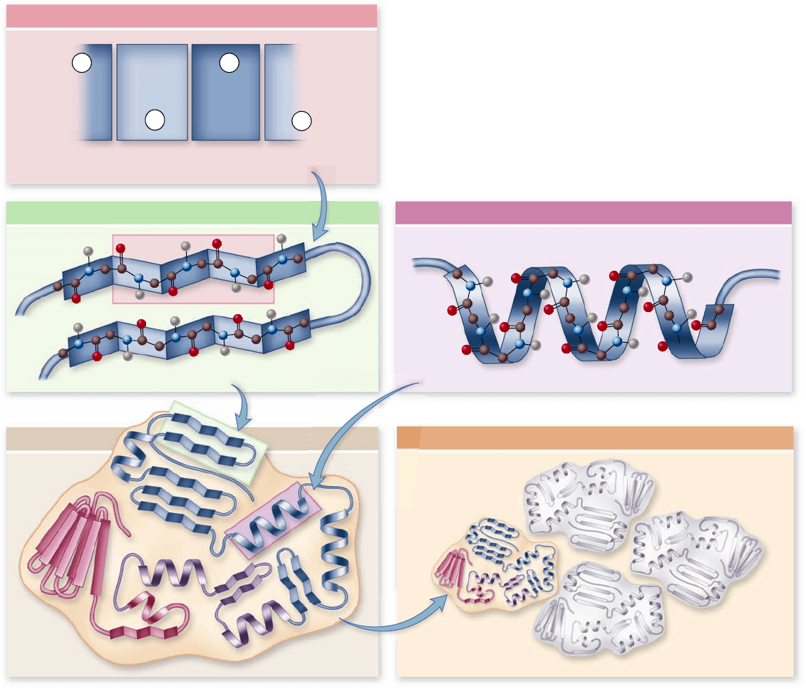
Figure 3.22
Levels of protein structure. The primary
structure of a protein is its amino acid sequence. Secondary structure
results from hydrogen bonds forming between nearby amino acids.
This produces two different kinds of structures: beta (β)-pleated
sheets, and coils called alpha (α)-helices. The tertiary structure is the
nal 3-D shape of the protein. This determines how regions of
secondary structure are then further folded in space to form the nal
shape of the protein. Quaternary structure is only found in proteins
with multiple polypeptides. In this case the nal structure of the
protein is the arrangement of the multiple polypeptides in space.
chapter
3
The Chemical Building Blocks of Life
49www.ravenbiology.com
rav32223_ch03_033-058.indd 49rav32223_ch03_033-058.indd 49 11/6/09 12:43:42 PM11/6/09 12:43:42 PM
Apago PDF Enhancer
Helix-turn-Helix
motif
b-a-b
motif
Motifs
Domain 1
Domain 2Domain 3
Domains
Motifs and domains are additional
structural characteristics
To directly determine the sequence of amino acids in a protein
is a laborious task. Although the process has been automated, it
remains slow and difficult.
The ability to sequence DNA changed this situation
rather suddenly. Sequencing DNA was a much simpler process,
and even before it was automated, the number of known se-
quences rose quickly. With the advent of automation, the known
sequences increased even more dramatically. Today the entire
sequence of hundreds of bacterial genomes and more than a
dozen animal genomes, including that of humans, has been de-
termined. Because the DNA sequence is directly related to
amino acid sequence in proteins, biologists now have a large
database of protein sequences to compare and analyze. This
new information has also stimulated thought about the logic of
the genetic code and whether underlying patterns exist in pro-
tein structure. Our view of protein structure has evolved with
this new information. Researchers still view the four-part hier-
archical structure as important, but two new terms have entered
the biologist’s vocabulary: motif and domain.
Motifs
As biologists discovered the 3-D structure of proteins (an
even more laborious task than determining the sequence),
they noticed similarities between otherwise dissimilar pro-
teins. These similar structures are called motifs, or sometimes
“supersecondary structure.” The term motif is borrowed from
the arts and refers to a recurring thematic element in music
or design.
One very common protein motif is the β-α-β motif,
which creates a fold or crease; the so-called “Rossmann fold” at
the core of nucleotide-binding sites in a wide variety of pro-
teins. A second motif that occurs in many proteins is the β bar-
rel, which is a β sheet folded around to form a tube. A third type
of motif, the helix-turn-helix, consists of two α helices sepa-
rated by a bend. This motif is important because many proteins
use it to bind to the DNA double helix (figure 3.23; see also
chapter 16 ).
Motifs indicate a logic to structure that investigators still
do not understand. Do they simply represent a reuse by evolu-
tion of something that already works, or are they an optimal
solution to a problem, such as how to bind a nucleotide? One
way to think about it is that if amino acids are letters in the
language of proteins, then motifs represent repeated words or
phrases. Motifs have been useful in determining the function of
unknown proteins. Databases of protein motifs are used to
search new unknown proteins. Finding motifs with known
functions may allow an investigator to infer the function of a
new protein.
Domains
Domains of proteins are functional units within a larger struc-
ture. They can be thought of as substructure within the tertiary
structure of a protein (see figure 3.23). To continue the meta-
phor: Amino acids are letters in the protein language, motifs
are words or phrases, and domains are paragraphs.
Most proteins are made up of multiple domains that
perform different parts of the protein’s function. In many
cases, these domains can be physically separated. For example,
transcription factors (discussed in chapter 16 ) are proteins
that bind to DNA and initiate its transcription. If the DNA-
binding region is exchanged with a different transcription fac-
tor, then the specificity of the factor for DNA can be changed
without changing its ability to stimulate transcription. Such
“domain-swapping” experiments have been performed with
many transcription factors, and they indicate, among other
things, that the DNA-binding and activation domains are
functionally separate.
These functional domains of proteins may also help the
protein to fold into its proper shape. As a polypeptide chain
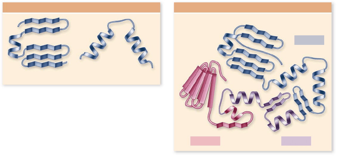
Figure 3.23
Motifs and domains. The elements of
secondary structure can combine, fold, or crease to form motifs.
These motifs are found in different proteins and can be used to
predict function. Proteins also are made of larger domains, which
are functionally distinct parts of a protein. The arrangement of
these domains in space is the tertiary structure of a protein.
50
part
I
The Molecular Basis of Life
rav32223_ch03_033-058.indd 50rav32223_ch03_033-058.indd 50 11/6/09 12:43:43 PM11/6/09 12:43:43 PM
Apago PDF Enhancer
ATP
Misfolded
protein
Chaperone
protein
Cap
Chance for protein to refold
Correctly
folded
protein
ADP + P
folds, the domains take their proper shape, each more or less
independently of the others. This action can be demonstrated
experimentally by artificially producing the fragment of a
polypeptide that forms the domain in the intact protein, and
showing that the fragment folds to form the same structure as
it exhibits in the intact protein. A single polypeptide chain con-
nects the domains of a protein, like a rope tied into several ad-
jacent knots.
Domains can also correspond to the structure of the genes
that encode them. Later, in chapter 15 , you will see that genes
in eukaryotes are often in pieces within the genome, and these
pieces, called exons, sometimes encode the functional domains
of a protein. This finding led to the idea of evolution acting by
shuffling protein-encoding domains.
The process of folding relies
on chaperone proteins
Until recently, scientific investigators thought that newly made
proteins fold spontaneously, randomly trying out different con-
figurations as hydrophobic interactions with water shoved non-
polar amino acids into the protein’s interior until the final
structure was arrived at. We now know this view is too simple.
Protein chains can fold in so many different ways that trial and
error would simply take too long. In addition, as the open chain
folds its way toward its final form, nonpolar “sticky” interior
portions are exposed during intermediate stages. If these inter-
mediate forms are placed in a test tube in an environment iden-
tical to that inside a cell, they stick to other, unwanted protein
partners, forming a gluey mess.
How do cells avoid having their proteins clump into a
mass? A vital clue came in studies of unusual mutations that pre-
vent viruses from replicating in bacterial cells. It turns out that
the virus proteins produced inside the cells could not fold prop-
erly. Further study revealed that normal cells contain chaperone
proteins, which help other proteins to fold correctly.
Molecular biologists have now identified many proteins
that act as molecular chaperones. This class of proteins has
multiple subclasses, and representatives have been found in es-
sentially every organism that has been examined. Furthermore,
these proteins seem to be essential for viability as well, illustrat-
ing their fundamental importance. Many are heat shock pro-
teins, produced in greatly increased amounts when cells are
exposed to elevated temperature. High temperatures cause
proteins to unfold, and heat shock chaperone proteins help the
cell’s proteins to refold properly.
One class of these proteins, called chaperonins, has been
extensively studied. In the bacterium Escherichia coli (E. coli), one
example is the essential protein GroE chaperonin. In mutants
in which the GroE chaperonin is inactivated, fully 30% of the
bacterial proteins fail to fold properly. Chaperonins associate to
form a large macromolecular complex that resembles a cylin-
drical container. Proteins can move into the container, and the
container itself can change its shape considerably (figure 3.24).
Experiments have shown that an improperly folded protein can
enter the chaperonin and be refolded. Although we don’t know
exactly how this happens, it seems to involve changes in the
hydrophobicity of the interior of the chamber.
The flexibility of the structure of chaperonins is amazing.
We tend to think of proteins as being fixed structures, but this
is clearly not the case for chaperonins and this flexibility is nec-
essary for their function. It also illustrates that even domains
that may be very widely separated in a very large protein are
still functionally connected. The folding process within a chap-
eronin harnesses the hydrolysis of ATP to power these changes
in structure necessary for function. This entire process can oc-
cur in a cyclic manner until the appropriate structure is achieved.
Cells use these chaperonins both to accomplish the original
folding of some proteins and to restore the structure of incor-
rectly folded ones.
Some diseases may result
from improper folding
Chaperone protein deficiencies may be implicated in certain dis-
eases in which key proteins are improperly folded. Cystic fibrosis
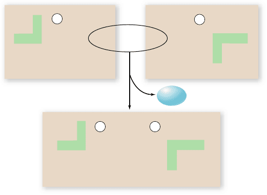
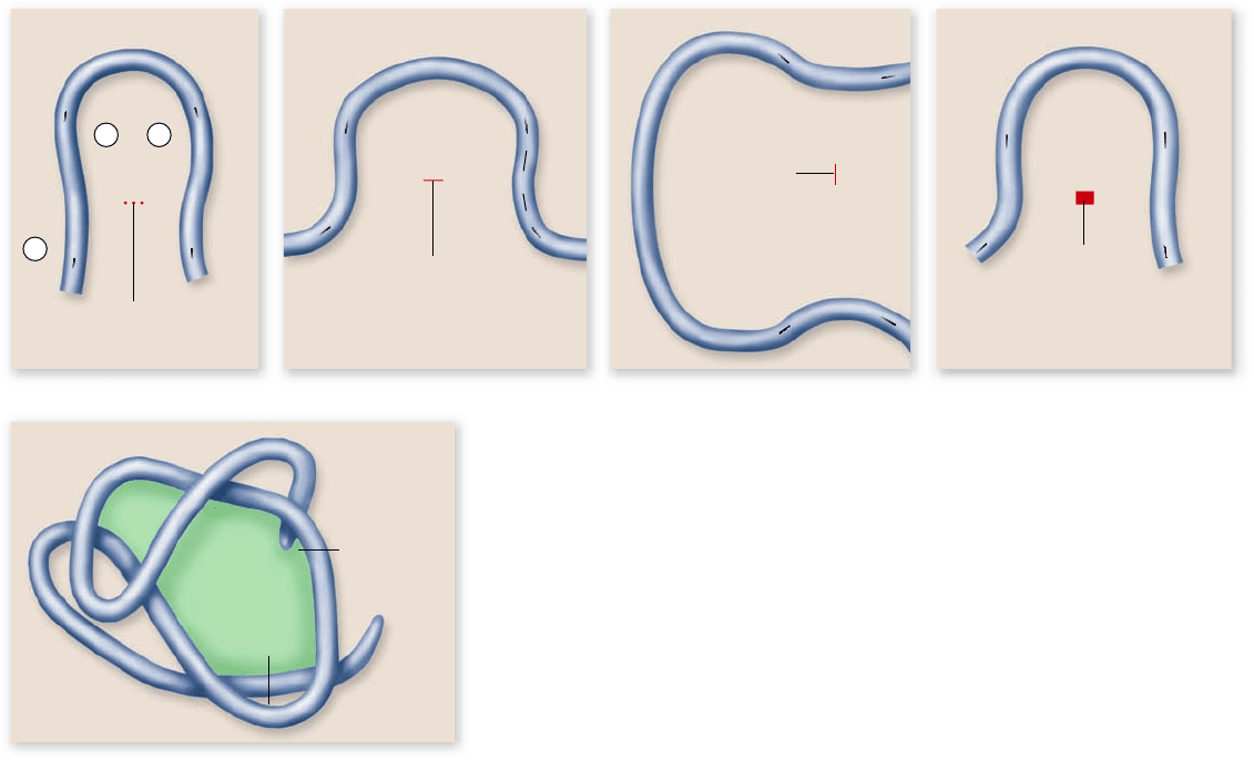
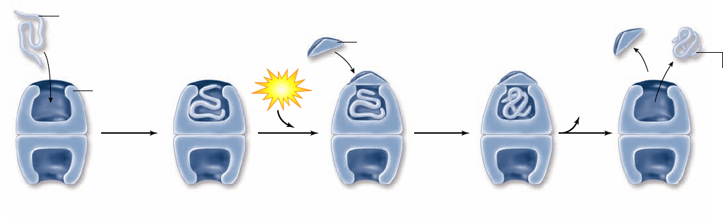
Figure 3.24
How one type of chaperone protein works. This barrel-shaped chaperonin is from the GroE family of chaperone
proteins. It is composed of two identical rings each with seven identical subunits, each of which has three distinct domains. An incorrectly
folded protein enters one chamber of the barrel, and a cap seals the chamber. Energy from the hydrolysis of ATP fuels structural alterations
to the chamber, changing it from hydrophobic to hydrophilic. This change allows the protein to refold. After a short time, the protein is
ejected, either folded or unfolded, and the cycle can repeat itself.
chapter
3
The Chemical Building Blocks of Life
51www.ravenbiology.com
rav32223_ch03_033-058.indd 51rav32223_ch03_033-058.indd 51 11/6/09 12:43:44 PM11/6/09 12:43:44 PM