Raven P.H., Johnson G.B., Mason K.A. Biology (Ninth Edition)
Подождите немного. Документ загружается.

Apago PDF Enhancer
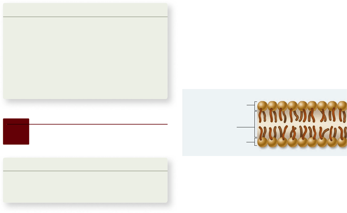
Polar hydrophilic heads
Polar hydrophilic heads
Nonpolar
hydrophobic tails
Extracellular fluid
Intracellular fluid (cytosol)
in a medium and quick frozen with liquid nitrogen. The frozen
tissue is then “tapped” with a knife, causing a crack between the
phospholipid layers of membranes. Proteins, carbohydrates,
pits, pores, channels, or any other structure affiliated with the
membrane will pull apart (whole, usually) and stick with one or
the other side of the split membrane.
Next, a very thin coating of platinum is evaporated onto
the fractured surface, forming a replica or “cast” of the surface.
After the topography of the membrane has been preserved
in the cast, the actual tissue is dissolved away, and the cast is
examined with electron microscopy, creating a textured and
three-dimensional view of the membrane.
Learning Outcomes Review 5.1
Cellular membranes contain four components: (1) a phospholipid bilayer,
(2) transmembrane proteins, (3) an internal protein network providing
structural support, and (4) cell-surface markers composed of glycoproteins
and glycolipids. The fl uid mosaic model of membrane structure includes
both the fl uid nature of the membrane and the mosaic composition
of proteins fl oating in the phospholipid bilayer. Transmission electron
microscopy (TEM) and scanning electron microscopy (SEM) have provided
evidence supporting the fl uid mosaic model.
■ If the plasma membrane were just a phospholipid
bilayer, how would this affect its function?
Phospholipids spontaneously form bilayers
The phosphate groups are charged, and other molecules at-
tached to them are polar or charged. This creates a huge change
in the molecule’s physical properties compared with a triglycer-
ide. The strongly polar phosphate end is hydrophilic, or “water-
loving,” while the fatty acid end is strongly nonpolar and
hydrophobic, or “water-fearing.” The two nonpolar fatty acids
extend in one direction, roughly parallel to each other, and the
polar phosphate group points in the other direction. To repre-
sent this structure, phospholipids are often diagrammed as a po-
lar head with two dangling nonpolar tails, as in figure 5.1c.
What happens when a collection of phospholipid mole-
cules is placed in water? The polar water molecules repel the
long, nonpolar tails of the phospholipids while seeking partners
for hydrogen bonding. Because of the polar nature of the water
molecules, the nonpolar tails of the phospholipids end up packed
closely together, sequestered as far as possible from water. Every
phospholipid molecule is oriented with its polar head toward wa-
ter and its nonpolar tails away. When two layers form with the
tails facing each other, no tails ever come in contact with water.
The resulting structure is the phospholipid bilayer. Phospholipid
bilayers form spontaneously, driven by the tendency of water
molecules to form the maximum number of hydrogen bonds.
The nonpolar interior of a lipid bilayer impedes the pas-
sage of any water-soluble substances through the bilayer, just as
a layer of oil impedes the passage of a drop of water. This bar-
rier to water-soluble substances is the key biological property
of the lipid bilayer.
The phospholipid bilayer is uid
A lipid bilayer is stable because water’s affinity for hydrogen
bonding never stops. Just as surface tension holds a soap bubble
together, even though it is made of a liquid, so the hydrogen
bonding of water holds a membrane together. Although water
continually drives phospholipid molecules into the bilayer con-
figuration, it does not have any effect on the mobility of phos-
pholipids relative to their lipid and nonlipid neighbors in the
bilayer. Because phospholipids interact relatively weakly with
one another, individual phospholipids and unanchored proteins
are comparatively free to move about within the membrane.
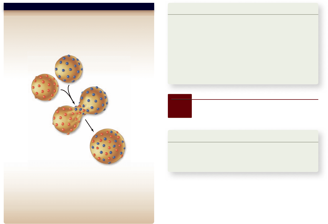
This can be demonstrated vividly by fusing cells and watching
their proteins intermix with time (figure 5.4).
Membrane uidity can change
The degree of membrane fluidity changes with the composi-
tion of the membrane itself. Much like triglycerides can be solid
or liquid at room temperature, depending on their fatty acid
5.2
Phospholipids: The
Membrane’s Foundation
Learning Outcomes
1. List the different components of phospholipids.
2. Explain how membranes form spontaneously.
Describe the factors involved in membrane fluidity.3.
Like the fat molecules (triglycerides) described in chapter 3 , a
phospholipid has a backbone derived from the three-carbon
polyalcohol glycerol. Attached to this backbone are one to three
fatty acids, long chains of carbon atoms ending in a car boxyl
(
–
COOH) group. A triglyceride molecule has three such
chains, one attached to each carbon in the backbone. Because
these chains are nonpolar, they do not form hydrogen bonds
with water, and triglycerides are not water-soluble.
A phospholipid, by contrast, has only two fatty acid chains
attached to its backbone. The third carbon of the glycerol car-
ries a phosphate group, thus the name phospholipid. An addi-
tional polar organic molecule is often added to the phosphate
group as well.
From this simple molecular framework, a large variety of
lipids can be constructed by varying the polar organic group
attached to the phosphate and the fatty acid chains attached to
the glycerol. Mammalian membranes, for example, contain
hundreds of chemically distinct species of lipids.
92
part
II
Biology of the Cell
rav32223_ch05_088-106.indd 92rav32223_ch05_088-106.indd 92 11/6/09 12:01:53 PM11/6/09 12:01:53 PM
Apago PDF Enhancer
Hypothesis: The plasma membrane is fluid, not rigid.
Prediction: If the membrane is fluid, membrane proteins may
diffuse laterally.
Test: Fuse mouse and human cells, then observe the distribution
of membrane proteins over time by labeling specific mouse and
human proteins.
Result: Over time, hybrid cells show increasingly intermixed proteins.
Conclusion: At least some membrane proteins can diffuse laterally in
the membrane.
Further Experiments: Can you think of any other explanation for these
observations? What if newly synthesized proteins were inserted into the
membrane during the experiment? How could you use this basic
experimental design to rule out this or other possible explanations?
SCIENTIFIC THINKING
Mouse
cell
Human
cell
Fuse
cells
Intermixed
membrane
proteins
Allow time for
mixing to occur
Figure 5.4
Test of membrane uidity.
composition, membrane fluidity can be altered by changing the
membrane’s fatty acid composition.
Saturated fats tend to make the membrane less fluid be-
cause they pack together well. Unsaturated fats make the mem-
brane more fluid—the “kinks” introduced by the double bonds
keep them from packing tightly. You saw this effect on fats and
oils earlier in chapter 3 . Most membranes also contain sterols
such as cholesterol, which can either increase or decrease mem-
brane fluidity, depending on the temperature.
Changes in the environment can have drastic effects on
the membranes of single-celled organisms such as bacteria. In-
creasing temperature makes a membrane more fluid, and de-
creasing temperature makes it less fluid. Bacteria have evolved
mechanisms to maintain a constant membrane fluidity despite
fluctuating temperatures. Some bacteria contain enzymes called
fatty acid desaturases that can introduce double bonds into fatty
acids in membranes. Genetic studies, involving either the inac-
tivation of these enzymes or the introduction of them into cells
that normally lack them, indicate that the action of these en-
zymes confers cold tolerance. At colder temperatures, the dou-
ble bonds introduced by fatty acid desaturase make the
membrane more fluid, counteracting the environmental effect
of reduced temperature.
Learning Outcomes Review 5.2
Biological membranes consist of a phospholipid bilayer. Each phospholipid
has a hydrophilic (phosphate) head and a hydrophobic (lipid) tail. In water,
phospholipid molecules spontaneously form a bilayer, with phosphate
groups facing out toward the water and lipid tails facing in, where they are
sequestered from water. Membrane fl uidity varies with composition and
conditions: unsaturated fats disturb packing of the lipid tails and make the
membrane more fl uid, as do higher temperatures.
■ Would a phospholipid bilayer form in a nonpolar
solvent?
5.3
Proteins: Multifunctional
Components
Learning Outcomes
1. List the functions of membrane proteins.
2. Explain how proteins can associate with the membrane.
Identify a transmembrane domain.3.
Cell membranes contain a complex assembly of proteins en-
meshed in the fluid soup of phospholipid molecules. This very
flexible organization permits a broad range of interactions with
the environment, some directly involving membrane proteins.
Proteins and protein complexes
perform key functions
Although cells interact with their environment through their
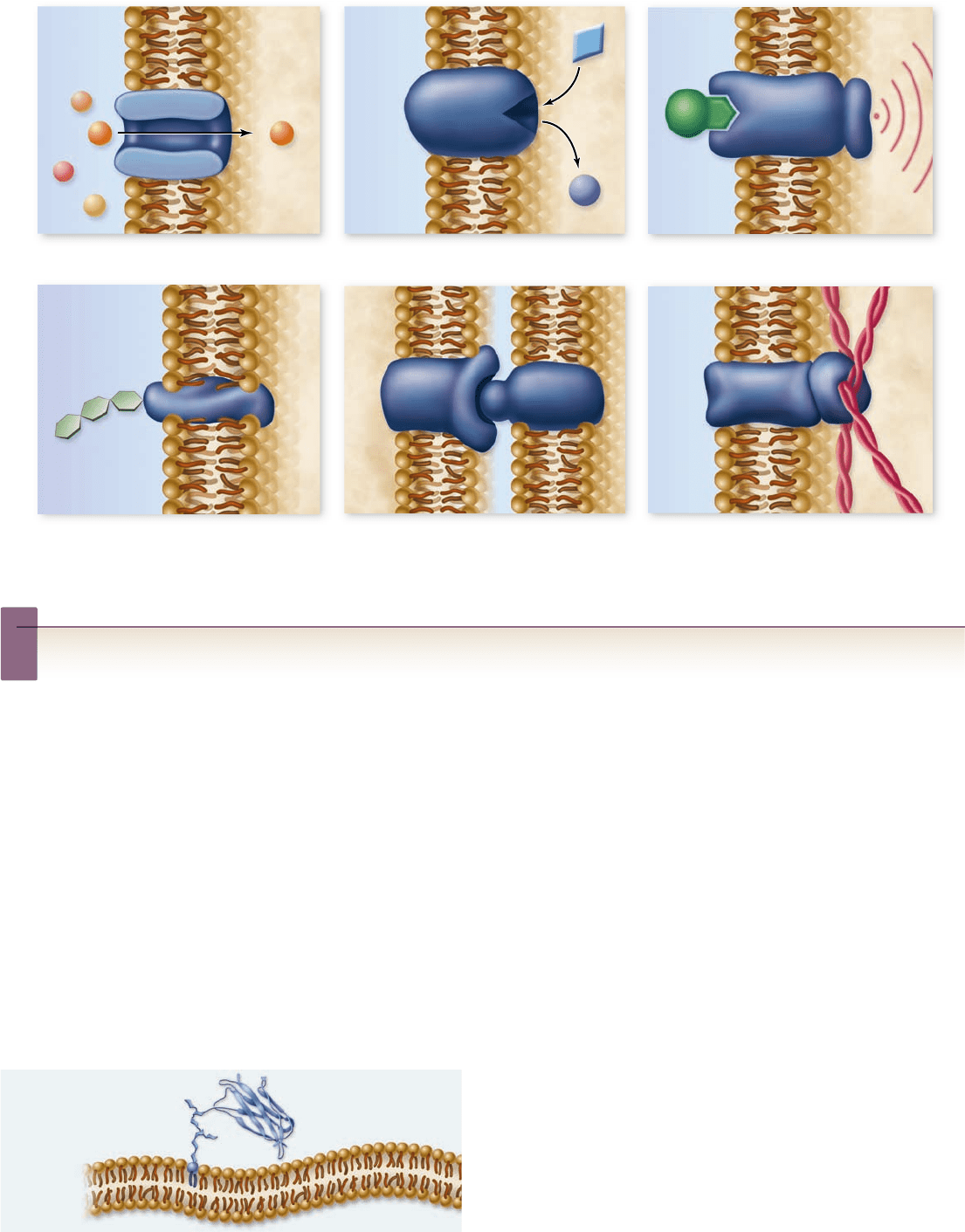
plasma membranes in many ways, we will focus on six key classes
of membrane protein in this chapter and in chapter 9 (figure 5.5).
Transporters.1. Membranes are very selective, allowing
only certain solutes to enter or leave the cell, either
through channels or carriers composed of proteins.
Enzymes.2. Cells carry out many chemical reactions on the
interior surface of the plasma membrane, using enzymes
attached to the membrane.
Cell-surface receptors.3. Membranes are exquisitely
sensitive to chemical messages, which are detected by
receptor proteins on their surfaces.
Cell-surface identity markers.4. Membranes carry
cell-surface markers that identify them to other cells. Most
cell types carry their own ID tags, speci c combinations of
cell-surface proteins and protein complexes such as
glycoproteins that are characteristic of that cell type.
Cell-to-cell adhesion proteins.5. Cells use speci c
proteins to glue themselves to one another. Some act by
forming temporary interactions, and others form a more
permanent bond. (See chapter 9 . )
Attachments to the cytoskeleton.6. Surface proteins that
interact with other cells are often anchored to the
cytoskeleton by linking proteins.
chapter
5
Membranes
93www.ravenbiology.com
rav32223_ch05_088-106.indd 93rav32223_ch05_088-106.indd 93 11/6/09 12:01:54 PM11/6/09 12:01:54 PM
Apago PDF Enhancer
Outside
cell
Inside
cell
Transporter Enzyme Cell-surface receptor
Cell-surface identity marker Cell-to-cell adhesion Attachment to the cytoskeleton
Protein anchored to
phospholipid
Figure 5.5
Functions of plasma membrane proteins. Membrane proteins act as transporters, enzymes, cell-surface receptors, and
cell-surface identity markers, as well as aiding in cell-to-cell adhesion and securing the cytoskeleton.
Inquiry question
?
According to the fluid mosaic model, membranes are held together by hydrophobic interactions. Considering the forces that some cells
may experience, why do membranes not break apart every time an animal moves?
Structural features of membrane
proteins relate to function
As we’ve just detailed, membrane proteins can serve a variety of
functions. These diverse functions arise from the diverse struc-
tures of these proteins, yet they also have common structural
features related to their role as membrane proteins.
The anchoring of proteins in the bilayer
Some membrane proteins are attached to the surface of the
membrane by special molecules that associate strongly with
phospholipids. Like a ship tied to a floating dock, these anchored
proteins are free to move about on the surface of the membrane
tethered to a phospholipid. The anchoring molecules are modi-
fied lipids that have (1) nonpolar regions that insert into the in-
ternal portion of the lipid bilayer and (2) chemical bonding
domains that link directly to proteins.
In contrast, other proteins actually span the lipid bilayer
(transmembrane proteins). The part of the protein that extends
through the lipid bilayer and that is in contact with the nonpolar
interior are α -helices or β-pleated sheets (see chapter 3 ) that con-
sist of nonpolar amino acids . Because water avoids nonpolar amino
acids, these portions of the protein are held within the interior of
the lipid bilayer. The polar ends protrude from both sides of the
membrane. Any movement of the protein out of the membrane, in
either direction, brings the nonpolar regions of the protein into
contact with water, which “shoves” the protein back into the inte-
rior. These forces prevent the transmembrane proteins from sim-
ply popping out of the membrane and floating away.
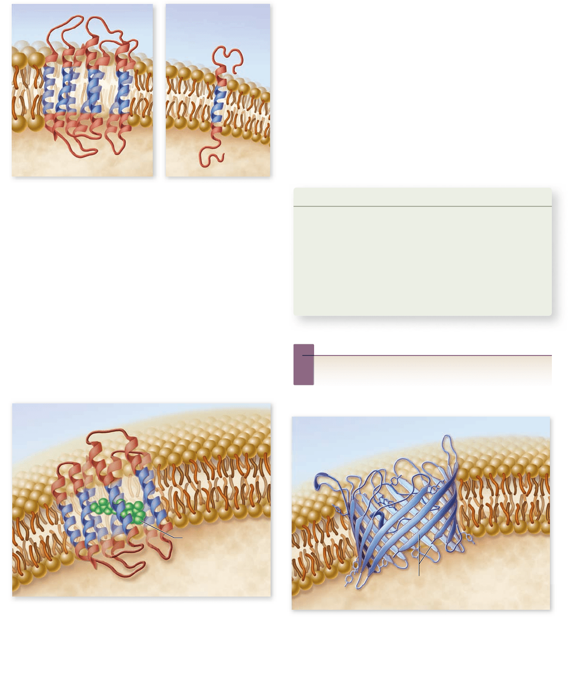
Transmembrane domains
Cell membranes contain a variety of different transmembrane pro-
teins, which differ in the way they traverse the lipid bilayer. The
primary difference lies in the number of times that the protein
crosses the membrane. Each membrane-spanning region is called a
transmembrane domain. These domains are composed of hydro-
phobic amino acids usually arranged into α helices (figure 5.6).
Proteins need only a single transmembrane domain to be
anchored in the membrane, but they often have more than one
such domain. An example of a protein with a single transmem-
brane domain is the linking protein that attaches the spectrin net-
work of the cytoskeleton to the interior of the plasma membrane.
94
part
II
Biology of the Cell
rav32223_ch05_088-106.indd 94rav32223_ch05_088-106.indd 94 11/6/09 12:01:55 PM11/6/09 12:01:55 PM
Apago PDF Enhancer
a. b.
Retinal chromophore
b-pleated sheets
Figure 5.7
Bacteriorhodopsin. This transmembrane
protein mediates photosynthesis in the archaean Halobacterium
salinarium. The protein traverses the membrane seven times with
hydrophobic helical strands that are within the hydrophobic center
of the lipid bilayer. The helical regions form a structure across the
bilayer through which protons are pumped by the retinal
chromophore (green) using energy from light.
Figure 5.6
Transmembrane domains. Integral membrane
proteins have at least one hydrophobic transmembrane domain
(shown in blue) to anchor them in the membrane. a. Receptor
protein with seven transmembrane domains. b. Protein with
single transmembrane domain.
Figure 5.8
A pore protein. The bacterial transmembrane
protein porin creates large open tunnels called pores in the outer
membrane of a bacterium. Sixteen strands of β-pleated sheets run
antiparallel to one another, creating a so-called β barrel in the
bacterial outer cell membrane. The tunnel allows water and other
materials to pass through the membrane.
Inquiry question
?
Based only on amino acid sequence, how would you
recognize an integral membrane protein?
Biologists classify some types of receptors based on the
number of transmembrane domains they have, such as
G protein–coupled receptors with seven membrane-span-
ning domains (chapter 9 ) . These receptors respond to exter-
nal molecules, such as epinephrine, and initiate a cascade of
events inside the cell.
Another example is bacteriorhodopsin, one of the key
transmembrane proteins that carries out photosynthesis in
halophilic (salt-loving) archaea. It contains seven nonpolar
helical segments that traverse the membrane, forming a struc-
ture within the membrane through which protons pass during
the light-driven pumping of protons (figure 5.7).
Pores
Some transmembrane proteins have extensive nonpolar regions
with secondary configurations of β-pleated sheets instead of
α helices (chapter 3 ) . The β sheets form a characteristic motif,
folding back and forth in a cylinder so the sheets arrange them-
selves like a pipe through the membrane. This forms a polar
environment in the interior of the β sheets spanning the mem-
brane. This so-called β barrel, open on both ends, is a common
feature of the porin class of proteins that are found within the
outer membrane of some bacteria. The openings allow mole-
cules to pass through the membrane (figure 5.8).
Learning Outcomes Review 5.3
Proteins in the membrane confer the main diff erences between
membranes of diff erent cells. Their functions include transport, enzymatic
action, reception of extracellular signals, cell-to-cell interactions, and cell
identity markers. Peripheral proteins can be anchored in the membrane by
modifi ed lipids. Integral membrane proteins span the membrane and have
one or more hydrophobic regions, called transmembrane domains, that
anchor them.
■ Why are transmembrane domains hydrophobic?
chapter
5
Membranes
95www.ravenbiology.com
rav32223_ch05_088-106.indd 95rav32223_ch05_088-106.indd 95 11/6/09 12:01:58 PM11/6/09 12:01:58 PM
Apago PDF Enhancer
a. b. c. d.
Figure 5.9
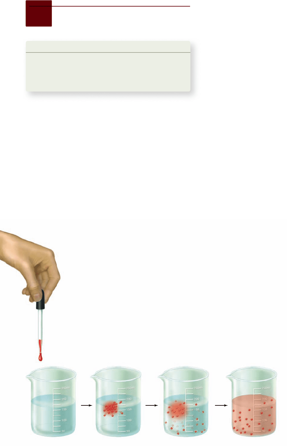
Di usion. If a drop of colored ink is
dropped into a beaker of water (a) its molecules
dissolve (b) and diffuse (c). Eventually, diffusion
results in an even distribution of ink molecules
throughout the water (d).
5.4
Passive Transport Across
Membranes
Learning Outcomes
1. Compare simple diffusion and facilitated diffusion.
2. Differentiate between channel proteins and
carrier proteins.
Explain the movement of water by osmosis.3.
Many substances can move in and out of the cell without the
cell’s having to expend energy. This type of movement is termed
passive transport. Some ions and molecules can pass through
the membrane fairly easily and do so because of a concentration
gradient —a difference between the concentration on the inside
of the membrane and that on the outside. Some substances also
move in response to a gradient, but do so through specific chan-
nels formed by proteins in the membrane.
Transport can occur by simple di usion
Molecules and ions dissolved in water are in constant random
motion. This random motion causes a net movement of these
substances from regions of high concentration to regions of
lower concentration, a process called diffusion (figure 5.9).
Net movement driven by diffusion will continue until the
concentration is the same in all regions. Consider what happens
when you add a drop of colored ink to a bowl of water. Over
time the ink becomes dispersed throughout the solu-
tion. This is due to diffusion of the ink molecules.
In the context of cells, we are usually concerned
with differences in concentration of molecules
across the plasma membrane. We need to con-
sider the relative concentrations both inside
and outside the cell, as well as how readily a
molecule can cross the membrane.
The major barrier to crossing a biological membrane is
the hydrophobic interior that repels polar molecules but not
nonpolar molecules. If a concentration difference exists for a
nonpolar molecule, it will move across the membrane until the
concentration is equal on both sides. At this point, movement
in both directions still occurs, but there is no net change in ei-
ther direction. This includes molecules like O
2
and nonpolar
organic molecules such as steroid hormones.
The plasma membrane has limited permeability to small
polar molecules and very limited permeability to larger polar mol-
ecules and ions. The movement of water, one of the most impor-
tant polar molecules, is discussed in its own section later on.
Proteins allow membrane di usion
to be selective
Many important molecules required by cells cannot easily
cross the plasma membrane. These molecules can still enter
the cell by diffusion through specific channel proteins or car-
rier proteins embedded in the plasma membrane, provided
there is a higher concentration of the molecule outside the cell
than inside. We call this process of diffusion mediated by a
membrane protein facilitated diffusion. Channel proteins
have a hydrophilic interior that provides an aqueous channel
through which polar molecules can pass when the channel is
open. Carrier proteins, in contrast to channels, bind specifi-
cally to the molecule they assist, much like an enzyme binds to
its substrate. These channels and carriers are usually selective
for one type of molecule, and thus the cell membrane is said to
be selectively permeable.
Facilitated diffusion of ions through channels
You saw in chapter 2 that atoms with an unequal number of
protons and electrons have an electric charge and are called
ions. Those that carry a positive charge are called cations and
those that carry a negative charge are called anions.
Because of their charge, ions interact well with polar
molecules such as water, but are repelled by nonpolar mole-
cules such as the interior of the plasma membrane. There-
fore, ions cannot move between the cytoplasm of a cell and
the extracellular fluid without the assistance of membrane
transport proteins.
Ion channels possess a hydrated interior that spans the
membrane. Ions can diffuse through the channel in either direc-
tion, depending on their relative concentration across the mem-
brane (figure 5.10). Some channel proteins can be opened or closed
in response to a stimulus. These
channels are called gated channels ,
and depending on the nature of
the channel, the stimulus can be
either chemical or electrical.
Three conditions determine
the direction of net movement
of the ions: (1) their relative
concentrations on either side of
the membrane, (2) the voltage
difference across the membrane
and for the gated channels, and
96
part
II
Biology of the Cell
rav32223_ch05_088-106.indd 96rav32223_ch05_088-106.indd 96 11/6/09 12:02:04 PM11/6/09 12:02:04 PM
Apago PDF Enhancer
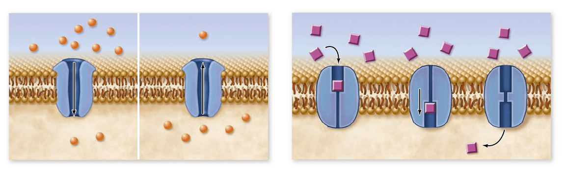
Extracellular fluid Extracellular fluid
Cytoplasm
a.
b.
Cytoplasm
Extracellular fluid
Cytoplasm
Figure 5.10
Facilitated di usion. Diffusion can be facilitated by membrane proteins. a. The movement of ions through a channel is
shown. On the left the concentration is higher outside the cell, so the ions move into the cell. On the right the situation is reversed. In both
cases, transport continues until the concentration is equal on both sides of the membrane. At this point, ions continue to cross the membrane in
both directions, but there is no net movement in either direction. b. Carrier proteins bind speci cally to the molecules they transport. In this
case, the concentration is higher outside the cell, so molecules bind to the carrier on the outside. The carrier’s shape changes, allowing the
molecule to cross the membrane. This is reversible, so net movement continues until the concentration is equal on both sides of the membrane.
(3) the state of the gate (open or closed). A voltage difference is
an electrical potential difference across the membrane called a
membrane potential. Changes in membrane potential form the
basis for transmission of signals in the nervous system and some
other tissues. (We discuss this topic in detail in chapter 45. )
Each type of channel is specific for a particular ion, such as
calcium (Ca
2+
), sodium (Na
+
), potassium (K
+
), or chloride (Cl
–
),
or in some cases, for more than one cation or anion. Ion chan-
nels play an essential role in signaling by the nervous system.
Facilitated diffusion by carrier proteins
Carrier proteins can help transport both ions and other solutes,
such as some sugars and amino acids, across the membrane.
Transport through a carrier is still a form of diffusion and there-
fore requires a concentration difference across the membrane.
Carriers must bind to the molecule they transport, so the
relationship between concentration and rate of transport dif-
fers from that due to simple diffusion. As concentration in-
creases, transport by simple diffusion shows a linear increase in
rate of transport. But when a carrier protein is involved, a con-
centration increase means that more of the carriers are bound
to the transported molecule. At high enough concentrations all
carriers will be occupied, and the rate of transport will be con-
stant. This means that the carrier exhibits saturation .
This situation is somewhat like that of a stadium (the cell)
where a crowd must pass through turnstiles to enter. If there are
unoccupied turnstiles, you can go right through, but when all are
occupied, you must wait. When ticket holders are passing through
the gates at maximum speed, the rate at which they enter cannot
increase, no matter how many are waiting outside.
Facilitated diffusion in red blood cells
Several examples of facilitated diffusion can be found in the
plasma membrane of vertebrate red blood cells (RBCs). One
RBC carrier protein, for example, transports a different mole-
cule in each direction: chloride ion (Cl
–
) in one direction and
bicarbonate ion (HCO
3
–
) in the opposite direction. As you will
learn in chapter 51 , this carrier is important in the uptake and
release of carbon dioxide.
The glucose transporter is a second vital facilitated diffu-
sion carrier in RBCs. Red blood cells keep their internal con-
centration of glucose low through a chemical trick: They
immediately add a phosphate group to any entering glucose
molecule, converting it to a highly charged glucose phosphate
that can no longer bind to the glucose transporter, and there-
fore cannot pass back across the membrane. This maintains a
steep concentration gradient for unphosphorylated glucose, fa-
voring its entry into the cell.
The glucose transporter that assists the entry of glucose
into the cell does not appear to form a channel in the mem-
brane. Instead, this transmembrane protein appears to bind to
a glucose molecule and then to flip its shape, dragging the glu-
cose through the bilayer and releasing it on the inside of the
plasma membrane. After it releases the glucose, the transporter
reverts to its original shape and is then available to bind the
next glucose molecule that comes along outside the cell.
Osmosis is the movement
of water across membranes
The cytoplasm of a cell contains ions and molecules, such as
sugars and amino acids, dissolved in water. The mixture of these
substances and water is called an aqueous solution. Water is
termed the solvent, and the substances dissolved in the water
are solutes. Both water and solutes tend to diffuse from re-
gions of high concentration to ones of low concentration; that
is, they diffuse down their concentration gradients.
When two regions are separated by a membrane, what
happens depends on whether the solutes can pass freely through
that membrane. Most solutes, including ions and sugars, are
not lipid-soluble and, therefore, are unable to cross the lipid
bilayer. The concentration gradient of these solutes can lead to
the movement of water.
chapter
5
Membranes
97www.ravenbiology.com
rav32223_ch05_088-106.indd 97rav32223_ch05_088-106.indd 97 11/6/09 12:02:05 PM11/6/09 12:02:05 PM
Apago PDF Enhancer
Semipermeable
membrane
Urea molecule
Water
molecules
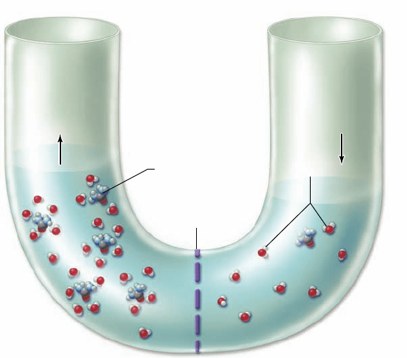
Figure 5.11
Osmosis. Concentration differences in charged
or polar molecules that cannot cross a semipermeable membrane
result in movement of water, which can cross the membrane. Water
molecules form hydrogen bonds with charged or polar molecules
creating a hydration shell around them in solution. A higher
concentration of polar molecules (urea) shown on the left side of
the membrane leads to water molecules gathering around each urea
molecule. These water molecules are no longer free to diffuse
across the membrane. The polar solute has reduced the
concentration of free water molecules, creating a gradient. This
causes a net movement of water by diffusion from right to left in
the U -tube, raising the level on the left and lowering the level on
the right.
Osmosis
Water molecules interact with dissolved solutes by forming hy-
dration shells around the charged solute molecules. When a
membrane separates two solutions with different concentra-
tions of solutes, the concentrations of free water molecules on
the two sides of the membrane also differ. The side with higher
solute concentration has tied up more water molecules in hy-
dration shells and thus has fewer free water molecules.
As a consequence of this difference, free water molecules
move down their concentration gradient, toward the higher
solute concentration. This net diffusion of water across a mem-
brane toward a higher solute concentration is called osmosis
(figure 5.11).
The concentration of all solutes in a solution determines
the osmotic concentration of the solution. If two solutions
have unequal osmotic concentrations, the solution with the
higher concentration is hypertonic (Greek hyper, “more than”),
and the solution with the lower concentration is hypotonic
(Greek hypo, “less than”). When two solutions have the same
osmotic concentration, the solutions are isotonic (Greek iso,
“equal”). The terms hyperosmotic, hypoosmotic, and isosmotic are
also used to describe these conditions.
A cell in any environment can be thought of as a plasma
membrane separating two solutions: the cytoplasm and the ex-
tracellular fluid. The direction and extent of any diffusion of
water across the plasma membrane is determined by comparing
the osmotic strength of these solutions. Put another way, water
diffuses out of a cell in a hypertonic solution (that is, the cyto-
plasm of the cell is hypotonic, compared with the extracellular
fluid). This loss of water causes the cell to shrink until the os-
motic concentrations of the cytoplasm and the extracellular
fluid become equal.
Aquaporins: Water channels
The transport of water across the membrane is complex. Stud-
ies on artificial membranes show that water, despite its polarity,
can cross the membrane, but this flow is limited. Water flow in
living cells is facilitated by aquaporins, which are specialized
channels for water.
A simple experiment demonstrates this. If an amphibian
egg is placed in hypotonic spring water (the solute concentra-
tion in the cell is higher than that of the surrounding water), it
does not swell. If aquaporin mRNA is then injected into the
egg, the channel proteins are expressed and appear in the egg’s
plasma membrane. Water can now diffuse into the egg, causing
it to swell.
More than 11 different kinds of aquaporins have been
found in mammals. These fall into two general classes: those
that are specific for only water, and those that allow other small
hydrophilic molecules, such as glycerol or urea, to cross the
membrane as well. This latter class explains how some mem-
branes allow the easy passage of small hydrophilic substances.
The human genetic disease, hereditary (nephrogenic) di-
abetes insipidus (NDI), has been shown to be caused by a non-
functional aquaporin protein. This disease causes the excretion
of large volumes of dilute urine, illustrating the importance of
aquaporins to our physiology.
Osmotic pressure
What happens to a cell in a hypotonic solution? (That is, the
cell’s cytoplasm is hypertonic relative to the extracellular fluid.)
In this situation, water diffuses into the cell from the extracel-
lular fluid, causing the cell to swell. The pressure of the cyto-
plasm pushing out against the cell membrane, or hydrostatic
pressure, increases. The amount of water that enters the cell
depends on the difference in solute concentration between the
cell and the extracellular fluid. This is measured as osmotic
pressure, defined as the force needed to stop osmotic flow.
If the membrane is strong enough, the cell reaches an
equilibrium, at which the osmotic pressure, which tends to
drive water into the cell, is exactly counterbalanced by the hy-
drostatic pressure, which tends to drive water back out of the
cell. However, a plasma membrane by itself cannot withstand
large internal pressures, and an isolated cell under such condi-
tions would burst like an overinflated balloon (figure 5.12).
Accordingly, it is important for animal cells, which only
have plasma membranes, to maintain osmotic balance. In con-
trast, the cells of prokaryotes, fungi, plants, and many protists
are surrounded by strong cell walls, which can withstand high
internal pressures without bursting.
98
part
II
Biology of the Cell
rav32223_ch05_088-106.indd 98rav32223_ch05_088-106.indd 98 11/6/09 12:02:11 PM11/6/09 12:02:11 PM
Apago PDF Enhancer
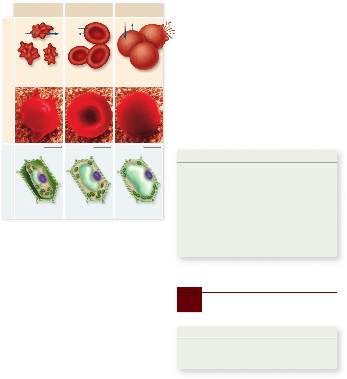
Hypertonic
Solution
Isotonic
Solution
Hypotonic
Solution
Human Red Blood Cells Plant Cells
Shriveled cells Normal cells
Cells swell and
eventually burst
Cell body shrinks
from cell wall
Flaccid cell Normal turgid cell
0.55 µm 0.55 µm 0.55 µm
Figure 5.12
How solutes create osmotic pressure. In a
hypertonic solution, water moves out of the cell, causing the cell to
shrivel. In an isotonic solution, water diffuses into and out of the cell
at the same rate, with no change in cell size. In a hypotonic solution,
water moves into the cell. Direction and amount of water movement
is shown with blue arrows (top). As water enters the cell from a
hypotonic solution, pressure is applied to the plasma membrane until
the cell ruptures. Water enters the cell due to osmotic pressure from
the higher solute concentration in the cell. Osmotic pressure is
measured as the force needed to stop osmosis. The strong cell wall of
plant cells can withstand the hydrostatic pressure to keep the cell
from rupturing. This is not the case with animal cells.
Maintaining osmotic balance
Organisms have developed many strategies for solving the di-
lemma posed by being hypertonic to their environment and
therefore having a steady influx of water by osmosis.
Extrusion. Some single-celled eukaryotes, such as the protist
Paramecium, use organelles called contractile vacuoles to
remove water. Each vacuole collects water from various
parts of the cytoplasm and transports it to the central part
of the vacuole, near the cell surface. The vacuole possesses
a small pore that opens to the outside of the cell. By
contracting rhythmically, the vacuole pumps out
(extrudes) through this pore the water that is
continuously drawn into the cell by osmotic forces.
Isosmotic Regulation. Some organisms that live in the ocean
adjust their internal concentration of solutes to match
that of the surrounding seawater. Because they are
isosmotic with respect to their environment, no net ow
of water occurs into or out of these cells.
Many terrestrial animals solve the problem in a
similar way, by circulating a uid through their bodies that
bathes cells in an isotonic solution. The blood in your body,
for example, contains a high concentration of the protein
albumin, which elevates the solute concentration of the
blood to match that of your cells’ cytoplasm.
Turgor. Most plant cells are hypertonic to their immediate
environment, containing a high concentration of solutes
in their central vacuoles. The resulting internal
hydrostatic pressure, known as turgor pressure, presses
the plasma membrane rmly against the interior of the
cell wall, making the cell rigid. Most green plants depend
on turgor pressure to maintain their shape, and thus they
wilt when they lack suf cient water.
Learning Outcomes Review 5.4
Passive transport involves diff usion, which requires a concentration
gradient. Hydrophobic molecules can diff use directly through the membrane
(simple diff usion). Polar molecules and ions can also diff use through the
membrane, but only with the aid of a channel or carrier protein (facilitated
diff usion). Channel proteins assist by forming a hydrophilic passageway
through the membrane, whereas carrier proteins bind to the molecule they
assist. Water passes through the membrane and through aquaporins in
response to solute concentration diff erences inside and outside the cell. This
process is called osmosis.
■ If you require intravenous (IV) medication in the
hospital, what should the concentration of solutes in the
IV solution be relative to your blood cells?
5.5
Active Transport Across
Membranes
Learning Outcomes
1. Differentiate between active transport and diffusion.
2. Describe the function of the Na
+
/K
+
pump.
Explain the energetics of coupled transport.3.
Diffusion, facilitated diffusion, and osmosis are passive trans-
port processes that move materials down their concentration
gradients, but cells can also actively move substances across a
cell membrane up their concentration gradients. This process
requires the expenditure of energy, typically from ATP, and is
therefore called active transport.
chapter
5
Membranes
99www.ravenbiology.com
rav32223_ch05_088-106.indd 99rav32223_ch05_088-106.indd 99 11/6/09 12:02:13 PM11/6/09 12:02:13 PM
Apago PDF Enhancer
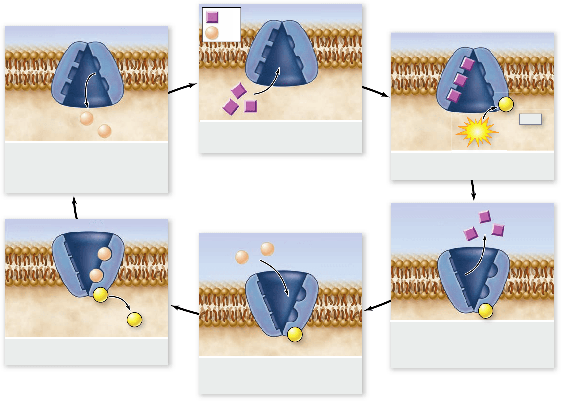
1. Carrier in membrane binds
intracellular sodium.
2. ATP phosphorylates protein with
bound sodium.
3. Phosphorylation causes
conformational change in protein,
reducing its affinity for Na
;
. The Na
;
then diffuses out.
4. This conformation has higher affinity
for K
;
. Extracellular potassium binds
to exposed sites.
5. Binding of potassium causes
dephosphorylation of protein.
6. Dephosphorylation of protein triggers
change back to original conformation,
with low affinity for K
;
. K
;
diffuses into
the cell, and the cycle repeats.
Extracellular
Intracellular
ATP
ADP
P
P
P
P
P
+
Na
;
K
;
Figure 5.13
The sodium–potassium pump. The protein carrier known as the sodium–potassium pump transports sodium (Na
+
) and
potassium (K
+
) across the plasma membrane. For every three Na
+
transported out of the cell, two K
+
are transported into it. The sodium–
potassium pump is fueled by ATP hydrolysis. The af nity of the pump for Na
+
and K
+
is changed by adding or removing phosphate (P), which
changes the conformation of the protein.
Active transport uses energy to move
materials against a concentration gradient
Like facilitated diffusion, active transport involves highly selec-
tive protein carriers within the membrane that bind to the
transported substance, which could be an ion or a simple
molecule, such as a sugar, an amino acid, or a nucleotide. These
carrier proteins are called uniporters if they transport a single
type of molecule and symporters or antiporters if they trans-
port two different molecules together. Symporters transport
two molecules in the same direction, and antiporters transport
two molecules in opposite directions. These terms can also be
used to describe facilitated diffusion carriers.
Active transport is one of the most important functions
of any cell. It enables a cell to take up additional molecules
of a substance that is already present in its cytoplasm in con-
centrations higher than in the extracellular fluid. Active
transport also enables a cell to move substances out of its
cytoplasm and into the extracellular fluid, despite higher ex-
ternal concentrations.
The use of energy from ATP in active transport may be
direct or indirect. Let’s first consider how ATP is used directly
to move ions against their concentration gradients.
The sodium–potassium pump
runs directly on ATP
More than one-third of all of the energy expended by an animal
cell that is not actively dividing is used in the active transport of
sodium (Na
+
) and potassium (K
+
) ions. Most animal cells have a
low internal concentration of Na
+
, relative to their surround-
ings, and a high internal concentration of K
+
. They maintain
these concentration differences by actively pumping Na
+
out of
the cell and K
+
in.
100
part
II
Biology of the Cell
rav32223_ch05_088-106.indd 100rav32223_ch05_088-106.indd 100 11/6/09 12:02:15 PM11/6/09 12:02:15 PM
Apago PDF Enhancer
Outside
of cell
Inside
of cell
Na
;
/ K
;
pump
Na
;
Glucose
Coupled
transport
protein
K
;
ADP+P
i
ATP
The remarkable protein that transports these two ions
across the cell membrane is known as the sodium– potassium
pump (figure 5.13). This carrier protein uses the energy stored
in ATP to move these two ions. In this case, the energy is used
to change the conformation of the carrier protein, which
changes its affinity for either Na
+
ions or K
+
ions. This is an
excellent illustration of how subtle changes in the structure of a
protein affect its function.
The important characteristic of the sodium–potassium
pump is that it is an active transport mechanism, transport-
ing Na
+
and K
+
from areas of low concentration to areas of
high concentration. This transport is the opposite of passive
transport by diffusion; it is achieved only by the constant
expenditure of metabolic energy. The sodium–potassium
pump works through the following series of conformational
changes in the transmembrane protein (summarized in
figure 5.13):
Step 1. Three Na
+
bind to the cytoplasmic side of the protein,
causing the protein to change its conformation.
Step 2. In its new conformation, the protein binds a molecule
of ATP and cleaves it into adenosine diphosphate (ADP)
and phosphate (P
i
). ADP is released, but the phosphate
group is covalently linked to the protein. The protein is
now phosphorylated.
Step 3. The phosphorylation of the protein induces a second
conformational change in the protein. This change
translocates the three Na
+
across the membrane, so they
now face the exterior. In this new conformation, the
protein has a low affinity for Na
+
, and the three bound
Na
+
break away from the protein and diffuse into the
extracellular fluid.
Step 4. The new conformation has a high affinity for K
+
, two
of which bind to the extracellular side of the protein as
soon as it is free of the Na
+
.
Step 5. The binding of the K
+
causes another conformational
change in the protein, this time resulting in the
hydrolysis of the bound phosphate group.
Step 6. Freed of the phosphate group, the protein reverts to its
original shape, exposing the two K
+
to the cytoplasm.
This conformation has a low affinity for K
+
, so the two
bound K
+
dissociate from the protein and diffuse into the
interior of the cell. The original conformation has a high
affinity for Na
+
. When these ions bind, they initiate
another cycle.
In every cycle, three Na
+
leave the cell and two K
+
enter. The
changes in protein conformation that occur during the cycle
are rapid, enabling each carrier to transport as many as 300 Na
+
per second. The sodium–potassium pump appears to exist in all
animal cells, although cells vary widely in the number of pump
proteins they contain.
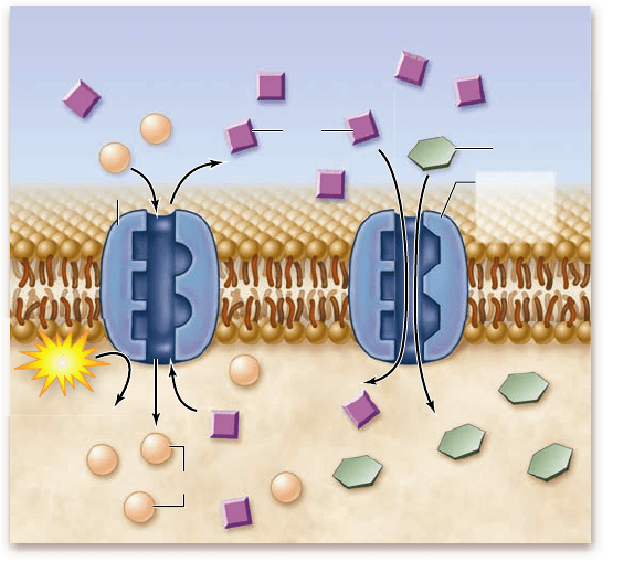
Coupled transport uses ATP indirectly
Some molecules are moved against their concentration gradi-
ent by using the energy stored in a gradient of a different mol-
ecule. In this process, called coupled transport, the energy released
as one molecule moves down its concentration gradient is cap-
tured and used to move a different molecule against its gradi-
ent. As you just saw, the energy stored in ATP molecules can be
used to create a gradient of Na
+
and K
+
across the membrane.
These gradients can then be used to power the transport of
other molecules across the membrane.
As one example, let’s consider the active transport of
glucose across the membrane in animal cells. Glucose is such
an important molecule that there are a variety of transporters
for it, one of which was discussed earlier under passive trans-
port. In a multicellular organism, intestinal epithelial cells can
have a higher concentration of glucose inside the cell than
outside, so these cells need to be able to transport glucose
against its concentration gradient. This requires energy and a
different transporter than the one involved in facilitated dif-
fusion of glucose.
The active glucose transporter uses the Na
+
gradient pro-
duced by the sodium–potassium pump as a source of energy to
power the movement of glucose into the cell. In this system,
both glucose and Na
+
bind to the transport protein, which al-
lows Na
+
to pass into the cell down its concentration gradient,
capturing the energy and using it to move glucose into the cell.
In this kind of cotransport, both molecules are moving in the
same direction across the membrane; therefore the transporter
is a symporter (figure 5.14).
Figure 5.14
Coupled transport. A membrane protein
transports Na
+
into the cell, down its concentration gradient, at the
same time it transports a glucose molecule into the cell. The
gradient driving the Na
+
entry allows sugar molecules to be
transported against their concentration gradient. The Na
+
gradient
is maintained by the Na
+
/K
+
pump. ADP = adenosine diphosphate;
ATP = adenosine triphosphate; P
i
= inorganic phosphate
chapter
5
Membranes
101www.ravenbiology.com
rav32223_ch05_088-106.indd 101rav32223_ch05_088-106.indd 101 11/6/09 12:02:21 PM11/6/09 12:02:21 PM