Raven P.H., Johnson G.B., Mason K.A. Biology (Ninth Edition)
Подождите немного. Документ загружается.


Apago PDF Enhancer
TABLE 4.4
Cell-to-Cell Connections and Cell Identity
Type of Connection Structure Function Example
Surface markers Variable, integral proteins or glycolipids in
plasma membrane
Identify the cell MHC complexes, blood groups, antibodies
Tight junctions Tightly bound, leakproof, brous protein
seal that surrounds cell
Organizing junction; holds cells together
such that materials pass through but not
between the cells
Junctions between epithelial cells in the
gut
Anchoring junction
(Desmosome)
Intermediate laments of cytoskeleton
linked to adjoining cells through cadherins
Anchoring junction; binds cells together Epithelium
Anchoring junction
(Adherens junction)
Transmembrane brous proteins Anchoring junction; connects extracellular
matrix to cytoskeleton
Tissues with high mechanical stress, such
as the skin
Communicating junction
(Gap junction)
Six transmembrane connexon proteins
creating a pore that connects cells
Communicating junction; allows passage of
small molecules from cell to cell in a tissue
Excitable tissue such as heart muscle
Communicating junction
(Plasmodesmata)
Cytoplasmic connections between gaps in
adjoining plant cell walls
Communicating junction between plant
cells
Plant tissues
Learning Outcomes Review 4.7
Cell movement involves proteins. These can either be internal in the case
of crawling cells that use actin and myosin, or external in the case of cells
powered by cilia or fl agella. Eukaryotic cilia and fl agella are diff erent from
prokaryotic fl agella because they are composed of bundles of microtubules
in a 9 + 2 array. They undulate rather than rotate.
Plant cells have a cellulose-based cell wall. Animal cells lack a cell wall. In
animal cells, the cytoskeleton is linked to a web of glycoproteins called the
extracellular matrix.
■ What cellular roles are performed by microtubules and
microfilaments and not intermediate filaments?
4.8
Cell-to-Cell Interactions
Learning Outcomes
Differentiate between types of cell junctions.1.
Describe the roles of surface proteins.2.
In multicellular organisms, not only must cells be able to com-
municate with one another, they must also be organized in spe-
cific ways. With the exception of a few primitive types of
organisms, the hallmark of multicellular life is the organization
of highly specialized groups of cells into tissues, such as blood and
muscle. Remarkably, each cell within a tissue performs the func-
tions of that tissue and no other, even though all cells of the body
are derived from a single fertilized cell and contain the same ge-
netic information—all of the genes found in the genome.
This kind of tissue organization requires that cells have
both identity and specific kinds of cell-to-cell connections. As
an organism develops, the cells acquire their identities by care-
fully controlling the expression of those genes, turning on the
specific set of genes that encode the functions of each cell type.
How do cells sense where they are? How do they “know” which
type of tissue they belong to? Table 4.4 provides a summary of
the kinds of connections seen between cells that are explored in
the following sections.
Surface proteins give cells identity
One key set of genes functions to mark the surfaces of cells, iden-
tifying them as being of a particular type. When cells make con-
tact, they “read” each other’s cell surface markers and react
accordingly. Cells that are part of the same tissue type recognize
each other, and they frequently respond by forming connections
between their surfaces to better coordinate their functions.
Glycolipids
Most tissue-specific cell surface markers are glycolipids, that is, lip-
ids with carbohydrate heads. The glycolipids on the surface of red
blood cells are also responsible for the A, B, and O blood types.
MHC proteins
One example of the function of cell surface markers is the rec-
ognition of “self ” and “nonself ” cells by the immune system.
This function is vital for multicellular organisms, which need
to defend themselves against invading or malignant cells. The
immune system of vertebrates uses a particular set of markers
to distinguish self from nonself cells, encoded by genes of the
82
part
II
Biology of the Cell
rav32223_ch04_059-087.indd 82rav32223_ch04_059-087.indd 82 11/6/09 12:24:16 PM11/6/09 12:24:16 PM
Apago PDF Enhancer
Intercellular space
Cytoskeletal filaments
anchored to plaque
Cytoplasmic
protein plaque
Cadherin
Adjacent plasma
membranes
Adjacent plasma
membranes
Intercellular
space
Tight junction
proteins
a.
b.
c.
Intercellular space
Adjacent plasma
membranes
Two adjacent connexons
forming an open channel
between cells
Connexon
Channel (diameter 1.5 nm)
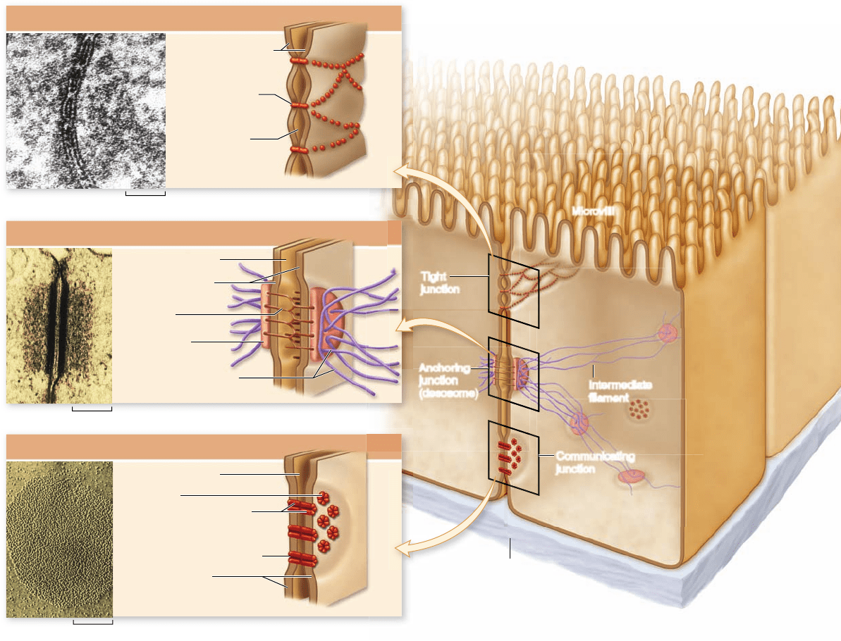
Microvilli
Tight
junction
Anchoring
junction
(desmosome)
Communicating
junction
Intermediate
filament
Basal lamina
Tight junction
Anchoring junction (desmosome)
Communicating junction
1.4 μm
2.5 μm
0.1 μm
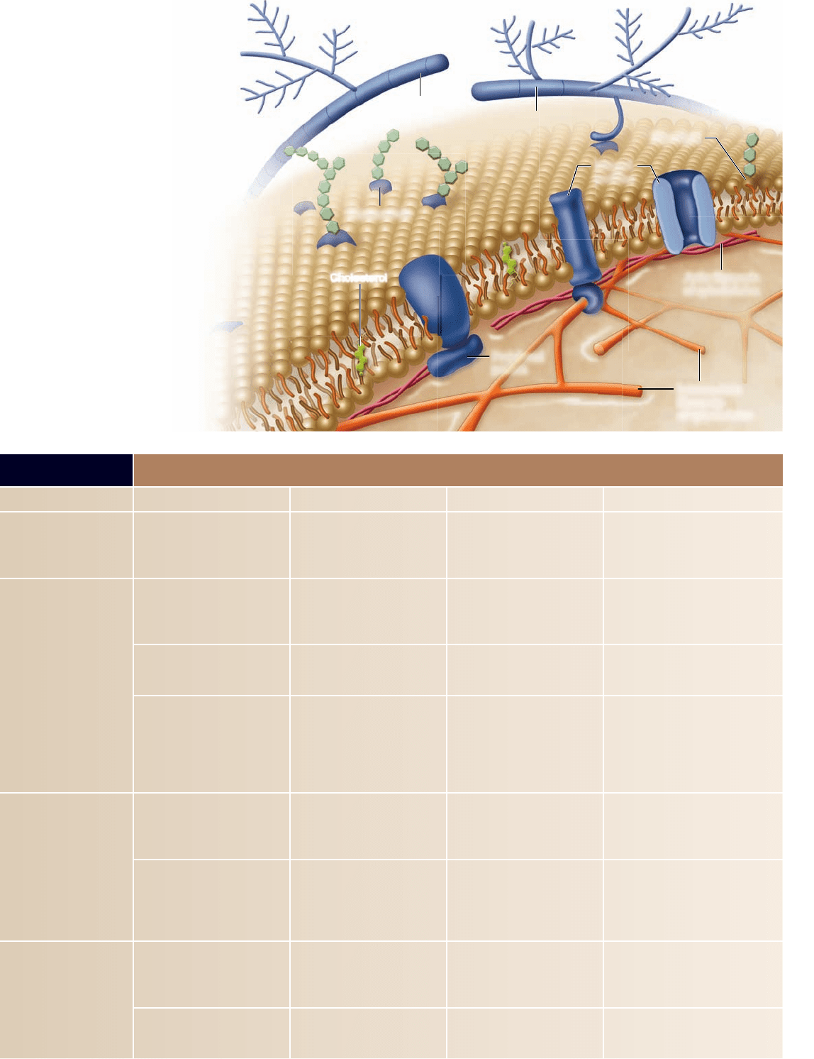
Figure 4.27
An overview of cell junction types. Here, the diagram of gut epithelial cells on the right illustrates the comparative
structures and locations of common cell junctions. The detailed models on the left show the structures of the three major types of cell
junctions: (a) tight junction; (b) anchoring junction, the example shown is a desmosome; (c) communicating junction, the example shown is a
gap junction.
major histocompatibility complex (MHC ). Cell recognition in the
immune system is covered in chapter 52 .
Cell connections mediate cell-to-cell adhesion
Most cells in a multicellular organism are in physical contact
with other cells at all times, usually as members of organized
tissues such as those in a leaf or those in your lungs, heart, or
gut. These cells and the mass of other cells clustered around
them form long-lasting or permanent connections with one an-
other called cell junctions.
The nature of the physical connections between the cells
of a tissue in large measure determines what the tissue is like.
Indeed, a tissue’s proper functioning often depends critically on
how the individual cells are arranged within it. Just as a house
cannot maintain its structure without nails and cement, so a tis-
sue cannot maintain its characteristic architecture without the
appropriate cell junctions.
Cell junctions are divided into three categories, based on
their functions: tight, anchoring, and communicating junctions
(figure 4.27).
Tight junctions
Tight junctions connect the plasma membranes of adjacent
cells in a sheet. This sheet of cells acts as a wall within the organ,
keeping molecules on one side or the other (figure 4.27a).
Creating sheets of cells.
The cells that line an animal’s di-
gestive tract are organized in a sheet only one cell thick. One
surface of the sheet faces the inside of the tract, and the other
chapter
4
Cell Structure
83www.ravenbiology.com
rav32223_ch04_059-087.indd 83rav32223_ch04_059-087.indd 83 11/6/09 12:24:17 PM11/6/09 12:24:17 PM
Apago PDF Enhancer
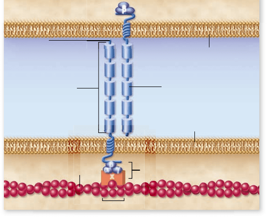
β
β
α
γ
x
Intercellular space
Extracellular
domains of
cadherin protein
Adjoining cell membrane
Plasma membrane
Cytoplasm
Cytoplasm
Cadherin of
adjoining cell
Actin
0.01 μm
COOH
Intracellular
attachment proteins
NH
2
Figure 4.28
A cadherin-mediated junction. The cadherin
molecule is anchored to actin in the cytoskeleton and passes through
the membrane to interact with the cadherin of an adjoining cell.
faces the extracellular space, where blood vessels are located.
Tight junctions encircle each cell in the sheet, like a belt
cinched around a person’s waist. The junctions between neigh-
boring cells are so securely attached that there is no space be-
tween them for leakage. Hence, nutrients absorbed from the
food in the digestive tract must pass directly through the cells
in the sheet to enter the bloodstream because they cannot pass
through spaces between cells.
Partitioning the sheet.
The tight junctions between the
cells lining the digestive tract also partition the plasma mem-
branes of these cells into separate compartments. Transport
proteins in the membrane facing the inside of the tract carry
nutrients from that side to the cytoplasm of the cells. Other
proteins, located in the membrane on the opposite side of the
cells, transport those nutrients from the cytoplasm to the ex-
tracellular uid, where they can enter the bloodstream.
For the sheet to absorb nutrients properly, these proteins
must remain in the correct locations within the fluid membrane.
Tight junctions effectively segregate the proteins on opposite sides
of the sheet, preventing them from drifting within the membrane
from one side of the sheet to the other. When tight junctions are
experimentally disrupted, just this sort of migration occurs.
Anchoring junctions
Anchoring junctions mechanically attach the cytoskeleton of
a cell to the cytoskeletons of other cells or to the extracellular
matrix. These junctions are most common in tissues subject to
mechanical stress, such as muscle and skin epithelium.
Cadherin and intermediate laments.
Desmosomes connect
the cytoskeletons of adjacent cells ( gure 4.27b), and hemides-
mosomes anchor epithelial cells to a basement membrane.
Proteins called cadherins, most of which are single-pass trans-
membrane glycoproteins, create the critical link. Proteins link
the short cytoplasmic end of a cadherin to the intermediate
laments in the cytoskeleton. The other end of the cadherin
molecule pro jects outward from the plasma membrane, join-
ing directly with a cadherin protruding from an adjacent cell
similar to a rm handshake, binding the cells together. Con-
nections between proteins tethered to the intermediate la-
ments are much more secure than connections between
free- oating membrane proteins.
Cadherin and actin laments.
Cadherins can also connect
the actin frameworks of cells in cadherin-mediated junctions
( gure 4.28). When they do, they form less stable links be-
tween cells than when they connect intermediate laments.
Many kinds of actin-linking cadherins occur in different tis-
sues. For example, during vertebrate development, the migra-
tion of neurons in the embryo is associated with changes in the
type of cadherin expressed on their plasma membranes.
Integrin-mediated links.
Anchoring junctions called adhe-
rens junctions connect the actin laments of one cell with
those of neighboring cells or with the extracellular matrix.
The linking proteins in these junctions are members of a large
superfamily of cell-surface receptors called integrins that bind
to a protein component of the extracellular matrix. At least 20
different integrins exist each with a differently shaped bind-
ing domain.
Communicating junctions
Many cells communicate with adjacent cells through direct
connections called communicating junctions. In these junctions, a
chemical or electrical signal passes directly from one cell to an
adjacent one. Communicating junctions permit small molecules
or ions to pass from one cell to the other. In animals, these di-
rect communication channels between cells are called gap junc-
tions, and in plants, plasmodesmata.
Gap junctions in animals.
Gap junctions are composed of
structures called connexons, complexes of six identical transmem-
brane proteins (see gure 4.27c). The proteins in a connexon are ar-
ranged in a circle to create a channel through the plasma membrane
that protrudes several nanometers from the cell surface. A gap junc-
tion forms when the connexons of two cells align perfectly, creating
an open channel that spans the plasma membranes of both cells.
Gap junctions provide passageways large enough to per-
mit small substances, such as simple sugars and amino acids, to
pass from one cell to the next. Yet the passages are small enough
to prevent the passage of larger molecules, such as proteins.
Gap junction channels are dynamic structures that can open
or close in response to a variety of factors, including Ca
2+
and H
+
ions. This gating serves at least one important function. When a
cell is damaged, its plasma membrane often becomes leaky. Ions in
high concentrations outside the cell, such as Ca
2+
, flow into the
damaged cell and close its gap junction channels. This isolates the
cell and so prevents the damage from spreading to other cells.
Plasmodesmata in plants.
In plants, cell walls separate every
cell from all others. Cell–cell junctions occur only at holes or
gaps in the walls, where the plasma membranes of adjacent
cells can come into contact with one another. Cytoplasmic
connections that form across the touching plasma membranes
are called plasmodesmata (singular, plasmodesma) ( gure 4.29).
The majority of living cells within a higher plant are con-
nected to their neighbors by these junctions.
84
part
II
Biology of the Cell
rav32223_ch04_059-087.indd 84rav32223_ch04_059-087.indd 84 11/6/09 12:24:26 PM11/6/09 12:24:26 PM

Apago PDF Enhancer
Plasmodesma
Central
tubule
Smooth
ER
Cell 1 Cell 2
Primary
cell wall
Middle lamella
Plasma
membrane
Figure 4.29
Plasmodesmata. Plant cells can communicate
through specialized openings in their cell walls, called plasmodesmata,
where the cytoplasm of adjoining cells are connected.
Chapter Review
4.1 Cell Theory
Cell theory is the unifying foundation of cell biology.
All organisms are composed of one or more cells. Cells arise only by
division of preexisting cells.
Cell size is limited.
Cell size is constrained by the diffusion distance. As cell size increases,
diffusion becomes inef cient.
Microscopes allow visualization of cells and components.
Magni cation gives better resolution than is possible with the naked
eye. Staining with chemicals enhances contrast of structures.
All cells exhibit basic structural similarities.
All cells have centrally located DNA, a semi uid cytoplasm, and an
enclosing plasma membrane.
4.2 Prokaryotic Cells (see gure 4.3)
Prokaryotic cells have relatively simple organization.
Prokaryotic cells contain DNA and ribosomes,
but they lack a
nucleus, an internal membrane system, and membrane-bounded
organelles. A rigid cell wall surrounds the plasma membrane.
Bacterial cell walls consist of peptidoglycan.
Peptidoglycan is composed of carbohydrate cross-linked with
short peptides.
Archaea lack peptidoglycan.
Archaeal cell walls do not contain peptidoglycan, and they have
unique plasma membranes.
Some prokaryotes move by means of rotating agella.
Prokaryotic agella rotate because of proton transfer across the
plasma membrane.
4.3 Eukaryotic Cells (see gures 4.6 and 4.7)
Eukaryotic cells have a membrane-bounded nucleus, an
endomembrane system,
and many different organelles.
The nucleus acts as the information center.
The nucleus is surrounded by an envelope of two phospholipid
bilayers; the outer layer is contiguous with the ER. Pores allow
exchange of small molecules. The nucleolus is a region of the
nucleoplasm where rRNA is transcribed and ribosomes are assembled.
In most prokaryotes, DNA is organized into a single circular
chromosome. In eukaryotes, numerous chromosomes are present.
Ribosomes are the cell’s protein synthesis machinery.
Ribosomes translate mRNA to produce polypeptides. They are found
in all cell types.
4.4 The Endomembrane System
The endoplasmic reticulum (ER) creates channels and passages within
the cytoplasm (see gure 4.10 ).
The rough ER is a site of protein synthesis.
The rough ER (RER), studded with ribosomes, synthesizes and
modi es proteins and manufactures membranes.
The smooth ER has multiple roles.
The smooth endoplasmic reticulum (SER) lacks ribosomes; it is
involved in carbohydrate and lipid synthesis and detoxi cation.
Plasmodesmata function much like gap junctions in ani-
mal cells, although their structure is more complex. Unlike gap
junctions, plasmodesmata are lined with plasma membrane and
contain a central tubule that connects the endoplasmic reticu-
lum of the two cells.
Learning Outcomes Review 4.8
Cell connections fall into three basic categories: (1) Tight junctions help
to make sheets of cells that form watertight seals; (2) anchoring junctions
provide strength and fl exibility; and (3) communicating junctions,
including gap junctions in animals and plasmodesmata in plants,
allow passage of some materials between cells. Cells in multicellular
organisms are usually organized into tissues, requiring that cells have
distinct identity and connections. Cell identity is conferred by surface
glycoproteins, which include the MHC proteins that are important in the
immune system.
■ How do cell junctions help to form tissues?
chapter
4
Cell Structure
85www.ravenbiology.com
rav32223_ch04_059-087.indd 85rav32223_ch04_059-087.indd 85 11/6/09 12:24:28 PM11/6/09 12:24:28 PM
Apago PDF Enhancer
The Golgi apparatus sorts and packages proteins.
The Golgi apparatus receives vesicles from the ER, modi es and
packages macromolecules, and transports them (see gure 4.11 ) .
Lysosomes contain digestive enzymes.
Lysosomes break down macromolecules and recycle the components
of old organelles (see gure 4.13 ) .
Microbodies are a diverse category of organelles.
Plants use vacuoles for storage and water balance.
4.5 Mitochondria and Chloroplasts:
Cellular Generators
Mitochondria and chloroplasts have a double-membrane structure,
contain their own DNA, and can divide independently.
Mitochondria metabolize sugar to generate ATP.
The inner membrane of mitochondria is extensively folded into
layers called cristae. Proteins on the surface and in the inner
membrane carry out metabolism to produce ATP (see gure 4.16 ).
Chloroplasts use light to generate ATP and sugars.
Chloroplasts capture light energy via thylakoid membranes arranged in
stacks called grana, and use it to synthesize glucose (see gure 4.17 ) .
Mitochondria and chloroplasts arose by endosymbiosis.
The endosymbiont theory proposes that mitochondria and
chloroplasts were once prokaryotes engulfed by another cell.
4.6 The Cytoskeleton
The cytoskeleton consists of crisscrossed protein bers that support
the shape of the cell and anchor organelles (see gure 4.19 ) .
Three types of bers compose the cytoskeleton.
Actin laments, or micro laments, are long, thin polymers involved
in cellular movement. Microtubules are hollow structures that move
materials within a cell. Intermediate laments serve a wide variety
of functions.
Centrosomes are microtubule-organizing centers.
Centrosomes help assemble the nuclear division apparatus of animal
cells (see gure 4.20 ) .
The cytoskeleton helps move materials within cells.
Molecular motors move vesicles along microtubules, like a train on a
railroad track. Kinesin and dynein are two motor proteins.
4.7 Extracellular Structures
and Cell Movement
Some cells crawl.
Cell crawling occurs as actin polymerization forces the cell
membrane forward, while myosin pulls the cell body forward.
Flagella and cilia aid movement.
Eukaryotic agella have a 9 + 2 structure and arise from a basal body. Cilia
are shorter and more numerous than agella.
Plant cell walls provide protection and support.
Plants have cell walls composed of cellulose bers. The middle
lamella, between cell walls, holds adjacent cells together.
Animal cells secrete an extracellular matrix.
Glycoproteins are the main component of the extracellular matrix
(ECM) of animal cells.
4.8 Cell-to-Cell Interactions (see gure 4.27)
Surface proteins give cells identity.
Glycolipids and MHC proteins on cell surfaces help distinguish self
from nonself.
C
ell connections mediate cell-to-cell adhesion.
Cell junctions include tight junctions, anchoring junctions, and
communicating junctions. In animals, gap junctions allow the
passage of small molecules between cells. In plants, plasmodesmata
penetrate the cell wall and connect cells.
Review Questions
U N DERS TAN D
1. Which of the following statements is NOT part of the
cell theory?
a. All organisms are composed of one or more cells.
b. Cells come from other cells by division.
c. Cells are the smallest living things.
d. Eukaryotic cells have evolved from prokaryotic cells.
2. All cells have all of the following except
a. plasma membrane. c. cytoplasm.
b. genetic material. d. cell wall.
3. Eukaryotic cells are more complex than prokaryotic cells.
Which of the following are found only in a eukaryotic cell?
a. Cell wall
b. Plasma membrane
c. Endoplasmic reticulum
d. Ribosomes
4. Which of the following are differences between bacteria
and archaea?
a. The molecular architecture of their cell walls
b. The type of ribosomes found in each
c. Archaea have an internal membrane system that bacteria lack.
d. Both a and b
5. The cytoskeleton includes
a. microtubules made of actin laments.
b. micro laments made of tubulin.
c. intermediate laments made of twisted bers of vimentin
and keratin.
d. smooth endoplasmic reticulum.
6. The smooth endoplasmic reticulum is
a. involved in protein synthesis.
b. a site of protein glycosylation.
c. used to store a variety of ions.
d. the site of lipid and membrane synthesis.
86
part
II
Biology of the Cell
rav32223_ch04_059-087.indd 86rav32223_ch04_059-087.indd 86 11/6/09 12:24:30 PM11/6/09 12:24:30 PM

Apago PDF Enhancer
7. Plasmodesmata in plants and gap junctions in animals are
functionally similar in that
a. each is used to anchor layers of cells.
b. they form channels between cells that allow diffusion of
small molecules.
c. they form tight junctions between cells.
d. they are anchored to the extracellular matrix.
APPLY
1. The most important factor that limits the size of a cell is the
a. quantity of proteins and organelles a cell can make.
b. rate of diffusion of small molecules.
c. surface area-to-volume ratio of the cell.
d. amount of DNA in the cell.
2. All eukaryotic cells possess each of the following except
a. mitochondria. c. cytoskeleton.
b. cell wall. d. nucleus.
3. Which of these organelles is NOT associated with the
production or sorting of proteins in a cell?
a. Ribosomes
b. Smooth endoplasmic reticulum (SER)
c. Rough endoplasmic reticulum (RER)
d. Golgi apparatus
4. Different motor proteins like kinesin and myosin are similar
in that they can
a. interact with microtubules.
b. use energy from ATP to produce movement.
c. interact with actin.
d. do both a and b.
5. The protein sorting pathway involves the following organelles/
compartments in order:
a. SER, RER, transport vesicle, Golgi.
b. RER, lysosome, Golgi.
c. RER, transport vesicle, Golgi, nal destination.
d. Golgi, transport vesicle, RER, nal destination.
6. Chloroplasts and mitochondria have many common features
because both
a. are present in plant cells.
b. arose by endosymbiosis.
c. function to oxidize glucose.
d. function to produce glucose.
7. Eukaryotic cells are composed of three types of cytoskeletal
laments. How are these three laments similar?
a. They contribute to the shape of the cell.
b. They are all made of the same type of protein.
c. They are all the same size and shape.
d. They are all equally dynamic and exible.
S Y N THES IZE
1. The smooth endoplasmic reticulum is the site of synthesis of
the phospholipids that make up all the membranes of a cell—
especially the plasma membrane. Use the diagram of an animal
cell (see gure 4.6) to trace a pathway that would carry a
phospholipid molecule from the SER to the plasma membrane.
What endomembrane compartments would the phospholipids
travel through? How can a phospholipid molecule move
between membrane compartments?
2. Use the information provided in table 4.3 to develop a set of
predictions about the properties of mitochondria and chloroplasts
if these organelles were once free-living prokaryotic cells. How
do your predictions match with the evidence for endosymbiosis?
3. In evolutionary theory, homologous traits are those with a
similar structure and function derived from a common ancestor.
Analogous traits represent adaptations to a similar environment,
but from distantly related organisms. Consider the structure
and function of the agella found on eukaryotic and prokaryotic
cells. Are the agella an example of a homologous or analogous
trait? Defend your answer.
4. The protist, Giardia intestinalis , is the organism associated with
water-borne diarrheal diseases. Giardia is an unusual eukaryote
because it seems to lack mitochondria. Provide two possible
evolutionary scenarios for this in the context of the
endosymbiotic theory.
O N LIN E RES OURCE
www.ravenbiology.com
Understand, Apply, and Synthesize—enhance your study with
animations that bring concepts to life and practice tests to assess
your understanding. Your instructor may also recommend the
interactive eBook, individualized learning tools, and more.
chapter
4
Cell Structure
87www.ravenbiology.com
rav32223_ch04_059-087.indd 87rav32223_ch04_059-087.indd 87 11/6/09 12:24:30 PM11/6/09 12:24:30 PM

Apago PDF Enhancer
A
0.16
μ
m
Chapter Outline
5.1 The Structure of Membranes
5.2 Phospholipids: The Membrane’s Foundation
5.3 Proteins: Multifunctional Components
5.4 Passive Transport Across Membranes
5.5 Active Transport Across Membranes
5.6 Bulk Transport by Endocytosis and Exocytosis
Introduction
A cell’s interactions with the environment are critical, a give-and-take that never ceases. Without it, life could not exist. Living cells
are encased within a lipid membrane through which few water-soluble substances can pass. The membrane also contains protein
passageways that permit speci c substances to move into and out of the cell and allow the cell to exchange information with
its environment. Eukaryotic cells also contain internal membranes like those of the mitochondrion and endoplasmic reticulum
pictured here. We call the delicate skin of lipids with embedded protein molecules that encase the cell a plasma membrane. This
chapter examines the structure and function of this remarkable membrane.
Chapter
5
Membranes
CHAPTER
5.1
The Structure of Membranes
Learning Outcomes
1. Describe the components of biological membranes.
2. Explain the fluid mosaic model of membrane
structure.
The membranes that encase all living cells are two phospho-
lipid sheets that are only 5–10 nanometers thick; more than
10,000 of these sheets piled on one another would just equal
the thickness of this sheet of paper. Biologists established the
components of membranes— not only lipids, but also proteins
and other molecules—through biochemical assays, but the or-
ganization of the membrane components remained elusive.
We begin by considering the theories that have been ad-
vanced about membrane structure. We then look at the indi-
vidual components of membranes more closely.
rav32223_ch05_088-106.indd 88rav32223_ch05_088-106.indd 88 11/9/09 10:29:16 AM11/9/09 10:29:16 AM
Apago PDF Enhancer
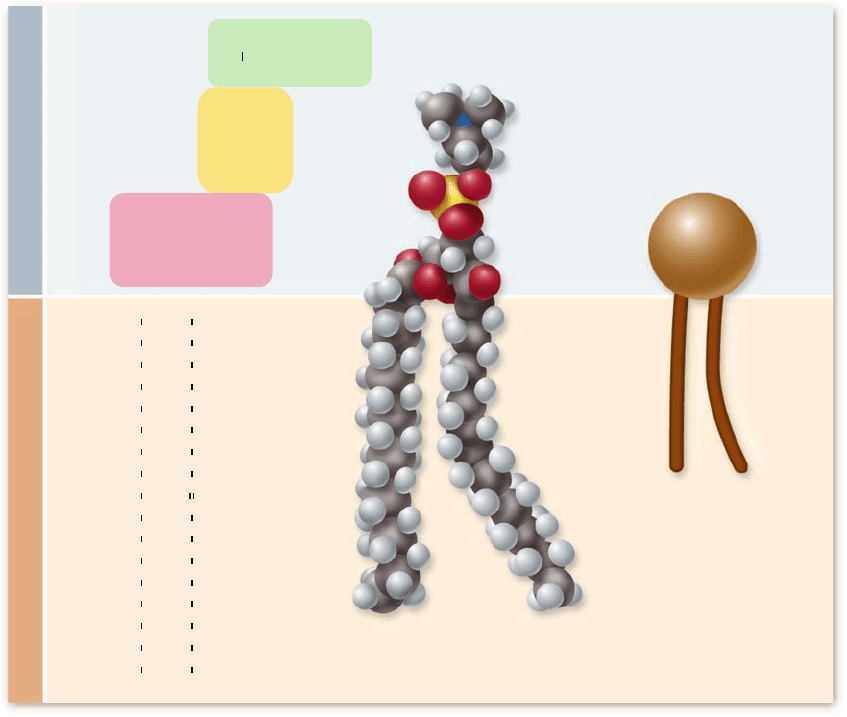
Polar Hydrophilic Heads Nonpolar Hydrophobic Tails
CH
2
J
N
;
(CH
3
)
3
CH
2
CH
2
H
2
C
O
O
O P
CH
2
CH
2
CH
2
CH
2
CH
2
CH
2
CH
2
CH
2
CH
2
CH
2
CH
2
CH
2
CH
2
CH
2
CH
2
CH
2
CH
3
CH
2
CH
2
CH
2
CH
2
CH
2
CH
2
CH
2
CH
CH
CH
2
CH
2
CH
2
CH
2
CH
2
CH
2
CH
2
CH
3
C
O O
O O
C
H
C
J J J J J J
J J
J
J
J
J
J
J J J J
J
J
J
J
J
J
O
:
a. Formula b. Space-filling model c. Icon
Figure 5.1
Di erent views of phospholipid structure. Phospholipids are composed of glycerol (pink) linked to two fatty acids and a
phosphate group. The phosphate group ( yello w) can have additional molecules attached, such as the positively charged choline ( g reen) shown.
Phosphatidylcholine is a common component of membranes, it is shown in (a) with its chemical formula, (b) as a space- lling model, and (c) as
the icon that is used in most of the gures in this chapter.
The uid mosaic model shows proteins
embedded in a uid lipid bilayer
The lipid layer that forms the foundation of a cell’s membranes
is a bilayer formed of phospholipids (figure 5.1). For many
years, biologists thought that the protein components of the
cell membrane covered the inner and outer surfaces of the
phospholipid bilayer like a coat of paint. An early model por-
trayed the membrane as a sandwich; a phospholipid bilayer be-
tween two layers of globular protein.
In 1972, S. Jonathan Singer and Garth J. Nicolson revised
the model in a simple but profound way: They proposed that
the globular proteins are inserted into the lipid bilayer, with
their nonpolar segments in contact with the nonpolar interior
of the bilayer and their polar portions protruding out from the
membrane surface. In this model, called the fluid mosaic model, a
mosaic of proteins floats in or on the fluid lipid bilayer like
boats on a pond (figure 5.2).
We now recognize two categories of membrane proteins
based on their association with the membrane. Integral mem-
brane proteins are embedded in the membrane, and peripheral
proteins are associated with the surface of the membrane.
Cellular membranes consist
of four component groups
A eukaryotic cell contains many membranes. Although they are
not all identical, they share the same fundamental architecture.
Cell membranes are assembled from four components (table 5.1):
Phospholipid bilayer.1. Every cell membrane is com-
posed of phospholipids in a bilayer. The other compo-
nents of the membrane are embedded within the bilayer,
which provides a exible matrix and, at the same time,
imposes a barrier to permeability. Animal cell mem-
branes also contain cholesterol, a steroid with a polar
hydroxyl group (
–
OH). Plant cells have a much lower
cholesterol content.
Transmembrane proteins.2. A major component of
every membrane is a collection of proteins that oat in
the lipid bilayer. These proteins have a variety of
functions, including transport and communication across
the membrane. Many integral membrane proteins are
not xed in position. They can move about, just as the
phospholipid molecules do. Some membranes are
chapter
5
Membranes
89www.ravenbiology.com
rav32223_ch05_088-106.indd 89rav32223_ch05_088-106.indd 89 11/6/09 12:01:47 PM11/6/09 12:01:47 PM
Apago PDF Enhancer
Extracellular
matrix protein
Glycolipid
Integral
proteins
Intermediate
filaments
of cytoskeleton
Peripheral
protein
Cholesterol
Actin filaments
of cytoskeleton
Glycoprotein
Glycoprotein
Figure 5.2
The uid mosaic
model of cell membranes.
Integral proteins protrude through the
plasma membrane, with nonpolar
regions that tether them to the
membrane’s hydrophobic interior.
Carbohydrate chains are often bound
to the extracellular portion of these
proteins, forming glycoproteins.
Peripheral membrane proteins are
associated with the surface of the
membrane. Membrane phospholipids
can be modi ed by the addition of
carbohydrates to form glycolipids.
Inside the cell, actin laments and
intermediate laments interact with
membrane proteins. Outside the cell,
many animal cells have an elaborate
extracellular matrix composed
primarily of glycoproteins.
TABLE 5.1
Components of the Cell Membrane
Component Composition Function How It Works Example
Phospholipid bilayer Phospholipid molecules Provides permeability barrier,
matrix for proteins
Excludes water-soluble molecules
from nonpolar interior of bilayer
and cell
Bilayer of cell is impermeable to
large water-soluble molecules, such
as glucose
Transmembrane proteins Carriers Actively or passively transport
molecules across membrane
Move speci c molecules through
the membrane in a series of
conformational changes
Glycophorin carrier for sugar transport;
sodium–potassium pump
Channels Passively transport molecules
across membrane
Create a selective tunnel that acts
as a passage through membrane
Sodium and potassium channels in
nerve, heart, and muscle cells
Receptors Transmit information into cell Signal molecules bind to cell-
surface portion of the receptor
protein. This alters the portion of
the receptor protein within the
cell, inducing activity
Speci c receptors bind peptide
hormones and neurotransmitters
Interior protein network Spectrins Determine shape of cell Form supporting sca old beneath
membrane, anchored to both
membrane and cytoskeleton
Red blood cell
Clathrins Anchor certain proteins to speci c
sites, especially on the exterior
plasma membrane in receptor-
mediated endocytosis
Proteins line coated pits and
facilitate binding to speci c
molecules
Localization of low-density lipoprotein
receptor within coated pits
Cell-surface markers Glycoproteins “Self” recognition Create a protein/carbohydrate
chain shape characteristic
of individual
Major histocompatibility complex
protein recognized by immune system
Glycolipid Tissue recognition Create a lipid/carbohydrate chain
shape characteristic of tissue
A, B, O blood group markers
90
part
II
Biology of the Cell
rav32223_ch05_088-106.indd 90rav32223_ch05_088-106.indd 90 11/6/09 12:01:47 PM11/6/09 12:01:47 PM
Apago PDF Enhancer
Cell 1
Cell 1
Cell 2
Cell 2
Plasma membrane of cell 1
Plasma membrane of cell 2
Cell 1
Cell 2
0.038 µm
0.15 µm
Exposed lower
half of lipid bilayer
Knife
External surface
of plasma
membrane
Exposed
lower half of
lipid bilayer
Cell
Medium
Fractured
upper half
of lipid bilayer
1. A cell frozen in
medium is cracked
with a knife blade.
2. The cell often
fractures through the
interior, hydrophobic
area of the lipid
bilayer, splitting the
plasma membrane
into two layers.
3. The plasma membrane separates such
that proteins and other embedded
membrane structures remain within one
or the other layers of the membrane.
4. The exposed membrane is
coated with platinum, which
forms a replica of the
membrane. The underlying
membrane is dissolved away,
and the replica is then viewed
with electron microscopy.
crowded with proteins, but in others, the proteins are
more sparsely distributed.
Interior protein network.3. Membranes are structurally
supported by intracellular proteins that reinforce the
membrane’s shape. For example, a red blood cell has a
characteristic biconcave shape because a scaffold made of
a protein called spectrin links proteins in the plasma
membrane with actin laments in the cell’s cytoskeleton.
Membranes use networks of other proteins to control
the lateral movements of some key membrane proteins,
anchoring them to speci c sites.
Cell-surface markers.4. As you learned in the preceding
chapter, membrane sections assemble in the endoplasmic
reticulum, transfer to the Golgi apparatus, and then are
transported to the plasma membrane. The ER adds chains
of sugar molecules to membrane proteins and lipids,
converting them into glycoproteins and glycolipids.
Different cell types exhibit different varieties of these
glycoproteins and glycolipids on their surfaces, which act
as cell identity markers.
Originally, it was believed that because of its fluidity,
the plasma membrane was uniform, with lipids and proteins
free to diffuse rapidly in the plane of the membrane. How-
ever, in the last decade evidence has accumulated suggesting
the plasma membrane is not homogeneous and contains
micro domains with distinct lipid and protein composition.
One type of microdomain, the lipid raft, is heavily enriched
with cholesterol, which fills space between the phospholip-
ids, packing them more tightly together than the surround-
ing membrane.
Although the distribution of membrane lipids is symmet-
rical in the ER where they are synthesized, this distribution is
asymmetrical in the plasma membrane, Golgi apparatus, and
endosomes. This is accomplished by enzymes that transport
lipids across the bilayer from one face to the other.
Electron microscopy has provided
structural evidence
Electron microscopy allows biologists to examine the deli-
cate, filmy structure of a cell membrane. We discussed two
types of electron microscopes in chapter 4: the transmission
electron microscope (TEM) and the scanning electron mi-
croscope (SEM). Both provide illuminating views of mem-
brane structure.
When examining cell membranes with electron mi cro s-
copy, specimens must be prepared for viewing. In one method
of preparing a specimen, the tissue of choice is embedded in a
hard epoxy matrix. The epoxy block is then cut with a micro-
tome, a machine with a very sharp blade that makes incredibly
thin, transparent “epoxy shavings” less than 1 μm thick that
peel away from the block of tissue.
These shavings are placed on a grid, and a beam of elec-
trons is directed through the grid with the TEM. At the high
magnification an electron microscope provides, resolution is
good enough to reveal the double layers of a membrane. False
color can be added to the micrograph to enhance detail.
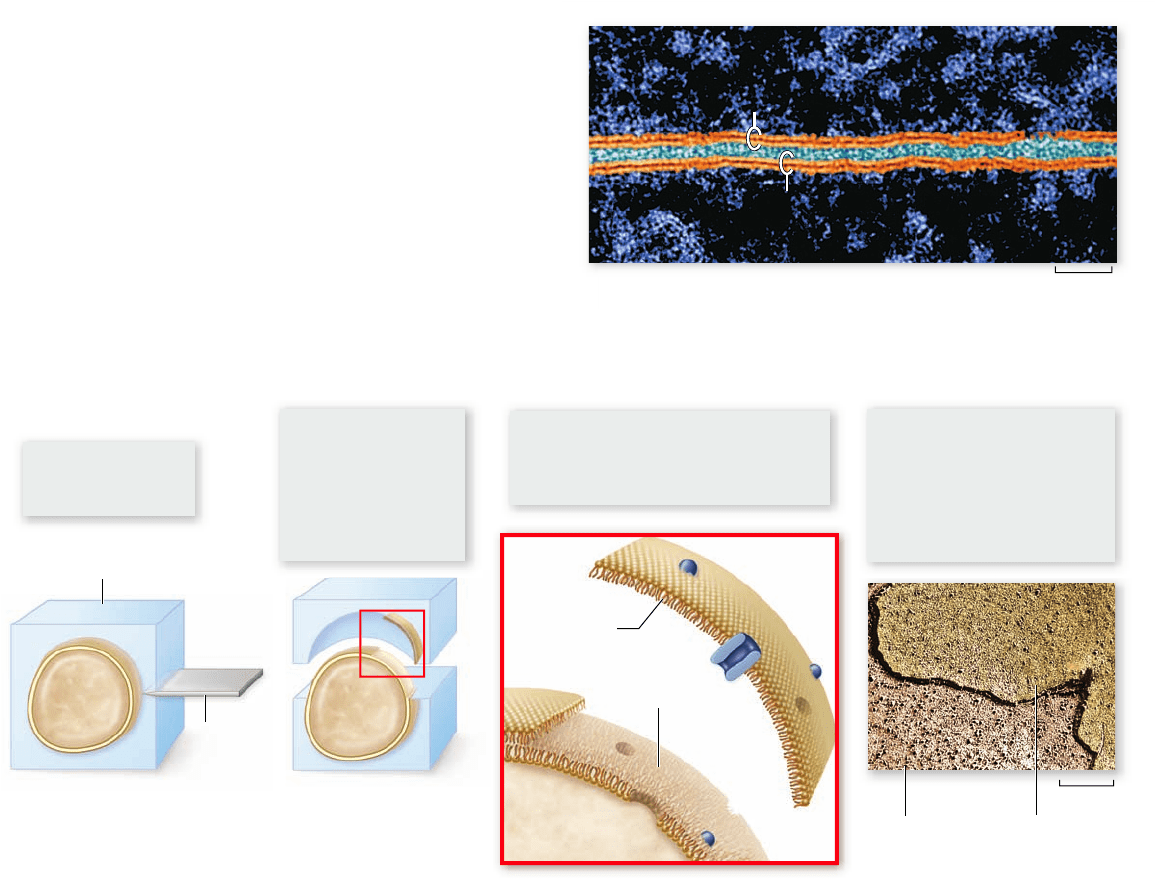
Figure 5.3
Viewing a plasma membrane with freeze-fracture microscopy.
Freeze-fracturing a specimen is another way to visualize
the inside of the membrane (figure 5.3). The tissue is embedded
chapter
5
Membranes
91www.ravenbiology.com
rav32223_ch05_088-106.indd 91rav32223_ch05_088-106.indd 91 11/6/09 12:01:51 PM11/6/09 12:01:51 PM