Raven P.H., Johnson G.B., Mason K.A. Biology (Ninth Edition)
Подождите немного. Документ загружается.

Apago PDF Enhancer
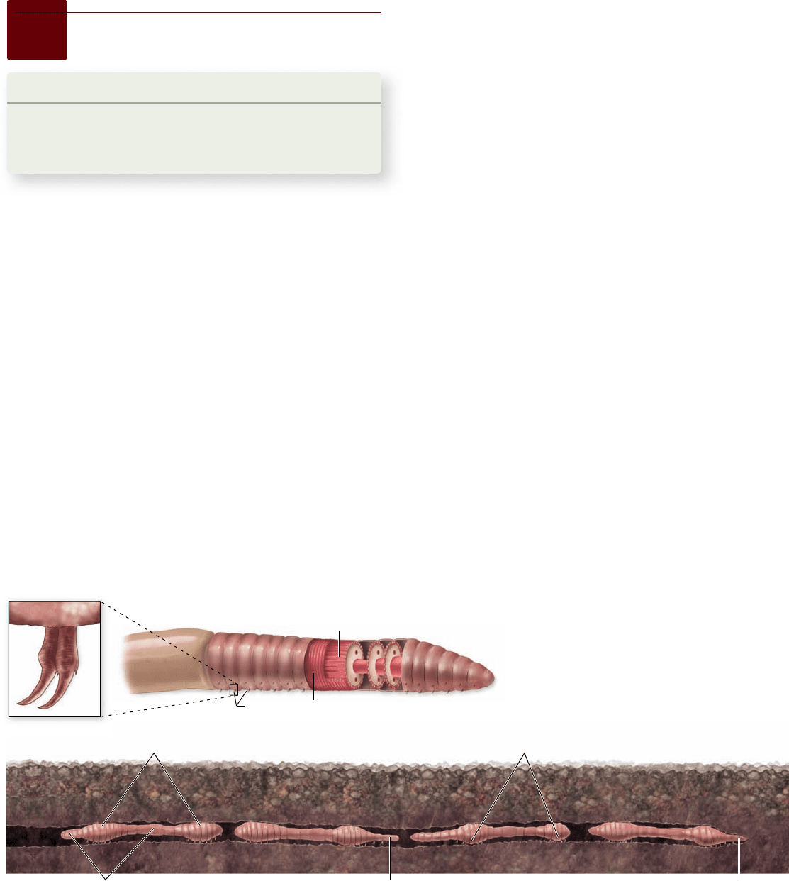
Anterior
Circular muscles
Chaetae
Longitudinal muscles
Circular muscles
contracted
Longitudinal muscles
contracted
Circular muscles contract, and
anterior end moves forward.
Chaetae lose attachment to ground.
Longitudinal muscles contract, and
segments catch up. Chaetae attach to
the ground and prevent backsliding.
Circular muscles
contract, and anterior
end moves forward.
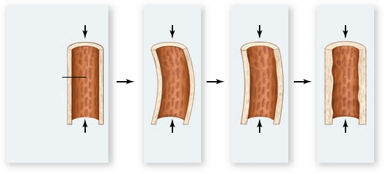
Figure 47.1
Locomotion in earthworms. The hydrostatic skeleton of the earthworm uses muscles to move uid within the
segmented body cavity, changing the shape of the animal. When circular muscles contract the pressure in the uid rises. At the same time
the longitudinal muscles relax, and the body becomes longer and thinner. When the longitudinal muscles contract and the circular muscles
relax, the chaetae of the worm’s lower surface extend to prevent backsliding. A wave of circular followed by longitudinal muscle contractions
down the body produces forward movement.
47.1
Types of Skeletal Systems
Learning Outcomes
Compare hydrostatic skeletons, exoskeletons, 1.
and endoskeletons.
Explain how animals with hydrostatic skeletons move.2.
Muscles have to pull against something to produce the changes
that cause movement. This necessary form of supporting struc-
ture is called a skeletal system. Zoologists commonly recog-
nize three types of skeletal systems in animals: hydrostatic
skeletons, exoskeletons, and endoskeletons.
Hydrostatic skeletons use water
pressure inside a body wall
Hydrostatic skeletons are found primarily in soft-bodied ter-
restrial invertebrates, such as earthworms and slugs, and soft-
bodied aquatic invertebrates, such as jellyfish, and squids.
Musculoskeletal action in earthworms
In these animals a fluid-filled central cavity is encompassed
by two sets of muscles in the body wall: circular muscles
that are repeated in segments and run the length of the
body, and longitudinal muscles that oppose the action of
the circular muscles.
Muscles act on the fluid in the body’s central space, which
represents the hydrostatic skeleton. As locomotion begins
(figure 47.1) the anterior circular muscles contract, pressing on
the inner fluid, and forcing the front of the body to become
thin as the body wall in this region extends forward.
On the underside of a worm’s body are short, bristle-like
structures called chaetae . When circular muscles act, the chaetae
of that region are pulled up close to the body and lose contact
with the ground. Circular-muscle activity is passed backward,
segment by segment, to create a backward wave of contraction.
As this wave continues, the anterior circular muscles now
relax, and the longitudinal muscles take over, thickening the
front end of the worm and allowing the chaetae to protrude and
regain contact with the ground. The chaetae now prevent that
body section from slipping backward. This locomotion process
proceeds as waves of circular muscle contraction are followed
by waves of longitudinal muscle effects.
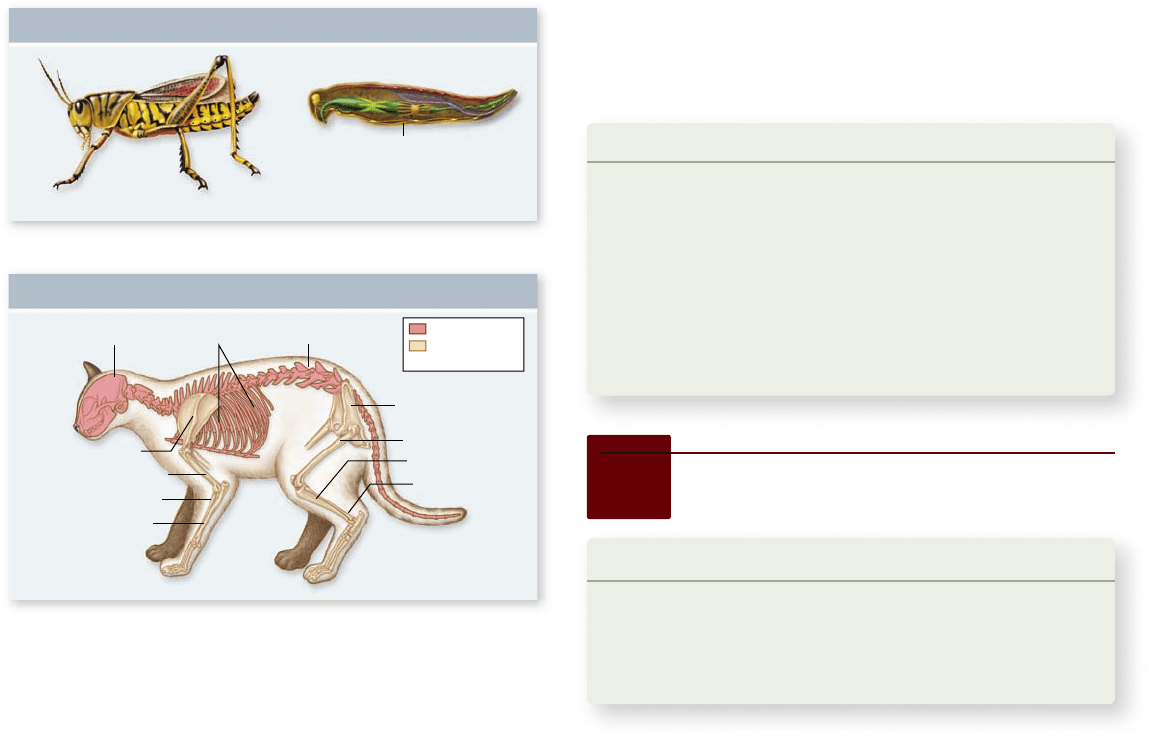
Exoskeletons consist of a rigid outer covering
Exoskeletons are a rigid, hard case that surrounds the body .
Arthropods, such as crustaceans and insects, have exoskeletons
made of the polysaccharide chitin (figure 47.2a) . As you learned
in earlier chapters, chitin is found in the cell walls of fungi and
some protists as well as in the exoskeletons of arthropods.
A chitinous exoskeleton resists bending and thus acts as
the skeletal framework of the body; it also protects the internal
organs and provides attachment sites for the muscles, which
lie inside the exoskeletal casing. But in order to grow, the
animal must periodically molt, shedding the exoskeleton (see
chapter 34 ). The animal is vulnerable to predation until the new
(slightly larger) exoskeleton forms. Molting crabs and lobsters
often hide until the process is completed.
Exoskeletons have other limitations. The chitinous
framework is not as strong as a bony, internal one. This fact
by itself would set a limit for insect size, but there is a more
important factor: Insects breathe through openings in their
body that lead into tiny tubes, and as insect size in-
creases beyond a certain limit, the ratio be-
tween the inside surface area of the tubes
and the volume of the body overwhelms
this sort of respiratory system. Finally,
when muscles are confined within an
962 part
VII
Animal Form and Function
rav32223_ch47_961-980.indd 962rav32223_ch47_961-980.indd 962 11/18/09 10:50:47 AM11/18/09 10:50:47 AM
Apago PDF Enhancer
Chitinous outer covering
Exoskeleton
Sagittal section
Vertebral column
Pelvis
Femur
Tibia
Fibula
Ulna
Radius
Humerus
Skull
Scapula
Ribs
Exoskeleton
Endoskeleton
a.
b.
axial skeleton
appendicular
skeleton
Figure 47.2
Exoskeleton and endoskeleton.
a. The hard, tough outer covering of an arthropod, such as this
grasshopper, is its exoskeleton and is composed of chitin.
b. Vertebrates, such as this cat, have endoskeletons formed of bone
and cartilage. Some of the major bony features are labeled.
especially if bone cells are present throughout the matrix, a
common condition. Bone, and to some extent cartilage, can
change and remodel itself in response to injury or to physi-
cal stresses.
Learning Outcomes Review 47.1
With a hydrostatic skeleton, muscle contraction puts pressure on the
fl uid inside the body, forcing the body to extend. Opposing muscles then
shorten the body to draw the animal forward. Invertebrate exoskeletons
consist of hard chitin; they must be shed and renewed (molting) for the
animal to grow. Endoskeletons are composed of fi brous dense connective
tissue along with cartilage or mineralized bone.
■ What limitations does an exoskeleton impose on
terrestrial invertebrates?
exoskeleton, they cannot enlarge in size and power with in-
creased use, as they can in animals with endoskeletons.
Endoskeletons are composed
of hard, internal structures
Endoskeletons, found in vertebrates and echinoderms, are rigid
internal skeletons that form the body’s framework and offer sur-
faces for muscle attachment. Echinoderms, such as sea urchins
and sand dollars, have skeletons made of calcite, a crystalline
form of calcium carbonate. This calcium compound is different
from that in bone, which is based on calcium phosphate.
Vertebrate skeletal tissues
The vertebrate endoskeleton (figure 47.2b) includes fibrous
dense connective tissue along with the more rigid special con-
nective tissues, cartilage or bone (see chapter 43). Cartilage is
strong and slightly flexible, a characteristic important in such
functions as padding the ends of bones where they come to-
gether in a joint. Although some large, active animals such as
sharks have totally cartilaginous skeletons, bone is the main
component in vertebrate skeletons. Bone is much stronger than
cartilage and much less flexible.
Unlike chitin, both cartilage and bone are living tis-
sues. Bone, particularly, can have high metabolic activity,
47.2
A Closer Look at Bone
Learning Outcomes
Compare intramembranous and endochondral 1.
development.
Describe how growth occurs in epiphyses.2.
Explain how bone remodeling occurs.3.
Bone is a hard but resilient tissue that is unique to vertebrate
animals. This connective tissue first appeared over 520 mya and
is now found in all vertebrates except cartilaginous fishes (see
chapter 35).
Bones can be classi ed by two
modes of development
Bone tissue itself can be of several types classified in a few dif-
ferent ways. The most common system is based on the way in
which bone develops.
Intramembranous development
In intramembranous development, bones form within a layer
of connective tissue. Many of the flat bones that make up the
exterior of the skull and jaw are intramembranous.
Typically, the site of the intramembranous bone-to-be
begins in a designated region in the dermis of the skin. Dur-
ing embryonic development, the dermis is formed largely of
mesenchyme—a loose tissue consisting of undifferentiated
mesenchyme cells and other cells that have arisen from them—
along with collagen fibers. Some of the undifferentiated mes-
enchyme cells differentiate to become specialized cells called
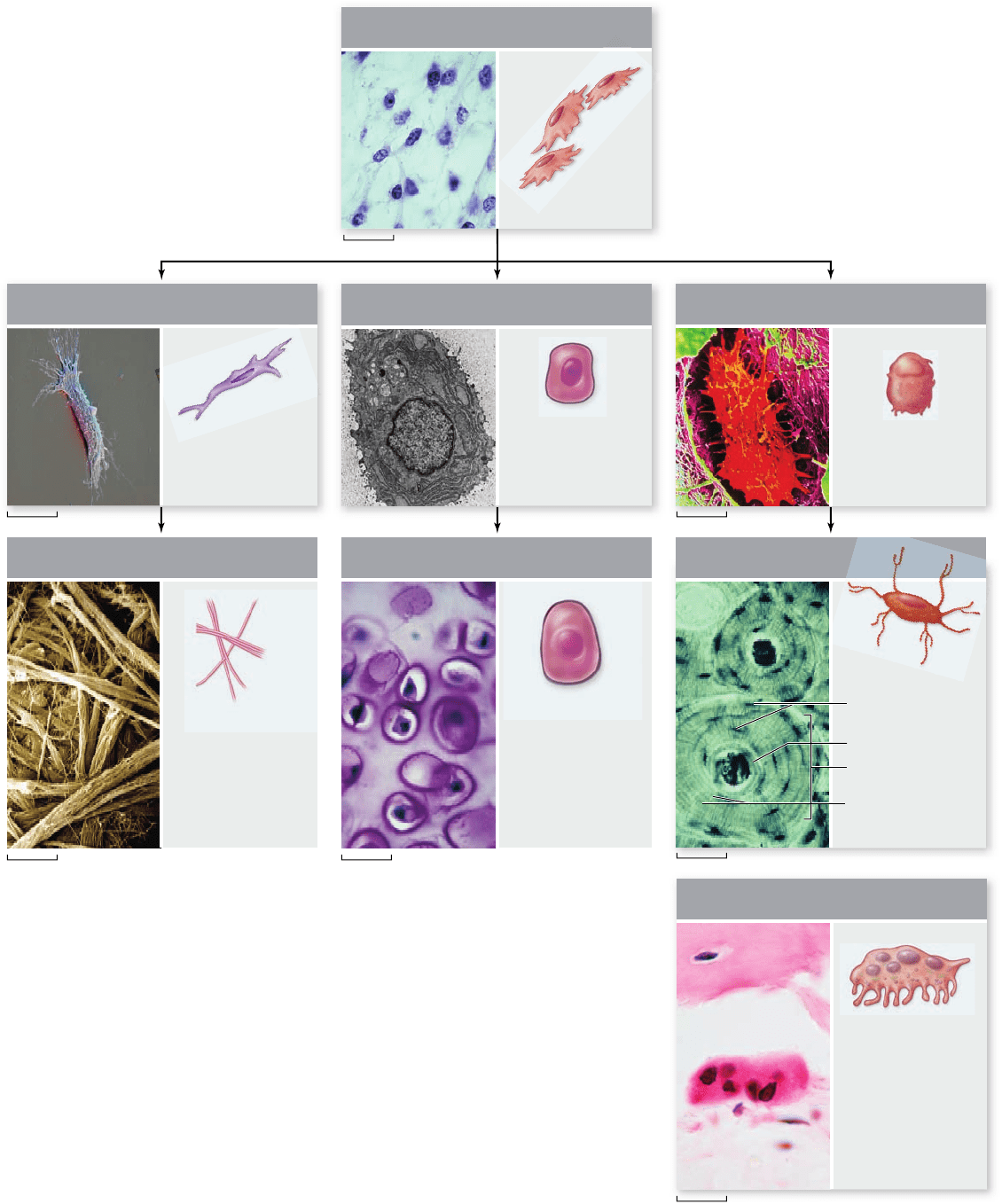
osteoblasts (figure 47.3). These osteoblasts arrange themselves
along the collagenous fibers and begin to secrete the enzyme
alkaline phosphatase, which causes calcium phosphate salts to
form in a crystalline configuration called hydroxyapatite. The
crystals merge along the fibers to encase them.
www.ravenbiology.com
chapter
47
The Musculoskeletal System
963
rav32223_ch47_961-980.indd 963rav32223_ch47_961-980.indd 963 11/18/09 10:50:50 AM11/18/09 10:50:50 AM
Apago PDF Enhancer
Chondrocyte
Collagen
(fibrous tissue)
Osteocyte
Haversian canal
Haversian system
Canaliculi
Osteocytes in
lacunae
ChondroblastFibroblast Osteoblasts
Osteoclast
Undifferentiated Mesenchymal Cells
2.1 µm
5.4 µm 10 µm
4.5 µm
100 µm
25 µm
40 µm
Figure 47.3
Cells involved in bone development. The lineage
of cell types involved in bone formation is depicted beginning with
undifferentiated mesenchyme cells, which give rise to a variety of cell
types with distinct functions. Fibroblasts produce collagen, chondroblasts
form cartilage and become chondrocytes (the cartilage cells), and
osteoblasts are bone-forming cells. When an osteoblast becomes trapped
in the bone matrix it is constructing, it becomes an osteocyte, or bone
cell. The osteocyte is shown with a section of bone with Haversian
systems and osteocytes between their lamellae. Osteocytes reside
in spaces called lacunae. Small canals (canaliculi) radiate out from
the central lacunar space, which contains the arms of the osteocyte.
Osteoclasts, bone-removing cells, are not derived from mesenchyme cells
but are formed by fusion of monocytes, a type of white blood cell.
964
part
VII
Animal Form and Function
rav32223_ch47_961-980.indd 964rav32223_ch47_961-980.indd 964 11/18/09 10:50:51 AM11/18/09 10:50:51 AM
Apago PDF Enhancer
Red marrow
in spongy bone
Capillary in
Haversian canal
Canaliculi Lamellae
Compact
bone
Growth plate
Compact
transition to
medullary
bone
Haversian
system
Outer layers
Sharpey’s fibers
Medullary cavity
Medullary
cavity
Periosteum
(osteoblasts
found here)
Lacunae containing
osteocytes
Medullary
bone
Shaft
Epiphysis
Epiphysis
The crystals give the bone its hardness, but without the
resilience afforded by collagen’s stretching ability, bone would
be rigid but dangerously brittle. Typical bones have roughly
equal volumes of collagen and hydroxyapatite, but hydroxyapa-
tite contributes about 65% to the bone’s weight.
As the osteoblasts continue to make bone crystals, some be-
come trapped in the bone matrix and undergo dramatic changes
in shape and function, now becoming cells called osteocytes (see
figure 47.3). They lie in tight spaces within the bone matrix called
lacunae. Little canals extending from the lacunae, called canaliculi,
permit contact of the starburst-like extensions of each osteocyte
with those of its neighbors (see figure 47.3). In this way, many cells
within bones can participate in intercellular communication.
As an intramembranous bone grows, it requires alterations of
shape. Imagine that you were modeling with clay, and you wanted
to take a tiny clay bowl and make it larger. Simply putting more
clay on the outside would not work; you would need to remove
clay from the inside to increase the bowl’s capacity as well. As bone
grows, it must also undergo a remodeling process, with matrix be-
ing added in some regions and removed in others. This is where
osteoclasts come in. These unusual cells are formed from the fusion
of monocytes, a type of white blood cell, to form large multinucle-
ate cells. Their function is to break down the bone matrix.
Endochondral development
Bones that form through endochondral development are typi-
cally those that are deeper in the body and form its architec-
tural framework. Examples include vertebrae, ribs, bones of
the shoulder and pelvis, long bones of the limbs, and the most
internal of the skull bones. Endochondral bones begin as tiny,
cartilaginous models that have the rough shape of the bones
that eventually will be formed. Bone development of this kind
consists of adding bone to the outside of the cartilaginous
model, while replacing the interior cartilage with bone.
Bone added to the outside of the model is produced in the
fibrous sheath that envelopes the cartilage. This sheath is tough
and made of collagen fibers, but it also contains undifferenti-
ated mesenchyme cells. Osteoblasts arise and sort themselves
out along the fibers in the deepest part of the sheath. Bone is
then formed between the sheath and the cartilaginous matrix.
This process is somewhat similar to what occurs in the dermis
in the production of intramembranous bone.
As the outer bone is formed, the interior cartilage begins
to calcify. The calcium source for this process seems to be the
cartilage cells themselves. As calcification continues, the inner
cartilaginous tissue breaks down into pieces of debris. Blood
vessels from the sheath, now called the periosteum, force their
way through the outer bony jacket, thus entering the interior of
the cartilaginous model, and cart off the debris. Again, trapped
osteoblasts transform into osteocytes, and osteoclasts for bone
remodeling arise from cell fusions in the same manner as occurs
in intramembranous bone. Growth in bone thickness occurs by
adding additional bone layers just beneath the periosteum.
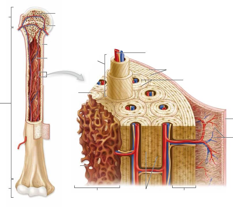
Endochondral bones increase in length in a different way,
unlike growth in intramembranous development. As an example,
consider a long bone such as a mammalian humerus (in humans,
the upper arm bone). Like many limb bones, it is formed of a
slender shaft with widened ends, called epiphyses (figure 47.4).
Figure 47.4
The structure of bone. A mammalian humerus is partly
opened to show its interior on the left. A section has been removed and magni ed
on the right to show the difference in structure between the outer compact bone
and the inner spongy bone that lines the medullary cavity. Details of basic layers,
Haversian canals, and osteocytes in lacunae can be seen here.
www.ravenbiology.com
chapter
47
The Musculoskeletal System
965
rav32223_ch47_961-980.indd 965rav32223_ch47_961-980.indd 965 11/18/09 10:51:08 AM11/18/09 10:51:08 AM
Apago PDF Enhancer
Force
Reaction
force
Force
Reaction
force
Force
Reaction
force
Force
Reaction
force
Medullary
cavity
a. b. c. d.
Figure 47.5
Model of stress and remodeling in a long
bone. This gure shows a diagrammatic section of a long bone,
such as a leg bone. The section is placed under a load or force,
which causes a reaction force from the ground the leg is standing
upon. a. Under a mild compressive load the bone does not bend.
b. If the load is large enough, and the bone is not suf ciently thick,
the bone will bend (the bending shown is exaggerated for clarity).
c. Osteoblasts are signaled by the stresses in the bending section to
produce additional bone. As the bone becomes thicker, the degree
of bending is reduced. d. When suf cient bone is added to prevent
signi cant bending, the production of new osteoblasts stops and no
more bone is added.
collagenous fibers but does possess other constituents in-
cluding mesenchyme cells.
Vascular bone usually has a special internal organization
called the Haversian system. Beneath the outer basic layers,
endochondral bone is constructed of concentric layers called
Haversian lamellae. These concentric tubes are laid down around
narrow channels called Haversian canals that run parallel to the
length of the bone. Haversian canals may contain nerve fibers
but always contain blood vessels that keep the osteocytes alive
even though they are entombed in the bony matrix.
The small vessels within the canals include both arte-
rioles and venules or capillaries, and they connect to larger
vessels that extend internally from both the periosteum and
endosteum and that run in canals perpendicular to the Haver-
sian canals.
Bone remodeling allows bone
to respond to use or disuse
It is easy to think of bones as being inert, especially since we
rarely encounter them except as the skeletons of dead animals.
But just as muscles, skin, and other body tissues may change
depending on the stresses of the environment, bone also is a
dynamic tissue that can change with demands made on it.
Mechanical stresses such as compression at joints, the
forces of muscles on certain portions and features of a bone,
and similar effects may all be remodeling factors that not only
shape the bone during its embryonic development, but after
birth as well. Depending on the directions and magnitudes of
forces impinging on a bone, it may thicken; the size and shape
of surface features to which muscles, tendons, or ligaments at-
tach may change in size and shape; even the direction of the
tiny bony struts that make up spongy bone may be altered.
Within the epiphyses are the epiphyseal growth plates that sepa-
rate the epiphyses from the shaft itself. As long as the bone is
growing in length, these growth plates are composed of cartilage
(see figure 47.4). The actual events taking place in the plates are
not simple, but they can be simply summarized.
1. During growth of a long bone, the cartilage of the growth
plates is actively growing in the lengthwise direction to
thicken the plate.
2. This growth pushes the epiphysis farther away from
the slender shaft portion, which effectively increases the
length of the bone.
3. At the same time, from the shaft’s side, a process of cartilage
calci cation encroaches on the cartilaginous growth plate,
so that the bony portion of the shaft elongates.
As long as the rate of new cartilage thickening stays ahead
of the creeping calcification, the bone continues to grow in length.
Eventually the cartilaginous expansion slows and is overtaken by
the calcification, which obliterates this region of growth.
Growth in length usually ceases in humans by late ado-
lescence. Although growth of the bone length is curtailed at
this time, growth in width is not. The diameter of the shaft
can be enhanced by bone addition just beneath the periosteum
throughout an individual’s life.
Bone structure may include
blood vessels and nerves
Developing bone often has an internal blood supply, which is es-
pecially evident in endochondral bones. The internal blood routes,
however, do not necessarily remain after the bones have com-
pleted development. In most mammals the endochondral bones
retain internal blood vessels and are called vascular bones. Vascu-
lar bone is also found in many reptiles and a few amphibians. Cellu-
lar bones contain osteocytes, and many such bones are also vascular.
This bone remains metabolically active (see figure 47.4 ).
In fishes and birds, bones are avascular. Typically avas-
cular bone does not contain osteocytes and is termed acellu-
lar bone. This type of bone is fairly inert except for its surface,
where the periosteum with its mesenchyme cells is capable of
repairing the bone.
Many bones, particularly the endochondral long bones, con-
tain a central cavity termed the medullary cavity. In many verte-
brates, the medullary cavity houses the bone marrow, important
in the manufacture of red and white blood cells. In such cases this
cavity is termed the marrow cavity. Not all medullary cavities
contain marrow, however. Light-boned birds, for example, have
huge interior cavities, but they are empty of marrow. Birds depend
on stem cells in other body locations to produce red blood cells.
Bone lining the medullary cavities differs from the
smooth, dense bone found closer to the outer surface. Based
on density and texture, bone falls into three categories: the
outer dense compact bone, the medullary bone that lines
the internal cavity, and spongy bone that has a honeycomb
structure and typically forms the epiphyses inside a thick
shell of compact bone. Both compact and spongy bone con-
tribute to a bone’s strength. Medullary cavities are lined
with thin tissues called the endosteum, which contains no
966 part
VII
Animal Form and Function
rav32223_ch47_961-980.indd 966rav32223_ch47_961-980.indd 966 11/18/09 10:51:10 AM11/18/09 10:51:10 AM
Apago PDF Enhancer
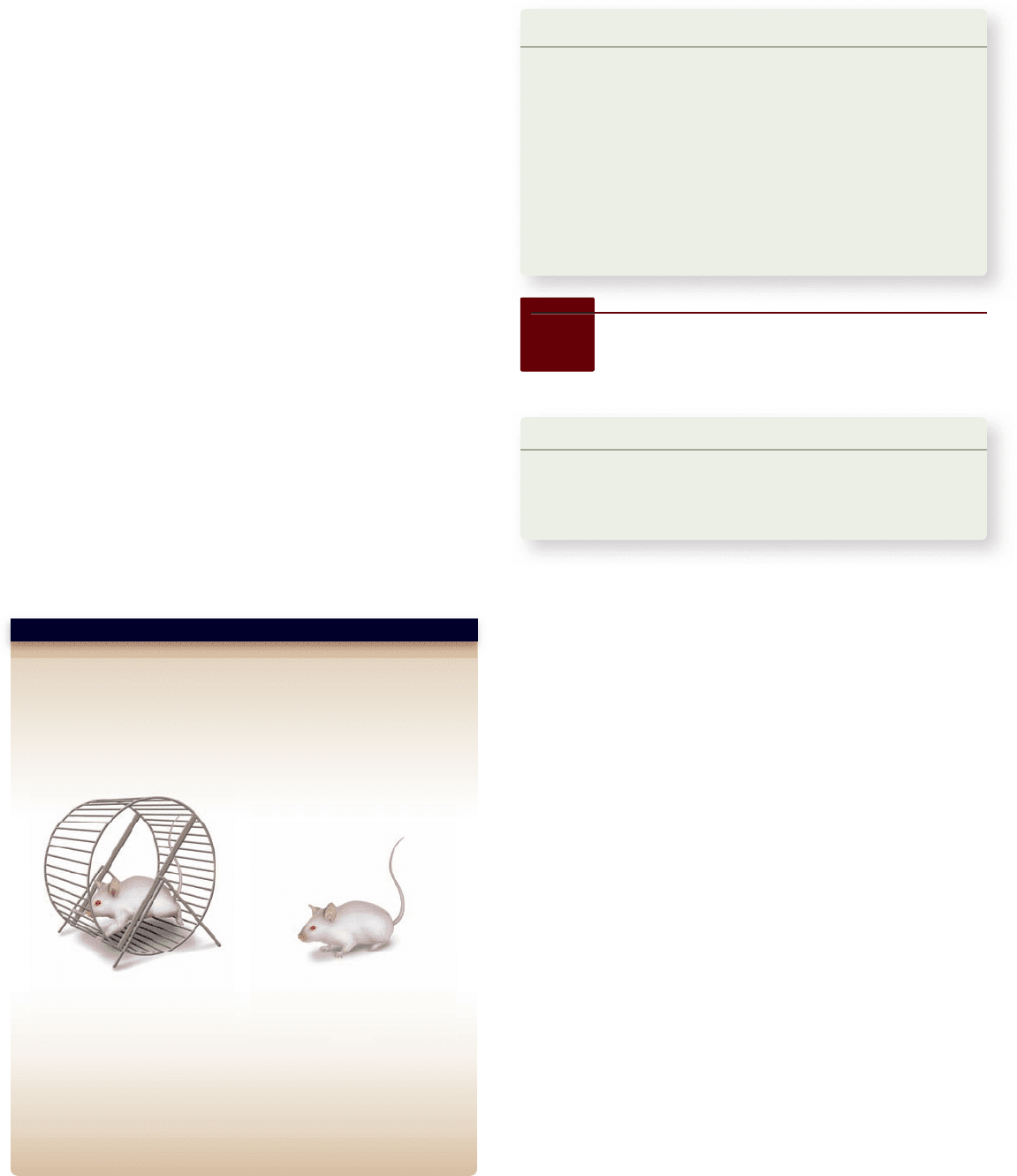
Hypothesis: Bone remodeling strengthens bones in response to
external pressures.
Prediction: Bones that are used in more strenuous activities will deposit
more bone and become stronger.
Test: Provide laboratory mice with an exercise wheel and make sure
they run for several hours a day; keep a control group without a wheel.
Result: After 10 weeks, the running mice developed thicker limb bones.
Further Experiments: Modern microelectronics allow the development
of stress sensors small enough to implant on the limb bone of a mouse.
With such sensors, experiments can quantify how much stress different
activities place on a bone and can more accurately investigate the
relationship between the direction and magnitude of forces placed on a
bone and the extent to which the bone remodels.
SCIENTIFIC THINKING
Mouse with exercise wheel Mouse without exercise wheel
Exercise and frequent use of muscles for a particular task
change more than just the muscles; blood vessels and fibrous
connective tissue increase, and the skeletal frame becomes
more robust through bone thickening and enhancement.
The phenomenon of remodeling is known for all bones,
but it is easiest to demonstrate in a long bone. Small forces may
not have much of an effect on the bone, but larger ones—if
frequent enough—can initiate remodeling (figure 47.5) . In the
example shown, larger compressive forces may tend to bend a
bone, even if the bend is imperceptible to the eye. This bend-
ing stress promotes bone formation that thickens the bone. As
the bone becomes thicker the amount of bending is reduced
(figure 47.5c). Further bone addition produces sufficient bone
thickness to entirely prevent significant bending (figure 47.5d).
Once this point has been attained, the bone addition stops. This
is another example of a negative-feedback system.
The effect of remodeling can be seen by examining bone
thickness in rodents forced to exercise. The continual stresses
placed on the limb bones cause additional bone to be deposited,
leading to thicker and stronger bone (figure 47.6) .
This phenomenon also has important medical implica-
tions. Osteoporosis, which is characterized by a loss of bone
mineral density, is a debilitating and potentially life-threatening
ailment that afflicts more than 25 million people in the United
States, affecting primarily postmenopausal women, but also
those suffering from malnutrition and a number of diseases.
One treatment is a regimen of weight-lifting to stimulate bone
deposition and thus counter the effects of osteoporosis.
Figure 47.6
The e ect of exercise on
bone remodeling.
47.3
Joints and Skeletal
Movement
Learning Outcomes
Define the different types of joints.1.
Explain how muscles produce movement at joints.2.
Describe how antagonistic muscles work at a joint.3.
Movements of the endoskeleton are powered by the skeletal
musculature. The skeletal movements that respond to mus-
cle action occur at joints, or articulations, where one bone
meets another.
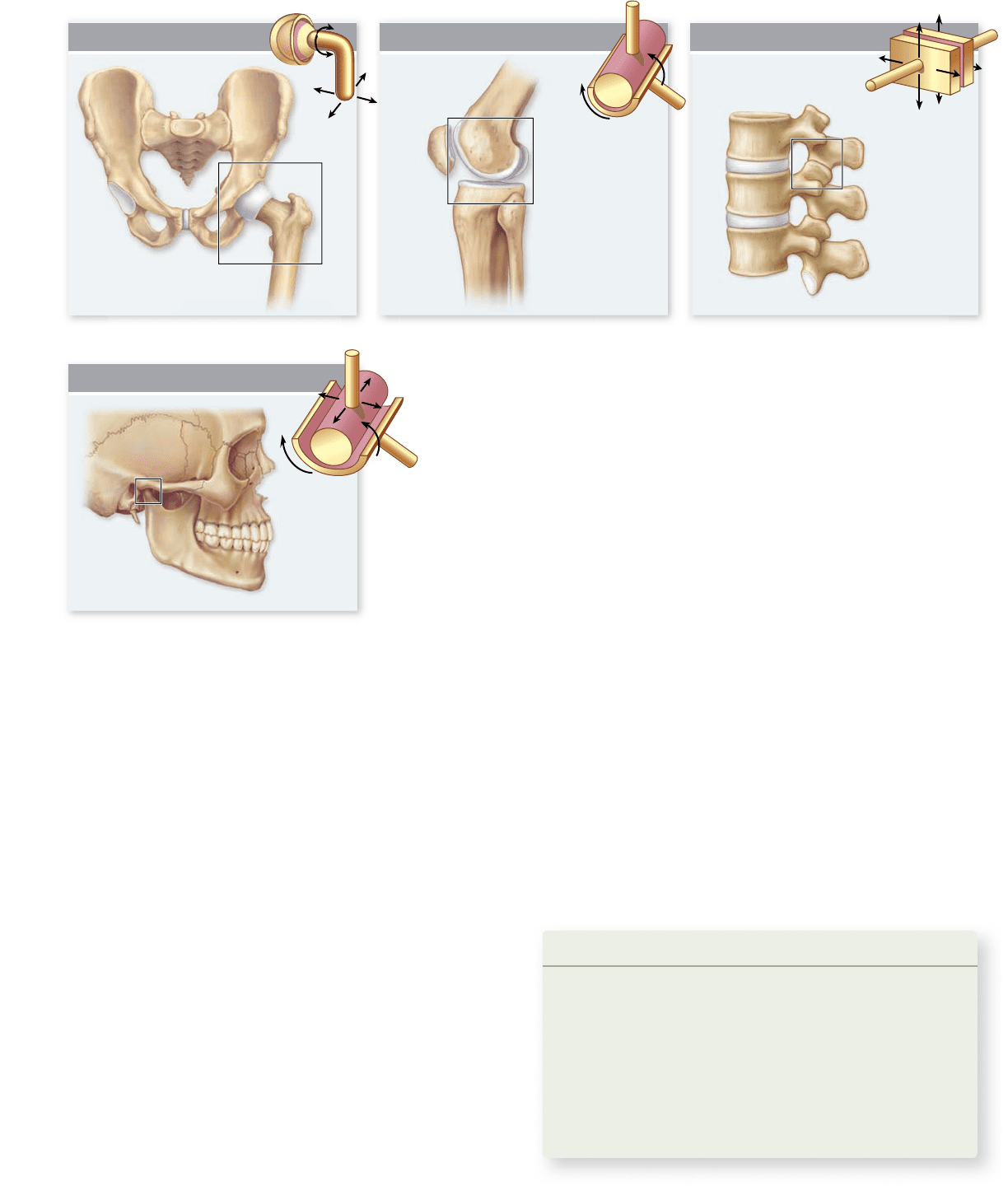
Moveable joints have di erent ranges
of motion, depending on type
Each movable joint within the skeleton has a characteristic
range of motion. Four basic joint movement patterns can be
distinguished: ball-and-socket, hinge, gliding, and combination.
Ball-and-socket joints are like those of the hip, where
the upper leg bone forms a ball fitting into a socket in the
pelvis. This type of joint can perform universal movement
in all directions, plus twisting of the ball (figure 47.7a) .
The simplest type of joint is the hinge joint, such as the
knee, where movement of the lower leg is restricted to rotate
forward or backward, but not side to side (figure 47.7b).
Gliding joints can be found in the skulls of a number of
nonmammalian vertebrates, but are also present between the
lateral vertebral projections in many of them and in mammals
as well (figure 47.7c). The vertebral projections are paired and
extend from the front and back of each vertebra. The projec-
tions in front are a little lower, and each can slip along the
undersurface of the posterior projection from the vertebra
just ahead of it. This sliding joint gives stability to the ver-
tebral column while allowing some flexibility of movement
between vertebrae.
Combination joints are, as you might suppose, those
that have movement characteristics of two or more joint
types. The typical mammalian jaw joint is a good example.
Learning Outcomes Review 47.2
Intramembranous bone forms within a layer of connective tissue;
endochondral bone originates with a cartilaginous model that is then
replaced with bone tissue. Epiphyses are cartilaginous growth plates
of endochondral bones. As the epiphyseal cartilage becomes calcified,
bone growth ceases. Bone remodeling occurs in response to repeated
stresses on bones from weight or muscle use, allowing bones
to adapt.
■ Why is vitamin D especially important for children and
the elderly?
www.ravenbiology.com
chapter
47
The Musculoskeletal System
967
rav32223_ch47_961-980.indd 967rav32223_ch47_961-980.indd 967 11/18/09 10:51:11 AM11/18/09 10:51:11 AM
Apago PDF Enhancer
Ball-and-Socket
Combination Joint
Hinge Joint Gliding Joint
a.
d.
b. c.
Figure 47.7
Patterns of joint movement. a. Ball-and-socket joints,
such as the hip joint, permit movement and twisting of the leg within the hip
socket. b. A hinge joint, as the term implies, allows movement in only one
plane. c. Gliding joints are well represented by the lateral vertebral joints (not
the central ones) that permit sliding of one surface on another. d. Combination
joints have features of more than one type of joint, such as the mammalian jaw
joint that allows both rotation and side-to-side sliding.
Most mammals chew food into small pieces. To chew food
well, the lower jaw needs to move from side to side to get
the best contact between upper and lower teeth. The lower
jaw can also slip forward and backward to some extent. At the
same time, the jaw joint must be shaped to allow the hinge-
like opening and closing of the mouth. The mammalian joint
conformation thus combines features from hinge and gliding
joints (figure 47.7d).
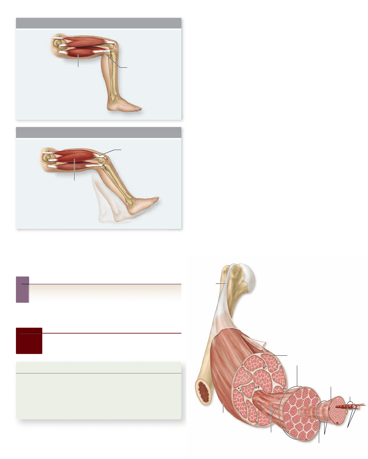
Skeletal muscles pull on bones
to produce movement at joints
Skeletal muscles produce movement of the skeleton when they
contract. Usually, the two ends of a skeletal muscle are attached
to different bones, although some may be attached to other
structures, such as skin. There are two means of bone attach-
ment: Muscle fibers may connect directly to the periosteum,
the bone’s fibrous covering, or sheets of muscle may be con-
nected to bone by a dense connective tissue strap or cord, called
a tendon that attaches to the periosteum (figure 47.8) .
One attachment of the muscle, the origin, remains rel-
atively stationary during a contraction. The other end, the
insertion, is attached to a bone that moves when the muscle
contracts. For example, contraction of the quadriceps muscles
of the leg causes the lower leg to rotate forward relative to the
upper leg section.
Typically, muscles are arranged so that any movement
produced by one muscle can be reversed by another. The leg
flexor muscles, called hamstrings (see figure 47.8), draw the
lower leg back and upward, bending the knee. Its movement
is countered by the quadriceps muscles. The important con-
cept is that two muscles or muscle groups can be mutually
antagonistic, with the action of one countered by the action
of the other.
Learning Outcomes Review 47.3
Types of joints include ball-and-socket, hinge, gliding, and combination
joints. Muscles, positioned across joints, cause movement of bones
relative to each other by contracting and exerting pulling force.
Antagonistic muscles oppose each other, a key feature since muscles can
only contract and cannot push.
■ In what ways does a bony endoskeleton overcome
the limitations of an exoskeleton for terrestrial
life forms?
968 part
VII
Animal Form and Function
rav32223_ch47_961-980.indd 968rav32223_ch47_961-980.indd 968 11/18/09 10:51:12 AM11/18/09 10:51:12 AM
Apago PDF Enhancer
Tendon
Skeletal
muscle
Bundle of
muscle fibers
Muscle fiber (cell)
Myofilaments
Myofibril
Plasma
membrane
Nuclei
Striations
Flexors
(hamstrings)
Tendon
Tendon
Extensors
(quadriceps)
Extension
Flexion
Figure 47.8
Flexor and extensor muscles of the leg.
Antagonistic muscles act in opposite ways. In humans, the
hamstrings, a group of three muscles, cause the lower leg to move
backward relative to the upper leg, whereas the quadriceps, a group
of four muscles, pull the lower leg forward.
Inquiry question
?
Would the antagonistic muscles work in the same way
in the legs of an animal with an exoskeleton, such as the
grasshopper in figure 47.2?
47.4
Muscle Contraction
Learning Outcomes
Explain the sliding filament mechanism of muscle 1.
contraction.
Describe the role of calcium in muscle contraction.2.
Differentiate between slow-twitch and fast-twitch 3.
muscle fibers.
This section concentrates on the skeletal muscle of verte-
brates. Vertebrate muscle has enjoyed the most attention
and is thus the best understood of animal muscular func-
Figure 47.9
The organization
of vertebrate skeletal muscle.
Each muscle is composed of many
bundles of muscle bers. Each ber is
composed of many myo brils, which
are each, in turn, composed
of myo laments.
tion. Each skeletal muscle contains numerous muscle fibers ,
as described in chapter 43. Each muscle fiber encloses a
bundle of 4 to 20 elongated structures called myofibrils.
Each myofibril, in turn, is composed of thick and thin
myofilaments (figure 47.9) .
Under a microscope, the myofibrils have alternat-
ing dark and light bands, which give skeletal muscle fi-
ber its striped appearance. The thick myofilaments are
stacked together to produce the dark bands, called A bands;
the thin filaments alone are found in the light bands, or
I bands.
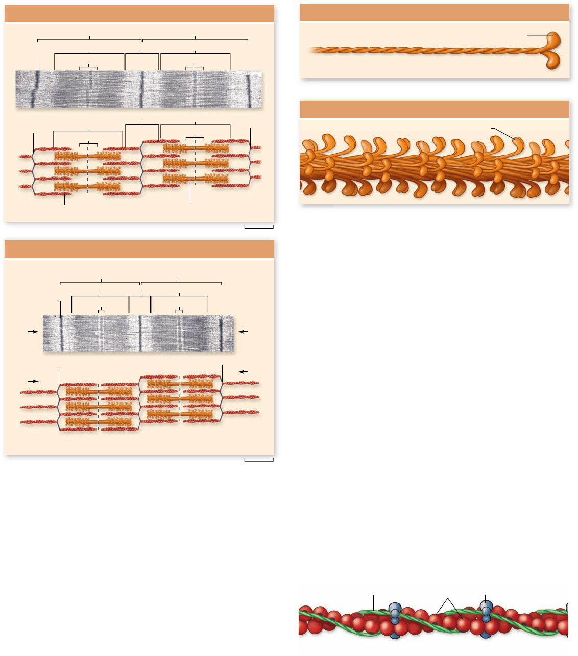
Each I band in a myofibril is divided in half by a disk of
protein called a Z line because of its appearance in electron mi-
crographs. The thin filaments are anchored to these disks. In an
electron micrograph of a myofibril (figure 47.10) , the structure
of the myofibril can be seen to repeat from Z line to Z line.
This repeating structure, called a sarcomere, is the smallest
subunit of muscle contraction.
Muscle bers contract as overlapping
laments slide together
The thin filaments overlap with thick filaments on each side
of an A band, but in a resting muscle, they do not project all
the way to the center of the A band. As a result, the center
of an A band (called an H band ) is lighter than the areas
on each side, which have interdigitating thick and thin fila-
ments. This appearance of the sarcomeres changes when the
muscle contracts.
www.ravenbiology.com
chapter
47
The Musculoskeletal System
969
rav32223_ch47_961-980.indd 969rav32223_ch47_961-980.indd 969 11/18/09 10:51:14 AM11/18/09 10:51:14 AM
Apago PDF Enhancer
Z line
H band
A band
A band
H band
H band
H band
A band I band
I band
Sarcomere Sarcomere
Z line
Z line
H band
A band
H band
A band I band
Sarcomere Sarcomere
Thin filaments (actin) Thick filaments (myosin)
Z line
Z line
Relaxed Muscle
Contracted Muscle
0.49 µm
0.45 µm
Z line
A band
Thin filament Actin molecules
Tropomyosin Troponin
Thick Filament
a.
b.
Myosin head
Myosin head
Myosin Molecule
Figure 47.10
The structure of sarcomeres in relaxed
and contracted muscles. Two sarcomeres are shown in
micrographs and as drawings of thick and thin laments. The
Z lines form the borders of each sarcomere and the A bands
represent thick laments. The thin laments are within the I bands
and extend into the A bands interdigitated with thick laments.
The H band is the lighter-appearing central region of the A band
containing only thick laments. The muscle on the top is shown
relaxed. In the contracted muscle in the bottom, the Z lines have
moved closer together, with the I bands and H bands becoming
shorter. The A band does not change in size as it contains the thick
laments, which do not change in length.
A muscle contracts and shortens because its myo fibrils
contract and shorten. When this occurs, the myo filaments do
not shorten; instead, the thick and thin myofilaments slide rela-
tive to each other (see figure 47.10). The thin filaments slide
deeper into the A bands, making the H bands narrower until,
at maximal shortening, they disappear entirely. This also makes
the I bands narrower, as the Z lines are brought closer together.
This is the sliding filament mechanism of contraction.
The sliding filament mechanism
Electron micrographs reveal cross-bridges that extend from the
thick to the thin filaments, suggesting a mechanism that might
cause the filaments to slide. To understand how this is accom-
plished requires examining the thick and thin filaments at a
molecular level. Biochemical studies show that each thick filament
is composed of many subunits of the protein myosin packed to-
gether. The myosin protein consists of two subunits, each shaped
like a golf club with a head region that protrudes from a long
filament, with the filaments twisted together. Thick filaments are
composed of many copies of myosin arranged with heads protrud-
ing from along the length of the fiber (figure 47.11). The myosin
heads form the cross-bridges seen in electron micrographs.
Each thin filament consists primarily of many globular ac-
tin proteins arranged into two fibers twisted into a double helix
(figure 47.12) . If we were able to see a sarcomere at the molecu-
lar level, it would have the structure depicted in figure 47.13 .
Figure 47.11
Thick laments are composed of myosin.
a. Each myosin molecule consists of two polypeptide chains shaped
like golf clubs and wrapped around each other; at the end of each
chain is a globular region referred to as the “head.” b. Thick
laments consist of myosin molecules combined into bundles from
which the heads protrude at regular intervals.
Figure 47.12
Thin laments are composed of globular
actin proteins. Two rows of actin proteins are twisted
together in a helix to produce the thin laments. Other proteins,
tropomyosin and troponin, associate with the strands of actin
and are involved in muscle contraction. These other proteins are
discussed later in the chapter.
970
part
VII
Animal Form and Function
rav32223_ch47_961-980.indd 970rav32223_ch47_961-980.indd 970 11/18/09 10:51:17 AM11/18/09 10:51:17 AM
Apago PDF Enhancer
Sarcomere
H band
A band I band
a.
b.
Z line
Thin filaments (actin) Thick filament (myosin) Cross-bridges
Myosin head
ATP hydrolysis Cross-bridge
formation
Power strokeATP binding,
actin release
Actin
ADP
P
i
a.
Cross-bridge
b.
c.
ATP
d.
Myosin
binding site
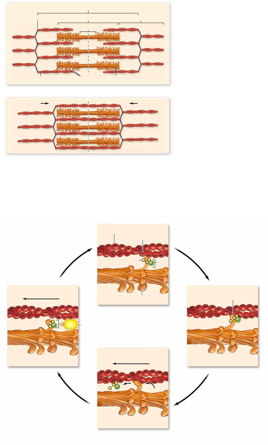
Myosin is a member of the class of protein called motor
proteins that are able to convert the chemical energy in ATP
into mechanical energy (see chapter 4). This occurs by a series
of events called the cross-bridge cycle (figure 47.14) . When
the myosin heads hydrolyze ATP into ADP and P
i
, the confor-
mation of myosin is changed, activating it for the later power
stroke. The ADP and P
i
both remain attached to the myosin
head, keeping it in this activated conformation. The analogy
to a mousetrap, set and ready to spring, is often made to de-
scribe this action. In this set position, the myosin head can bind
to actin, forming cross-bridges. When a myosin head binds to
actin, it releases the P
i
and undergoes another conformational
change, pulling the thin filament toward the center of the sar-
comere in the power stroke, at which point it loses the ADP (see
figures 47.13b, 47.14). At the end of the power stroke, the myo-
sin head binds to a new molecule of ATP, which displaces it from
actin. This cross-bridge cycle repeats as long as the muscle is
stimulated to contract. This sequence of events can be thought
of like pulling a rope hand-over-hand. The myosin heads are
the hands and the actin fibers the rope.
In death, the cell can no longer produce ATP, and there-
fore the cross-bridges cannot be broken—causing the muscle
stiffness of death called rigor mortis. A living cell, however, al-
ways has enough ATP to allow the myosin heads to detach from
actin. How, then, is the cross-bridge cycle arrested so that the
muscle can relax? We discuss the regulation of contraction and
relaxation next.
Figure 47.13
The interaction of thick and thin
laments in striated muscle sarcomeres. a. The heads on the
two ends of the thick laments are oriented in opposite directions
so that the cross-bridges pull the thin laments and the Z lines on
each side of the sarcomere toward the center. b. This sliding of the
laments produces muscle contraction.
Figure 47.14
The cross-
bridge cycle in muscle
contraction. a. Hydrolysis
of ATP by myosin causes
a conformational change
that moves the head into an
energized state. The ADP
and P
i
remain bound to the
myosin head, which can bind
to actin. b. Myosin binds to
actin forming a cross-bridge.
c. During the power stroke,
myosin returns to its original
conformation, releasing ADP
and P
i
. d. ATP binds to the
myosin head breaking the
cross-bridge. ATP hydrolysis
returns the myosin head to
its energized conformation,
allowing the cycle to
begin again.
www.ravenbiology.com
chapter
47
The Musculoskeletal System
971
rav32223_ch47_961-980.indd 971rav32223_ch47_961-980.indd 971 11/18/09 10:51:21 AM11/18/09 10:51:21 AM