Raven P.H., Johnson G.B., Mason K.A. Biology (Ninth Edition)
Подождите немного. Документ загружается.

Apago PDF Enhancer
Gastrovascular
cavity
Body stalk
Tentacle
Mouth
Food
Wastes
Nematode Earthworm
Mouth
Mouth
Pharynx
Pharynx
Intestine
Intestine
Anus
Anus
Crop
Gizzard
Salamander
Mouth
Esophagus
Intestine
Anus
Liver
Pancreas
Stomach
Cloaca
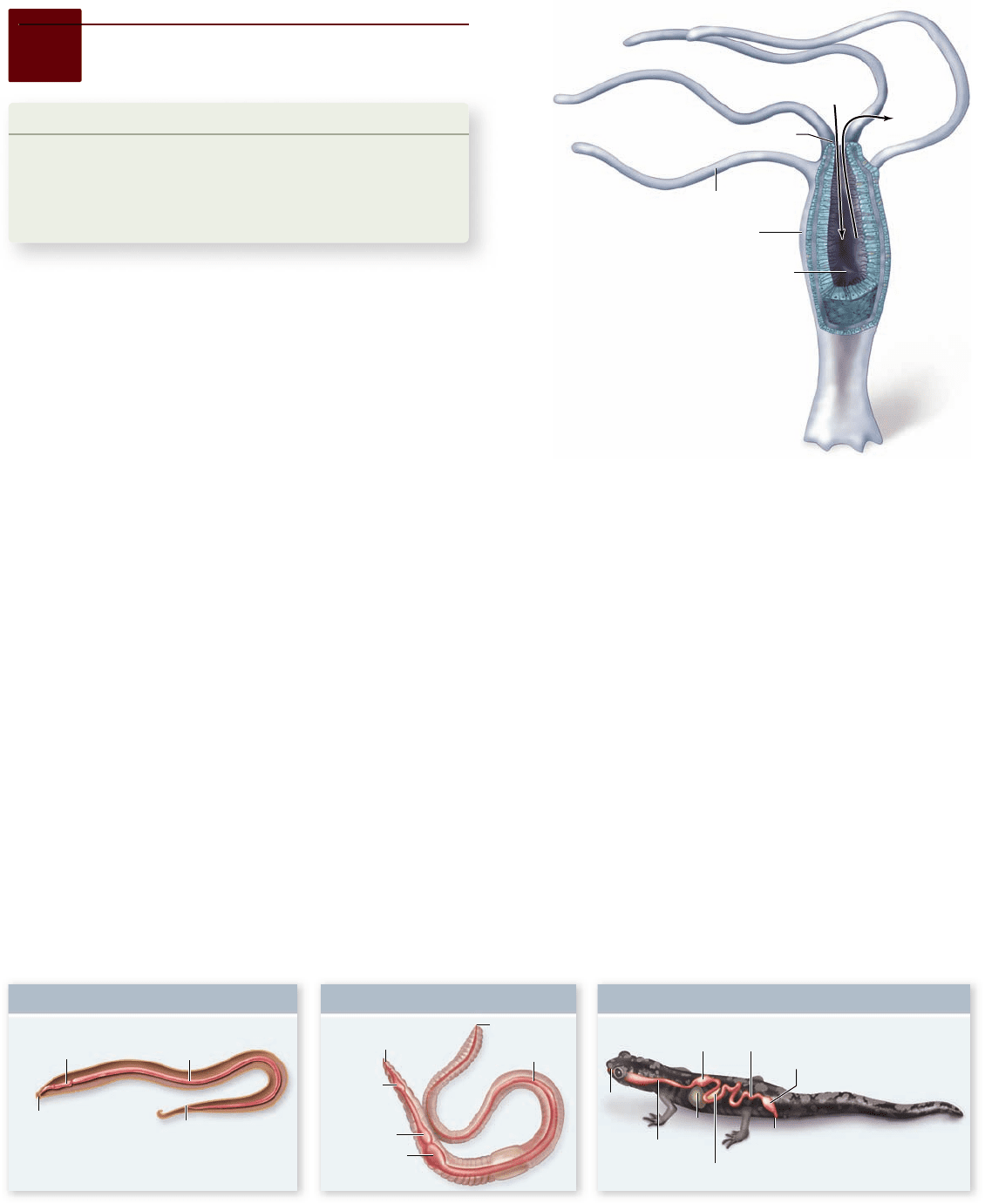
Figure 48.1
The gastrovascular
cavity of Hydra, a cnidarian. In
gastrovascular cavities, one common
opening serves as both the mouth and
the anus. There are no specialized
regions, and extracellular digestion
occurs throughout the cavity.
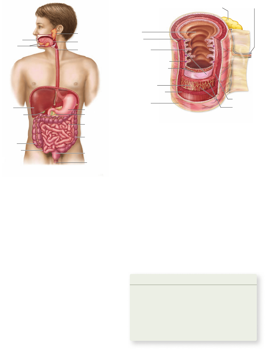
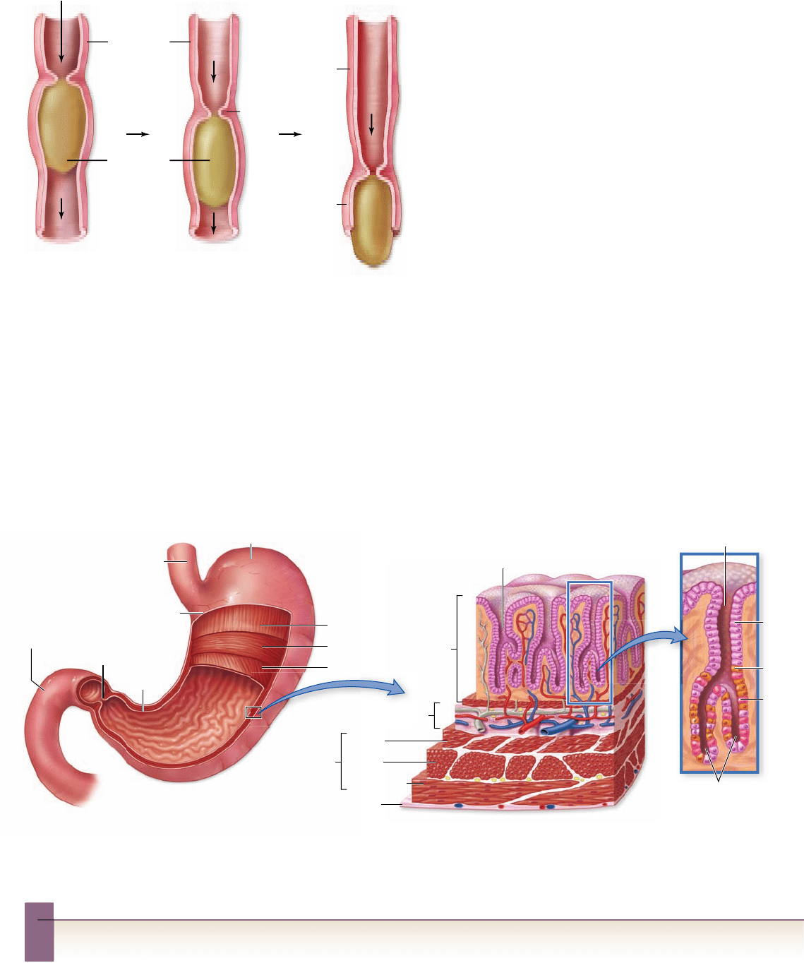
Figure 48.2
The one-way digestive tract of nematodes, earthworms, and vertebrates. One-way movement through the
digestive tract allows different regions of the digestive system to become specialized for different functions.
mentation. This fragmentation may occur through the chewing
action of teeth (in the mouth of many vertebrates) or the grind-
ing action of pebbles (in the gizzard of earthworms and birds).
Chemical digestion then occurs, breaking down the larger food
molecules of polysaccharides and disaccharides, fats, and pro-
teins into their smallest subunits.
Chemical digestion involves hydrolysis reactions that
liberate the subunit molecules—primarily monosaccharides,
amino acids, and fatty acids—from the food. These products
of chemical digestion pass through the epithelial lining of the
gut into the blood, in a process known as absorption. Any mol-
ecules in the food that are not absorbed cannot be used by the
animal. These waste products are excreted, or defecated, from
the anus.
Vertebrate digestive systems include highly
specialized structures molded by diet
In humans and other vertebrates, the digestive system consists
of a tubular gastrointestinal tract and accessory digestive or-
gans (figure 48.3).
48.1
Types of Digestive Systems
Learning Outcomes
Distinguish between incomplete and complete 1.
digestive systems.
List the components of the vertebrate digestive tract.2.
Describe the tissue layers of the gastrointestinal tract.3.
Heterotrophs are divided into three groups on the basis of their
food sources. Animals that eat plants exclusively are classified
as herbivores; common examples include algae-eating snails,
sapsucking insects, and vertebrates such as cattle, horses, rab-
bits, and sparrows. Animals that eat other animals, such as crabs,
squid, many insects, cats, eagles, trout, and frogs, are carnivores.
Animals that eat both plants and other animals are omnivores.
Humans are omnivores, as are pigs, bears, and crows.
Invertebrate digestive systems are bags or tubes
Single-celled organisms as well as sponges digest their food in-
tracellularly. Other multicellular animals digest their food ex-
tracellularly, within a digestive cavity. In this case, the digestive
enzymes are released into a cavity that is continuous with the
animal’s external environment. In cnidarians and in flatworms
such as planarians, the digestive cavity has only one opening
that serves as both mouth and anus (see chapter 33). There is
no specialization within this type of digestive system, called a
gastrovascular cavity, because every cell is exposed to all stages of
food digestion (figure 48.1) .
Specialization occurs when the digestive tract, or alimen-
tary canal, has a separate mouth and anus, so that transport of
food is one-way. The most primitive digestive tract is seen in
nematodes (phylum Nematoda), where it is simply a tubular
gut lined by an epithelial membrane. Earthworms (phylum An-
nelida) have a digestive tract specialized in different regions for
the ingestion, storage, fragmentation, digestion, and absorption
of food. All more complex animal groups, including all verte-
brates, show similar specializations (figure 48.2) .
The ingested food may be stored in a specialized region of
the digestive tract or it may first be subjected to physical frag-
982 part
VII
Animal Form and Function
rav32223_ch48_981-1000.indd 982rav32223_ch48_981-1000.indd 982 11/18/09 1:14:00 PM11/18/09 1:14:00 PM
Apago PDF Enhancer
Salivary gland
Salivary
glands
Liver
Oral cavity
Esophagus
Gallbladder
Pharynx
Cecum
Appendix
Anus
Rectum
Small intestine
Pancreas
Stomach
Large intestine
Blood vessel
Nerve
Myenteric
plexus
Submucosal plexus
Epithelial
tissue layer
Gland in submucosa
Longitudinal layer
Circular layer
Muscularis
Gland outside gastrointestinal tract
Mucosa
Lumen
Submucosa
Serosa
Figure 48.3
The human digestive system. The human
digestive system consists of the oral cavity, esophagus, stomach,
small intestine, large intestine, rectum, and anus; and is aided by
accessory organs.
Figure 48.4
The layers of the gastrointestinal tract.
The mucosa contains an epithelial lining; the submucosa is
composed of connective tissue; and the muscularis consists of
smooth muscles. Glands secrete substances via ducts into speci c
regions of the tract.
are secreted into the first region of the small intestine, the duo-
denum, where they aid digestion.
Tissues of the digestive tract
The tubular gastrointestinal tract of a vertebrate has a charac-
teristic layered structure (figure 48.4) . The innermost layer is
the mucosa, an epithelium that lines the interior, or lumen , of
the tract. The next major tissue layer, made of connective tis-
sue, is called the submucosa.
Just outside the submucosa is the muscularis, which con-
sists of a double layer of smooth muscles. The muscles in the
inner layer have a circular orientation and serve to constrict the
gut, whereas those in the outer layer are arranged longitudi-
nally and work to shorten it. Another epithelial tissue layer, the
serosa, covers the external surface of the tract. Nerve networks,
intertwined in plexuses between muscle layers, are located in the
submucosa and help regulate the gastrointestinal activities.
In the rest of this chapter, we focus on the details of the verte-
brate digestive system’s structure and function. We close the chap-
ter with discussion of nutrients that are essential to vertebrates.
Learning Outcomes Review 48.1
Incomplete digestive tracts have only one opening; complete digestive
tracts are fl ow-through, with a mouth and an anus. The digestive system
of vertebrates includes mouth and pharynx, esophagus, stomach, small
and large intestines, cloaca or rectum, anus, and accessory organs.
The layers of tissue that compose the tubular tract are the mucosa, the
submucosa, the muscularis, and the serosa.
■ What might be the advantages of a one-way
digestive system?
Overview of the digestive tract
The initial components of the gastrointestinal tract are the
mouth and the pharynx, which is the common passage of the
oral and nasal cavities. The pharynx leads to the esophagus, a
muscular tube that delivers food to the stomach, where some
preliminary digestion occurs.
From the stomach, food passes to the small intestine,
where a battery of digestive enzymes continues the digestive
process. The products of digestion, together with minerals
and water, are absorbed across the wall of the small intestine
into the bloodstream. What remains is emptied into the large
intestine, where some of the remaining water and minerals
are absorbed.
In most vertebrates other than mammals, the waste prod-
ucts emerge from the large intestine into a cavity called the cloaca
(see figure 48.2), which also receives the products of the urinary
and reproductive systems. In mammals, the urogenital products
are separated from the fecal material in the large intestine; the fe-
cal material enters the rectum and is expelled through the anus.
The accessory digestive organs include the liver, which
produces bile (a green solution that emulsifies fat), the gallblad-
der, which stores and concentrates the bile, and the pancreas.
The pancreas produces pancreatic juice, which contains digestive
enzymes and bicarbonate buffer. Both bile and pancreatic juice
www.ravenbiology.com
chapter
48
The Digestive System
983
rav32223_ch48_981-1000.indd 983rav32223_ch48_981-1000.indd 983 11/18/09 1:14:02 PM11/18/09 1:14:02 PM
Apago PDF Enhancer
Mou
MouMou
Mou
Mou
Mou
Mou
Mou
Mou
M
Mou
o
o
o
o
o
ou
Mou
Mo
o
o
o
o
Mou
o
Mou
Mou
Mou
o
M
M
Mo
o
Mo
Mou
o
ou
Mou
M
Mo
o
Mo
o
o
u
Mou
ou
Mou
Mou
Mou
ou
Mo
M
M
o
o
u
Mou
Mou
Mou
M
o
o
o
ou
M
M
o
o
o
Mo
u
u
M
M
Mo
M
o
M
o
o
ou
u
M
o
o
o
o
o
o
o
o
o
o
M
o
M
M
o
o
o
ou
u
M
o
M
o
th
th
th
th
th
th
th
t
th
th
t
t
th
th
t
t
t
th
th
th
th
th
th
th
t
th
h
th
th
th
t
th
t
h
t
t
h
th
th
h
h
t
th
th
t
h
E
E
Es
Eso
Es
so
Eso
so
o
Eso
so
Eso
o
o
o
E
so
Eso
o
E
Eso
so
so
so
o
Es
o
Eso
Es
Es
Eso
o
E
E
E
E
E
Eso
o
E
Eso
o
E
o
E
ph
phapha
h
pha
pha
pha
ph
h
pha
pha
pha
pha
ph
h
h
h
pha
ha
pha
pha
pha
ha
pha
pha
pha
p
p
ph
h
h
h
pha
pha
p
a
a
a
a
a
ph
p
ph
h
pp
p
p
p
gus
gu
gu
gus
us
gus
us
gus
g
gu
gus
g
s
gus
gus
us
gu
us
g
g
g
u
g
g
g
StoSto
t
t
o
Sto
Sto
o
o
o
o
o
mac
m
mac
mac
mac
mac
mac
mac
mac
mac
ac
mac
m
mac
mac
ma
c
m
a
m
c
m
h
hh
h
h
h
h
Giz
Giz
Gi
G
Gi
Giz
Giz
Giz
iz
z
z
z
G
G
z
Giz
G
G
Giz
z
Giz
Giz
z
G
z
z
z
za
ar
a
ar
ar
ar
z
za
a
a
z
zar
ar
z
z
z
ar
z
a
z
z
zar
z
a
z
a
ar
r
d
d
d
d
d
d
d
d
d
d
d
d
d
d
d
d
d
d
d
d
d
d
d
d
d
d
d
d
d
I
ntestine
Anu
s
C
C
C
Cro
Cro
Cro
o
o
o
C
C
C
C
Cro
Cro
C
C
Cro
C
C
C
C
p
p
p
p
Horse Lion Human
Molars
Premolars
Canines
Incisors
Herbivore Carnivore Omnivore
Figure 48.6
Patterns of dentition
depend on diet. Different vertebrates
(herbivore, carnivore, or omnivore) have evolved
speci c variations from a generalized pattern of
dentition depending on their diets.
Figure 48.5
The digestive tract of birds. Birds lack teeth
but have a muscular chamber called the gizzard that works to break
down food. Birds swallow gritty objects or pebbles that lodge in the
gizzard and pulverize food before it passes into the intestine. Food
is stored in the crop.
with fluid secretions. Carnivorous mammals have pointed teeth
that lack flat grinding surfaces. Such teeth are adapted for cut-
ting and shearing. Carnivores often tear off pieces of their prey
but have little need to chew them, because digestive enzymes
can act directly on animal cells. By contrast, grass-eating her-
bivores must pulverize the cellulose cell walls of plant tissue
before the bacteria in their rumens or cecae can digest them.
These animals have large, flat teeth with complex ridges well
suited to grinding.
Human teeth are specialized for eating both plant and
animal food. Viewed simply, humans are carnivores in the front
of the mouth and herbivores in the back (see figure 48.6). The
four front teeth in the upper and lower jaws are sharp, chisel-
shaped incisors used for biting. On each side of the incisors
are sharp, pointed teeth called cuspids (sometimes referred to
as “canine” teeth), which are used for tearing food. Behind the
canines are two premolars and three molars, all with flattened,
ridged surfaces for grinding and crushing food.
The mouth is a chamber for ingestion
and initial processing
Inside the mouth, the tongue mixes food with a mucous solu-
tion, saliva. In humans, three pairs of salivary glands secrete
saliva into the mouth through ducts in the mouth’s mucosal
lining. Saliva moistens and lubricates the food so that it is easier
to swallow and does not abrade the tissue of the esophagus as
it passes through .
Saliva also contains the hydrolytic enzyme salivary amy-
lase, which initiates the breakdown of the polysaccharide starch
into the disaccharide maltose. This digestion is usually minimal
in humans, however, because most people don’t chew their food
very long.
Stimulation of salivation
The secretions of the salivary glands are controlled by the ner-
vous system, which in humans maintains a constant flow of
about half a milliliter per minute when the mouth is empty of
food. This continuous secretion keeps the mouth moist.
The presence of food in the mouth triggers an increased
rate of secretion. Taste buds as well as olfactory (smell) neu-
rons send impulses to the brain, which responds by stimu-
lating the salivary glands (see chapter 46). The most potent
stimuli are acidic solutions; lemon juice, for example, can
increase the rate of salivation eightfold. The sight, sound,
or smell of food can stimulate salivation markedly in many
48.2
The Mouth and Teeth: Food
Capture and Bulk Processing
Learning Outcomes
Identify adaptive variation in vertebrate tooth shape.1.
Understand the role of the mouth in the digestive process.2.
Specializations of the digestive systems in different kinds of ver-
tebrates reflect the way these animals live. Birds, which lack teeth,
break up food in their two-chambered stomachs (figure 48.5) . In
one of these chambers, called the gizzard, small pebbles ingested
by the bird are churned together with the food by muscular ac-
tion. This churning grinds up the seeds and other hard plant ma-
terial into smaller chunks that can be digested more easily.
Vertebrate teeth are adapted to di erent
types of food items
Many vertebrates have teeth (figure 48.6), used for chewing, or
mastication, that break up food into small particles and mix it
984 part
VII
Animal Form and Function
rav32223_ch48_981-1000.indd 984rav32223_ch48_981-1000.indd 984 11/18/09 1:14:07 PM11/18/09 1:14:07 PM
Apago PDF Enhancer
Pharynx
Soft palate
Hard palate
Tongue
Epiglottis
Larynx
Trachea
Esophagus
Air
1. As f
ood moves to the
back of the mouth, the
soft palate seals off
the nasal cavity.
2. During swallowing, the larynx rises and is sealed off by
the epiglottis. This forces the bolus into the esophagus
and prevents entry into the trachea. As the bolus moves
into the esophagus the larynx relaxes.
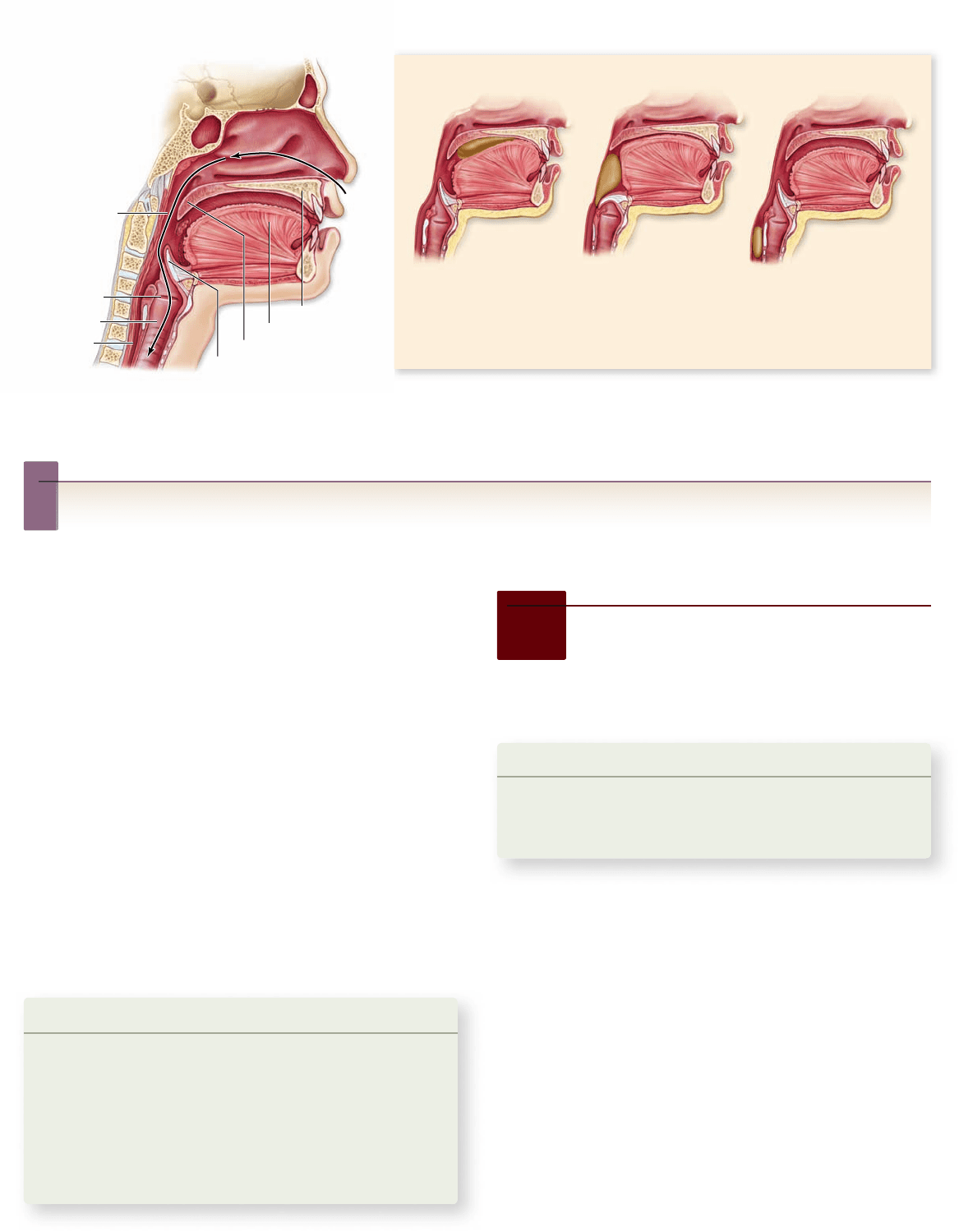
Figure 48.7
The mechanics of swallowing. Cross section through head and throat showing relevant structures (left). During
swallowing (right) the tongue pushes the palate upward, and the soft palate seals off the nasal cavity. Elevation of the larynx causes the
epiglottis to seal off the trachea, thus preventing food from entering the airway.
Inquiry question
?
What goes wrong to cause someone to choke?
animals; in humans, thinking or talking about food can also
have this effect.
Swallowing
Swallowing is initiated by voluntary action, then is continued
under involuntary control. When food is ready to be swallowed,
the tongue moves it to the back of the mouth. In mammals, the
process of swallowing begins when the soft palate elevates, push-
ing against the back wall of the pharynx (figure 48.7) . Elevation
of the soft palate seals off the nasal cavity and prevents food from
entering it. Pressure against the pharynx triggers an automatic,
involuntary response, the swallowing reflex. Because it is a reflex,
swallowing cannot be stopped once it is initiated.
Neurons within the walls of the pharynx send impulses
to the swallowing center in the brain. In response, electrical
impulses in motor neurons stimulate muscles to contract and
raise the larynx (voice box). This pushes the glottis, the open-
ing from the larynx into the trachea (windpipe), against a flap of
tissue called the epiglottis. These actions keep food out of the
respiratory tract, directing it instead into the esophagus.
Learning Outcomes Review 48.2
In vertebrates with teeth, tooth shape exhibits adaptations to diet:
herbivores have large, fl at teeth for grinding, whereas carnivores have
pointed teeth for tearing. The mouth serves as an initial processing
center, tasting ingested food, breaking it down, and beginning digestion
with saliva secretion prior to swallowing.
■ Which parts of the food ingestion process are voluntary
and which are involuntary?
48.3
The Esophagus and the
Stomach: The Early Stages
of Digestion
Learning Outcomes
Describe how food moves through the esophagus.1.
Explain what digestive processes take place in 2.
the stomach.
Swallowed food enters a muscular tube called the esophagus,
which connects the pharynx to the stomach. The esophagus ac-
tively moves a processed lump of food, called a bolus, through
the action of muscles. Food from a meal is stored in the stom-
ach where it undergoes early stages of digestion.
Muscular contractions of the esophagus
move food to the stomach
In adult humans, the esophagus is about 25 cm long; the up-
per third is enveloped in skeletal muscle for voluntary control
of swallowing, whereas the lower two-thirds is surrounded by
involuntary smooth muscle. The swallowing center stimu-
lates successive one-directional waves of contraction in these
muscles that move food along the esophagus to the stomach.
These rhythmic waves of muscular contraction are called
www.ravenbiology.com
chapter
48
The Digestive System
985
rav32223_ch48_981-1000.indd 985rav32223_ch48_981-1000.indd 985 11/18/09 1:14:08 PM11/18/09 1:14:08 PM
Apago PDF Enhancer
Esophagus
Food Bolus
Peristalic
movement
Contraction
Relaxation
Relaxation
Gastric pit
Gastric pit
Muscularis
Pyloric
sphincter
Longitudinal
Circular
Oblique
Muscularis
Longitudinal
Circular
Oblique
Serosa
Serosa
Submucosa
Mucosa
Gastric glands
Chief
cell
Parietal
cell
Mucous
cell
Esophagus
Stomach
Mucosa
Duodenum
Figure 48. 8
The esophagus and peristalsis. After
food has entered the esophagus, rhythmic waves of muscular
contraction, called peristalsis, move the food down to the stomach.
Figure 48.9
The stomach and duodenum. Food enters the stomach from the esophagus. A ring of smooth muscle called the pyloric
sphincter controls the entrance to the duodenum, the upper part of the small intestine. The epithelial walls of the stomach are dotted with
deep infoldings called gastric pits that contain gastric glands. The gastric glands consist of mucous cells, chief cells that secrete pepsinogen,
and parietal cells that secrete HCl. Gastric pits are the openings of the gastric glands.
Inquiry question
?
How does the digestive system keep from being digested by the gastric secretions it produces?
peristalsis (figure 48.8) ; they enable humans and other verte-
brates to swallow even if they are upside down.
In many vertebrates, the movement of food from the
esophagus into the stomach is controlled by a ring of circular
smooth muscle, or a sphincter, that opens in response to the pres-
sure exerted by the food. Contraction of this sphincter prevents
food in the stomach from moving back into the esophagus. Ro-
dents and horses have a true sphincter at this site, and as a result
they cannot regurgitate; humans lack a true sphincter. Normally,
the esophagus is closed off except during swallowing.
The stomach is a “holding station”
involved in acidic breakdown of food
The stomach (figure 48.9) is a saclike portion of the digestive
tract. Its inner surface is highly convoluted, enabling it to fold
up when empty and open out like an expanding balloon as it
fills with food. For example, the human stomach has a volume
of only about 50 mL when empty, but, it may expand to contain
2 to 4 L of food when full. Carnivores that engage in sporadic
gorging as an important survival strategy possess stomachs that
are able to distend even more.
Secretory systems
The stomach contains a third layer of smooth muscle for churn-
ing food and mixing it with gastric juice, an acidic secretion of
the tubular gastric glands of the mucosa (see figure 48.9). These
exocrine glands contain three kinds of secretory cells: mucus-
secreting cells, parietal cells, which secrete hydrochloric acid
(HCl), and chief cells, which secrete pepsinogen, the inactive
form of the protease (protein-digesting enzyme) pepsin.
Pepsinogen has 44 additional amino acids that block its
active site. HCl causes pepsinogen to unfold, exposing the active
site, which then acts to remove the 44 amino acids. This yields
the active protease, pepsin. This process of secreting an inac-
tive form that is then converted into an active enzyme outside
the cell prevents the chief cells from digesting themselves. In
the stomach, mucus produced by mucus-secreting cells serves
986 part
VII
Animal Form and Function
rav32223_ch48_981-1000.indd 986rav32223_ch48_981-1000.indd 986 11/18/09 5:02:10 PM11/18/09 5:02:10 PM

Apago PDF Enhancer
the same purpose, covering the interior walls and preventing
them from being digested.
In addition to producing HCl, the parietal cells of the
stomach also secrete intrinsic factor, a polypeptide needed for
the intestinal absorption of vitamin B
12
. Because this vitamin
is required for the production of red blood cells, people who
lack sufficient intrinsic factor develop a type of anemia (low red
blood cell count) called pernicious anemia.
Action of acid
The human stomach produces about 2 L of HCl and other
gastric secretions every day, creating a very acidic solution.
The concentration of HCl in this solution is about 10 milli-
molar (mM), equal to a pH of 2. Thus, gastric juice is about
250,000 times more acidic than blood, whose normal pH
is 7.4.
The low pH in the stomach helps denature food proteins,
making them easier to digest, and keeps pepsin maximally ac-
tive. Active pepsin hydrolyzes food proteins into shorter chains
of polypeptides that are not fully digested until the mixture
enters the small intestine. The mixture of partially digested
food and gastric juice is called chyme. In adult humans, only
proteins are partially digested in the stomach—no significant
digestion of carbohydrates or fats occurs there.
The acidic solution within the stomach also kills most of
the bacteria that are ingested with the food. The few bacteria
that survive the stomach and enter the intestine intact are able
to grow and multiply there, particularly in the large intestine.
In fact, vertebrates harbor thriving colonies of bacteria within
their intestines, and bacteria are a major component of feces.
As we discuss later, bacteria that live within the digestive tracts
of ruminants play a key role in the ability of these mammals to
digest cellulose.
Ulcers
Overproduction of gastric acid can occasionally eat a hole
through the wall of the stomach or the duodenum, causing a
peptic ulcer. Although we once blamed consumption of spicy
food, the most common cause of peptic ulcers is now thought
to be infection with the bacterium Heliobacter pylori.
H. pylori can grow on the lining of the human stom-
ach, surviving the acid pH by secreting substances that buf-
fer the pH of its immediate surroundings. Although infection
with H. pylori is common in the United States (about 20% of
people younger than 40 and 50% older than 60), most people
are asymp tom at ic. However, in some cases, infection by H. py-
lori can reduce or weaken the mucosal layer in the stomach or
duodenum, allowing acidic secretions to attack the underlying
epithelium. Antibiotic treatment of the infection can reduce
symptoms and often even cure the ulcer.
Leaving the stomach
Chyme leaves the stomach through the pyloric sphincter (see
figure 48.9) to enter the small intestine. This is where all termi-
nal digestion of carbohydrates, lipids, and proteins occurs and
where the products of digestion—amino acids, glucose, and so
on—are absorbed into the blood. Only some of the water in
chyme and a few substances, such as aspirin and alcohol, are
absorbed through the wall of the stomach.
Learning Outcomes Review 48.3
Peristaltic waves of contraction and relaxation of smooth muscle propel
food along the esophagus to the stomach. Gastric juice contains strong
hydrochloric acid and the enzyme pepsin, a protease that begins the
breakdown of proteins into shorter polypeptides. The acidic chyme is
then transferred through the pyloric sphincter into the small intestine.
■ Suppose you ate a chicken sandwich (chicken breast on
bread with mayonnaise). Which of these foods would
begin its breakdown in the stomach?
48.4
The Intestines: Breakdown,
Absorption, and Elimination
Learning Outcomes
Compare the structures of the small and large intestines.1.
Name the accessory organs and describe their roles.2.
Explain how absorbed nutrients move into the blood or 3.
lymph capillaries.
The capacity of the small intestine is limited, and its diges-
tive processes take time. Consequently, efficient digestion
requires that only relatively small amounts of chyme be in-
troduced from the stomach into the small intestine at any one
time. Coordination between gastric and intestinal activities is
regulated by neural and hormonal signals, which we will de-
scribe in section 48.6.
The structure of the small intestine is
specialized for digestion and nutrient uptake
The small intestine is approximately 4.5 m long in a living per-
son, but 6 m long at autopsy when all the muscles have relaxed.
The first 25 cm is the duodenum; the remainder of the small
intestine is divided into the jejunum and the ileum.
The duodenum receives acidic chyme from the stom-
ach, digestive enzymes and bicarbonate from the pancreas,
and bile from the liver and gallbladder. Enzymes in the pan-
creatic juice digest larger food molecules into smaller frag-
ments. This digestion occurs primarily in the duodenum
and jejunum.
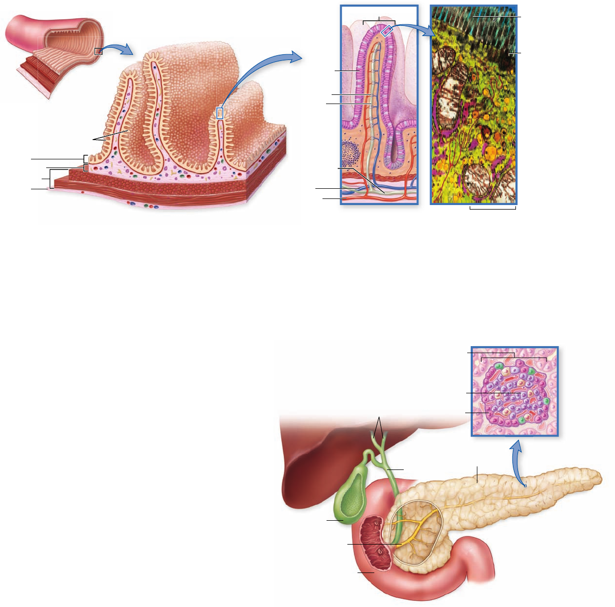
The epithelial wall of the small intestine is covered with tiny,
fingerlike projections called villi (singular, villus; figure 48.10) .
In turn, each epithelial cell lining the villi is covered on its apical
surface (the side facing the lumen) by many foldings of the plasma
membrane that form cytoplasmic extensions called microvilli.
These are quite tiny and can be seen clearly only with an electron
microscope. Under a light micrograph, the microvilli resemble the
www.ravenbiology.com
chapter
48
The Digestive System
987
rav32223_ch48_981-1000.indd 987rav32223_ch48_981-1000.indd 987 11/18/09 1:14:15 PM11/18/09 1:14:15 PM
Apago PDF Enhancer
2 μm
Small intestine
Villi
Villus
Mucosa
Serosa
Submucosa
Muscularis
Epithelial
cell
Capillary
Lacteal
Vein
Artery
Lymphatic
duct
Cell
membrane
Microvilli
From liver
Gallbladder
Pancreatic
duct
Pancreas
Common
bile duct
Duodenum
β cell
α cell
Pancreatic islet
(of Langerhans)
Figure 48.10
The small intestine. Successive enlargements show folded epithelium studded with villi that increase the surface area.
The micrograph shows an epithelial cell with numerous microvilli.
Figure 48.11
The pancreas. The pancreatic and bile
ducts empty into the duodenum. The pancreas secretes pancreatic
juice into the pancreatic duct. The pancreatic islets of Langerhans
secrete hormones into the blood; α cells secrete glucagon, and
β cells secrete insulin. The liver secretes bile, which consists of bile
pigments (waste products from the liver) and bile salts. Bile salts
play a role in the digestion of fats. Bile is concentrated and stored
in the gallbladder until it is needed in the duodenum on the arrival
of fatty food.
bristles of a brush, and for that reason the epithelial wall of the
small intestine is also called a brush border.
The villi and microvilli greatly increase the surface area
of the small intestine; in humans, this surface area is 300 m
2
—
about 3200 square feet, larger than a tennis court! It is over this
vast surface that the products of digestion are absorbed.
The microvilli also participate in digestion because a
number of digestive enzymes are embedded within the epi-
thelial cells’ plasma membranes, with their active sites exposed
to the chyme. These brush border enzymes include those that
hydrolyze the disaccharides lactose and sucrose, among others.
Many adult humans lose the ability to produce the brush bor-
der enzyme lactase and therefore cannot digest lactose (milk
sugar), a rather common condition called lactose intolerance.
The brush border enzymes complete the digestive process that
started with the action of salivary amylase in the mouth.
Accessory organs secrete enzymes
into the small intestine
The main organs that aid digestion are the pancreas, liver, and
gallbladder. They empty their secretions, primarily enzymes,
through ducts directly into the small intestine.
Secretions of the pancreas
The pancreas (figure 48.11) , a large gland situated near the junc-
tion of the stomach and the small intestine, secretes pancreatic
fluid into the duodenum through the pancreatic duct; thus, the
pancreas functions as an exocrine gland. This fluid contains a host
of enzymes, including trypsin and chymotrypsin, which digest
proteins; pancreatic amylase, which digests starch; and lipase,
which digests fat. Like pepsin in the stomach, these enzymes are
released into the duodenum primarily as inactive enzymes and
are then activated by trypsin, which is first activated by a brush
border enzyme of the intestine.
Pancreatic enzymes digest proteins into smaller polypep-
tides, polysaccharides into shorter chains of sugars, and fats
into free fatty acids and monoglycerides. Digestion of proteins
and carbohydrates is then completed by the brush border en-
zymes. Pancreatic fluid also contains bicarbonate, which neu-
tralizes the HCl from the stomach and gives the chyme in the
duodenum a slightly alkaline pH. The digestive enzymes and
988 part
VII
Animal Form and Function
rav32223_ch48_981-1000.indd 988rav32223_ch48_981-1000.indd 988 11/18/09 1:14:16 PM11/18/09 1:14:16 PM
Apago PDF Enhancer
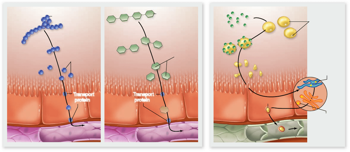
Lumen
of
small
intestine
Protein
Transport
protein
Transport
protein
Carbohydrate
Bile salts
Emulsified
droplets
Free fatty acids,
monoglycerides
Chylomicron
Fat globules
(triglycerides)
Monosaccharides
Amino acids
Blood capillary
Lymphatic
capillary
Epithelial
cell of
intestinal
villus
Resynthesis
of triglycerides
Triglycerides
get protein
cover
a. b.
Figure 48.12
Absorption of the products of digestion. a. Monosaccharides and amino acids are transported into blood
capillaries. b. Fatty acids and monoglycerides within the intestinal lumen are absorbed and converted within the intestinal epithelial cells into
triglycerides. These are then coated with proteins to form structures called chylomicrons, which enter lymphatic capillaries.
bicarbonate are produced by clusters of secretory cells known
as acini.
In addition to its exocrine role in digestion, the pan-
creas also functions as an endocrine gland, secreting several
hormones into the blood that control the blood levels of glu-
cose and other nutrients. These hormones are produced in
the islets of Langerhans, clusters of endocrine cells scattered
throughout the pancreas. The two most important pancreatic
hormones, insulin and glucagon, were described in chapter 46 ;
their actions are also discussed later on.
Liver and gallbladder
The liver is the largest internal organ of the body (see figure 48.3).
In an adult human, the liver weighs about 1.5 kg and is the size
of a football. The main exocrine secretion of the liver is bile, a
fluid mixture consisting of bile pigments and bile salts that is de-
livered into the duodenum during the digestion of a meal.
The bile pigments do not participate in digestion; they
are waste products resulting from the liver’s destruction of old
red blood cells and are ultimately eliminated with the feces. If
the excretion of bile pigments by the liver is blocked, the pig-
ments can accumulate in the blood and cause a yellow staining
of the tissues known as jaundice.
In contrast, the bile salts play a very important role in
preparing fats for subsequent enzymatic digestion. Because fats
are insoluble in water, they enter the intestine as drops within
the watery chyme. The bile salts, which are partly lipid-soluble
and partly water-soluble, work like detergents, dispersing the
large drops of fat into a fine suspension of smaller droplets.
This emulsification action produces a greater surface area of fat
for the action of lipase enzymes, and thus allows the digestion
of fat to proceed more rapidly.
After bile is produced in the liver, it is stored and concen-
trated in the gallbladder. The arrival of fatty food in the duode-
num triggers a neural and endocrine reflex that stimulates the
gallbladder to contract, causing bile to be transported through
the common bile duct and injected into the duodenum (these
reflexes are the topic of a later section). Gallstones are hardened
precipitates of cholesterol that form in some individuals. If these
stones block the bile duct, contraction of the gallbladder causes
intense pain, often felt in the back. In severe cases of blockage,
surgical removal of the gallbladder may be performed.
Absorbed nutrients move into blood
or lymph capillaries
After their enzymatic breakdown, proteins and carbohy-
drates are absorbed as amino acids and monosaccharides, re-
spectively. They are transported across the brush border into
the epithelial cells that line the intestine by a combination
of active transport and facilitated diffusion (figure 48.12a) .
Glucose is transported by coupled transport with Na
+
ions
(also called secondary active transport). Fructose, found in most
fruit, is transported by facilitated diffusion. Most amino acids
are transported by active transport using a variety of different
transporters. Some of these carrier proteins use cotransport
with Na
+
ions; others transport only amino acids. Once they
have entered epithelial cells across the apical membrane, these
monosaccharides and amino acids move through the cytoplasm
and are transported across the basolateral membrane and into
the blood capillaries within the villi.
The blood carries these products of digestion from the in-
testine to the liver via the hepatic portal vein. A portal vein con-
nects two beds of capillaries instead of returning to the heart. In
www.ravenbiology.com
chapter
48
The Digestive System
989
rav32223_ch48_981-1000.indd 989rav32223_ch48_981-1000.indd 989 11/18/09 1:14:19 PM11/18/09 1:14:19 PM
Apago PDF Enhancer
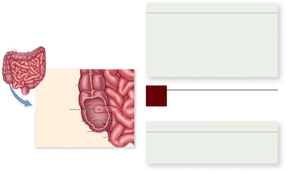
Ascending portion
of large intestine
Appendix
Last portion of
small intestine
Cecum
Ileocecal valve
Figure 48.13
The junction of the small and large
intestines in humans. The large intestine, or colon, starts with
the cecum, which is relatively small in humans compared with
that in other mammals. A vestigial structure called the appendix
extends from the cecum.
The large intestine is not as convoluted as the small intes-
tine, and its inner surface has no villi. Consequently, the large
intestine has less than 1/30 the absorptive surface area of the
small intestine. The function of the large intestine is to absorb
water, remaining electrolytes, and products of bacterial metab-
olism (including vitamin K). The large intestine prepares waste
material to be expelled from the body.
Many bacteria live and reproduce within the large intestine,
and the excess bacteria are incorporated into the refuse material,
called feces. Bacterial fermentation produces gas within the colon
at a rate of about 500 mL per day. This rate increases greatly
after the consumption of beans or other types of vegetables be-
cause the passage of undigested plant material (fiber) into the
large intestine provides substrates for bacterial fermentation.
The human colon has evolved to process food with a rela-
tively high fiber content. Diets that are low in fiber, which are com-
mon in the United States and other developed countries, result
in a slower passage of food through the colon. Low dietary fiber
content is thought to be associated with the level of colon cancer in
the United States, which is among the highest in the world.
Compacted feces, driven by peristaltic contractions of the
large intestine, pass from the large intestine into a short tube called
the rectum. From the rectum, the feces exit the body through
the anus. Two sphincters control passage through the anus. The
first is composed of smooth muscle and opens involuntarily in
response to pressure inside the rectum. The second, composed of
striated muscle, can be controlled voluntarily by the brain, thus
permitting a conscious decision to delay defecation.
Learning Outcomes Review 48.4
The small intestine is where most digestion takes place; its inner
surface is covered with villi that increase its absorptive surface area. The
large intestine absorbs water, electrolytes, and bacterial metabolites.
Digestion is accomplished by a combination of enzymes from the
pancreas and by bile salts released from the liver. Glucose and amino
acids are absorbed by active transport and facilitated diff usion. Fat is
absorbed by simple diff usion.
■ Why does fat not require transport to cross the
intestinal epithelium?
this case, the intestine is connected to the liver by the hepatic por-
tal vein, thus the liver receives blood-borne molecules from the
intestine. Because of the hepatic portal vein, the liver is the first
organ to receive most of the products of digestion, except for fat.
The products of fat digestion are absorbed by a differ-
ent mechanism (figure 48.12b). Fats (triglycerides) are hydro-
lyzed into fatty acids and monoglycerides by digestion. These
fatty acids and monoglycerides are nonpolar and can thus enter
epithelial cells by simple diffusion. Once inside the intestinal ep-
ithelial cells they are reassembled into triglycerides. The triglyc-
erides then combine with proteins to form small particles called
chylomicrons, which are too bulky to enter blood capillaries in
the intestine. Instead of entering the hepatic portal circulation,
the chylomicrons are absorbed into lymphatic capillaries (see
chapter 50 ), which empty their contents into the blood in veins
near the neck. Chylomicrons can make the blood plasma appear
cloudy if a sample of blood is drawn after a fatty meal.
The amount of fluid passing through the small intes-
tine in a day is startlingly large: approximately 9 L. However,
almost all of this fluid is absorbed into the body rather than
eliminated in the feces: About 8.5 L is absorbed in the small
intestine and an additional 350 mL in the large intestine. Only
about 50 g of solid and 100 mL of liquid leaves the body as
feces. The normal fluid absorption efficiency of the human
digestive tract approaches 99%, which is very high indeed.
The large intestine eliminates waste material
The large intestine, or colon, is much shorter than the small
intestine, occupying approximately the last meter of the di-
gestive tract; it is called “large” because of its larger diameter,
not its length. The small intestine empties directly into the
large intestine at a junction where two vestigial structures, the
cecum and the appendix, remain (figure 48.13) . No digestion
takes place within the large intestine, and only about 4% of
the absorption of fluids by the intestine
occurs there.
48.5
Variations in Vertebrate
Digestive Systems
Learning Outcomes
Explain how vertebrates digest cellulose.1.
Describe how rumination works.2.
Discuss convergent evolution at the molecular level 3.
in herbivores.
Animals lack the enzymes necessary to digest cellulose, but
the digestive tracts of some animals contain bacteria and
990 part
VII
Animal Form and Function
rav32223_ch48_981-1000.indd 990rav32223_ch48_981-1000.indd 990 11/18/09 1:14:21 PM11/18/09 1:14:21 PM
Apago PDF Enhancer
Anus
Spiral
loop
Esophagus
Rumen
Reticulum
Omasum
Abomasum
Cecum
Small
intestine
Large
intestine
Ruminant Herbivore
Four-chambered stomach with large rumen;
long small and large intestine
Anus
Anus
Cecum
Small
intestine
Large
intestine
Stomach
Esophagus
Stomach
Anus
Esophagus
Cecum
Stomach
Small
intestine
Large intestine
Small
intestine
Large
intestine
Esophagus
Nonruminant Herbivore
Simple stomach, large cecum
Insectivore
Short intestine, no cecum
Carnivore
Short intestine and colon, small cecum
protists that convert cellulose into substances the host can ab-
sorb. Although digestion by gastrointestinal microorganisms
plays a relatively small role in human nutrition, it is an essen-
tial element in the nutrition of many other kinds of animals,
including insects such as termites and cockroaches, and a few
groups of herbivorous mammals. The relationships between
these microorganisms and their animal hosts are mutually
beneficial and provide an excellent example of symbiosis (see
chapter 57 ).
Plant cellulose is particularly resistant to digestion. As
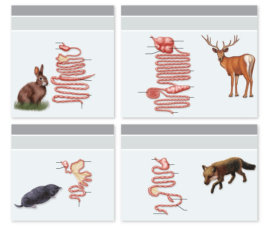
a result, herbivores tend to have much longer digestive tracts
than carnivores, allowing greater time for digestion to occur
(figure 48.14) . In addition, many herbivores have modified
their digestive tracts to enhance digestion of plant material.
Ruminants rechew regurgitated food
Ruminants have a four-chambered stomach (figure 48.15) .
The first three portions include the reticulum, the rumen,
and the omasum. These are followed by the true stomach,
the abomasum.
The rumen, which may hold up to 50 gallons, serves as a
fermentation vat where bacteria and protists convert cellulose
and other molecules into a variety of simpler compounds. The
location of the rumen at the front of the four chambers allows
the animal to regurgitate and rechew the contents of the ru-
men, an activity called rumination, or “chewing the cud.” This
Figure 48.14
The
digestive systems of
di erent mammals re ect
their diets. Herbivores, such
as rabbits and deer, require
long digestive tracts with
specialized compartments for
the breakdown of plant matter.
Diets composed of animal
matter, thus lacking cellulose,
are more easily digested;
insectivorous and carnivorous
mammals, such as voles and
foxes, respectively, have short
digestive tracts with few
specialized pouches.
breaks tougher fiber in the diet into smaller particles, increas-
ing the surface area for microbial attachment.
After chewing, the cud is swallowed for further microbial
digestion in the rumen, then passes to the omasum, and then
to the abomasum, where it is finally mixed with gastric juice.
This process leads to far more efficient digestion of cellulose in
ruminants than in mammalian herbivores such as horses, that
lack a rumen.
Foregut fermentation has evolved
convergently many times
Although the four-chambered stomach has only evolved once,
many other types of herbivores—including hippopotamuses,
langur monkeys, sloths, kangaroos, and hoatzins (a type of
bird)—have evolved large stomachs to enhance microbial fer-
mentation. In many cases, these species have evolved a variety
of other anatomical structures that serve to slow down the pas-
sage of food through the stomach, leading to increased time
for fermentation.
A remarkable case of convergent evolution at the mo-
lecular level is exhibited by ruminants and the langur monkey,
which subsists primarily on leaves. In most mammals, lysozymes
are enzymes found in saliva and tears, which attack invading
bacteria. However, in ruminants and langurs, lysozymes have
been modified to take on a new role, digesting bacteria in the
stomach. In both cases, five identical amino acid changes have
www.ravenbiology.com
chapter
48
The Digestive System
991
rav32223_ch48_981-1000.indd 991rav32223_ch48_981-1000.indd 991 11/18/09 1:14:23 PM11/18/09 1:14:23 PM