Raven P.H., Johnson G.B., Mason K.A. Biology (Ninth Edition)
Подождите немного. Документ загружается.

Apago PDF Enhancer
Earthworm
Closed Circulation
Water
Water
Water
Water Water
Mouth
Water
Incurrent
pore
Sponge Hydra Nematode
Gastrovascular
cavity
Gastrovascular
cavity
Gastrovascular cavity
Osculum
(excurrent
opening)
a.
b. c.
No Circulatory System
Grasshopper
Open Circulation
Anus
Hemolymph
Body
cavity
Heart Heart
Lateral
hearts
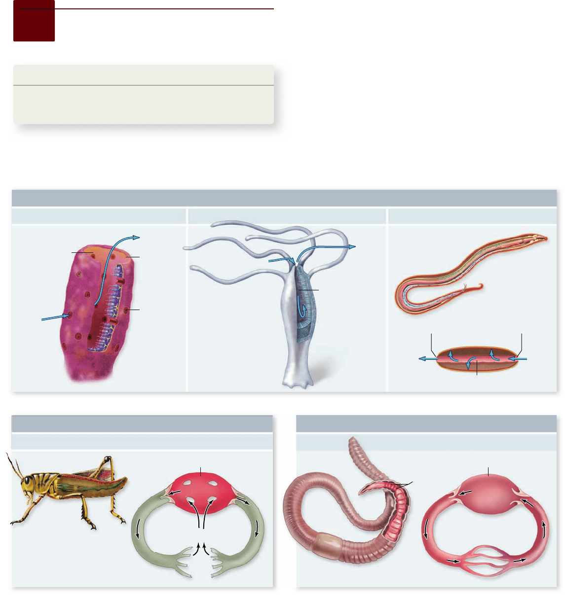
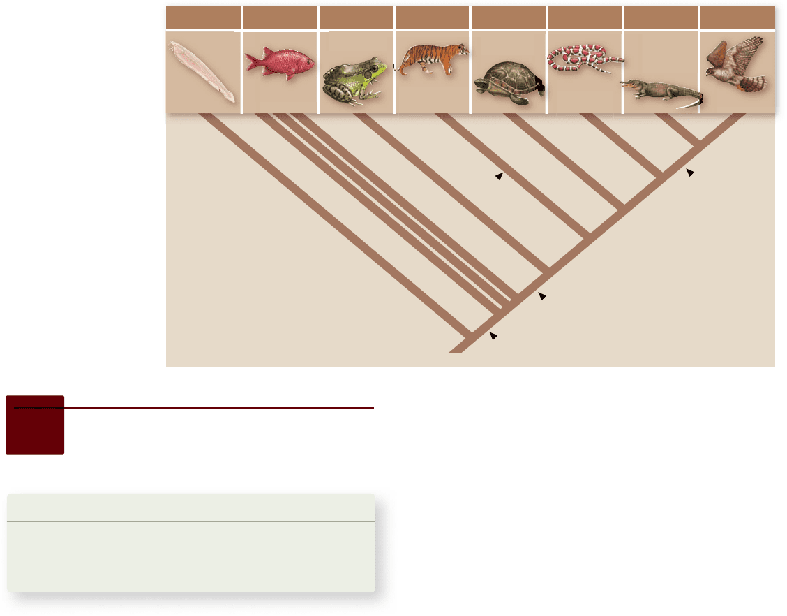
Figure 50.4
Circulatory systems of the animal kingdom. a. Sponges (left panel ) do not have a separate circulatory system. They
circulate water using many incurrent pores and one excurrent pore. The gastrovascular cavity of a hydra (middle panel ) serves as both a
digestive and a circulatory system, delivering nutrients directly to the tissue cells by diffusion from the digestive cavity. The nematode (right
panel ) is thin enough that the digestive tract can also be used as a circulatory system. Larger animals require a separate circulatory system to
carry nutrients to and wastes away from tissues. b. In the open circulation of an insect, hemolymph is pumped from a tubular heart into
cavities in the insect’s body; the hemolymph then returns to the blood vessels so that it can be recirculated. c. In the closed circulation of the
earthworm, blood pumped from the hearts remains within a system of vessels that returns it to the hearts. All vertebrates also have closed
circulatory systems.
50.2
Invertebrate Circulatory
Systems
Learning Outcomes
Distinguish between open and closed circulatory systems.1.
Define hemolymph.2.
The nature of the circulatory system in multicellular inverte-
brates is directly related to the size, complexity, and lifestyle of
the organism in question. Sponges and most cnidarians utilize
water from the environment as a circulatory fluid. Sponges
pass water through a series of channels in their bodies, and
Hydra and other cnidarians circulate water through a
gastrovascular cavity (figure 50.4a) . Because the body wall in
Hydra species is only two cell layers thick, each cell layer is in
direct contact with either the external environment or the gas-
trovascular cavity.
Pseudocoelomate invertebrates (roundworms, rotifers)
use the fluids of the body cavity for circulation. Most of these
invertebrates are quite small or are long and thin, and there-
fore adequate circulation is accomplished by movements of the
body against the body fluids, which are in direct contact with
the internal tissues and organs. Larger animals, however, have
tissues that are several cell layers thick, so that many cells are
1022
part
VII
Animal Form and Function
rav32223_ch50_1018-1037.indd 1022rav32223_ch50_1018-1037.indd 1022 11/19/09 11:44:35 AM11/19/09 11:44:35 AM
Apago PDF Enhancer
Systemic
capillaries
Respiratory
capillaries
Gills
Sinus
venosus
Ventricle
Conus
arteriosus
Atrium
Body
50.3
Vertebrate Circulatory Systems
Learning Outcomes
Trace the evolution of the chambered heart from 1.
lancelets to birds and mammals.
Delineate the flow of blood through the circulatory 2.
system in birds and mammals.
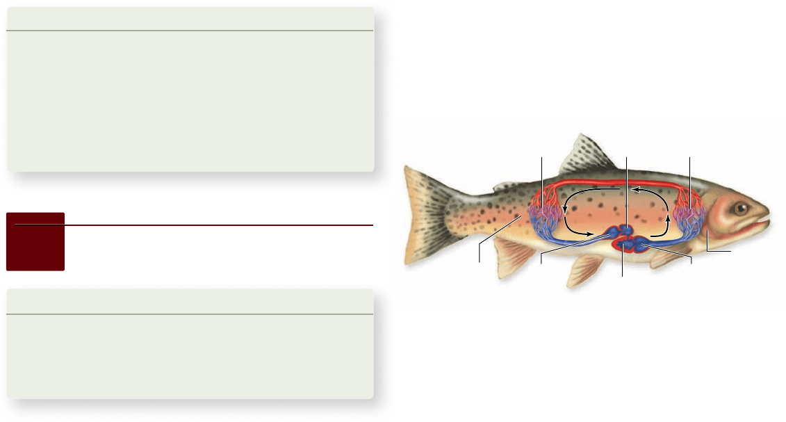
Figure 50.5
The heart and circulation of a sh. Diagram
of a sh heart, showing the structures in series with each other
(sinus venosus; atrium; ventricles; conus arteriosus) that form two
pumping chambers. Blood is pumped by the ventricle through the
gills and then to the body. Blood rich in oxygen (oxygenated) is
shown in red; blood low in oxygen (deoxygenated) is shown in blue.
too far away from the body surface or digestive cavity to di-
rectly exchange materials with the environment. Instead, oxy-
gen and nutrients are transported from the environment and
digestive cavity to the body cells by an internal fluid within a
circulatory system.
Open circulatory systems move
uids in a one-way path
The two main types of circulatory systems are open and closed.
In an open circulatory system , such as that found in most mol-
lusks and in arthropods (figure 50.4b), there is no distinction
between the circulating fluid and the extracellular fluid of the
body tissues. This fluid is thus called hemolymph.
In insects, a muscular tube, or heart, pumps hemolymph
through a network of channels and cavities in the body. The
fluid then drains back into the central cavity.
Closed circulatory systems
move uids in a loop
In a closed circulatory system , the circulating fluid, blood, is
always enclosed within blood vessels that transport it away from
and back to the heart (figure 50.4c). Some invertebrates, such as
cephalopod mollusks and annelids (see chapter 34), and all ver-
tebrates have a closed circulatory system.
In annelids such as earthworms, a dorsal vessel contracts
rhythmically to function as a pump. Blood is pushed through
five small connecting arteries, which also function as pumps, to
a ventral vessel, which transports the blood posteriorly until it
eventually reenters the dorsal vessel. Smaller vessels branch
from each artery to supply the tissues of the earthworm with
oxygen and nutrients and to remove waste products.
Learning Outcomes Review 50.2
In invertebrates, open circulatory systems pump hemolymph into tissues,
from which it then drains into a central cavity. Closed circulatory systems
move fl uid in a loop to and from a muscular pumping region such as a heart.
Hemolymph (invertebrates) is identical to the extracellular fl uid in
the tissues.
■ In the open circulatory system of insects, how does
hemolymph get back to the heart?
The evolution of large and complex hearts and closed circula-
tory systems put a premium on efficient circulation. In re-
sponse, vertebrates have evolved a remarkable set of adaptations
inextricably linking circulation and respiration, which has fa-
cilitated diversification throughout aquatic and terrestrial hab-
itats and permitted the evolution of large body size.
In shes, more e cient circulation
developed concurrently with gills
Chordates ancestral to the vertebrates are thought to have had
simple tubular hearts, similar to those now seen in lancelets (see
chapter 35). The heart was little more than a specialized zone
of the ventral artery that was more heavily muscled than the
rest of the arteries; it contracted in simple peristaltic waves.
The development of gills by fishes required a more effi-
cient pump, and in fishes we see the evolution of a true
chamber-pump heart. The fish heart is, in essence, a tube with
four structures arrayed one after the other to form two pump-
ing chambers (figure 50.5) . The first two structures—the sinus
venosus and atrium—form the first chamber; the second two,
the ventricle and conus arteriosus, form the second chamber.
The sinus venosus is the first to contract, followed by the atrium,
the ventricle, and finally the conus arteriosus.
Despite shifts in the relative positions of these structures,
this heartbeat sequence is maintained in all vertebrates. In fish,
the electrical impulse that produces the contraction is initiated
in the sinus venosus; in other vertebrates, the electrical impulse
is initiated by a structure homologous to the sinus venosus—
the sinoatrial (SA) node.
After blood leaves the conus arteriosus, it moves through
the gills, becoming oxygenated. Blood leaving the gills then
flows through a network of arteries to the rest of the body, fi-
nally returning to the sinus venosus. This simple loop has one
serious limitation: in passing through the capillaries in the
gills, blood pressure drops significantly. This slows circulation
from the gills to the rest of the body and can limit oxygen de-
livery to tissues.
chapter
50
The Circulatory System
1023www.ravenbiology.com
rav32223_ch50_1018-1037.indd 1023rav32223_ch50_1018-1037.indd 1023 11/19/09 11:44:36 AM11/19/09 11:44:36 AM
Apago PDF Enhancer
a. b.
Lungs
Body
Pulmonary
capillaries
Systemic
capillaries
Ventricle
Conus
arteriosus
Right atrium
Pulmonary vein
Pulmocutaneous
artery
Posterior vena cava
Truncus arteriosus
Aorta
Carotid artery
Systemic artery
Left atrium
Sinus venosus
Ventricle
Conus
arteriosus
Right atrium
Left atrium
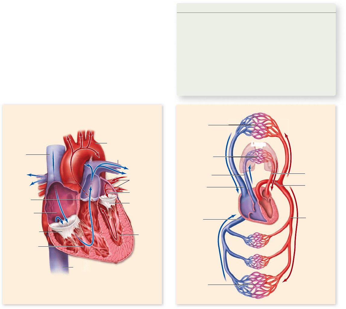
In amphibians and most reptiles, lungs
required a separate circulation
The advent of lungs in amphibians (see chapter 49) involved a
major change in the pattern of circulation, a second pumping
circuit. After blood is pumped by the heart through the pulmo-
nary arteries to the lungs, it does not go directly to the tissues of
the body. Instead, it is returned via the pulmonary veins to the
heart. Blood leaves the heart a second time to be circulated
through other tissues. This system is termed double circulation:
One system, the pulmonary circulation, moves blood between
heart and lungs, and another, the systemic circulation, moves
blood between the heart and the rest of the body.
Amphibian circulation
Optimally, oxygenated blood from lungs would go directly to
tissues, rather than being mixed in the heart with deoxygen-
ated blood returning from the body. The amphibian heart has
two structural features that significantly reduce this mixing
(figure 50.6) . First, the atrium is divided into two chambers:
The right atrium receives deoxygenated blood from the sys-
temic circulation, and the left atrium receives oxygenated
blood from the lungs. These two types of blood, therefore, do
not mix in the atria.
Because an amphibian heart has a single ventricle, the
separation of the pulmonary and systemic circulations is in-
complete. The extent of mixing when the contents of each
atrium enter the ventricle is reduced by internal channels cre-
ated by recesses in the ventricular wall. The conus arteriosus is
partially separated by a dividing wall, which directs deoxygen-
ated blood into the pulmonary arteries and oxygenated blood
into the aorta, the major artery of the systemic circulation.
Amphibians living in water can obtain additional oxygen
by diffusion through their skin. Thus, amphibians have a pulmo-
cutaneous circuit that sends blood to both the lungs and the skin.
Cutaneous respiration is also seen in many aquatic reptiles such
as turtles.
Reptilian circulation
Among reptiles, additional modifications have further reduced
the mixing of blood in the heart. In addition to having two sep-
arate atria, reptiles have a septum that partially subdivides the
ventricle. This separation is complete in one order of reptiles,
the crocodilians, which have two separate ventricles divided by
a complete septum (see the following section). Another change
in the circulation of reptiles is that the conus arteriosus has
become incorporated into the trunks of the large arteries leav-
ing the heart.
Figure 50.6
The heart and circulation of an amphibian. a. The frog has a three-chambered heart with two atria but only one
ventricle, which pumps blood both to the lungs and to the body. b. Despite the potential for mixing, the oxygenated and deoxygenated bloods
(red and blue lines, respectively) mix little as they are pumped to the body and lungs. Oxygenation of blood also occurs by gas exchange
through the skin.
1024
part
VII
Animal Form and Function
rav32223_ch50_1018-1037.indd 1024rav32223_ch50_1018-1037.indd 1024 11/19/09 11:44:38 AM11/19/09 11:44:38 AM
Apago PDF Enhancer
Head
Body
Lungs
Systemic
capillaries
Pulmonary
artery
Pulmonary
semilunar valve
Inferior
vena cava
Superior
vena cava
Aorta
Tricuspid valve
Right ventricle
Left
ventricle
Right atrium
Pulmonary
veins
Left atrium
Bicuspid
mitral
valve
Systemic
capillaries
Pulmonary
artery
Superior
vena cava
Inferior
vena cava
Artery
Pulmonary
vein
Aorta
Respiratory
capillaries
a. b.
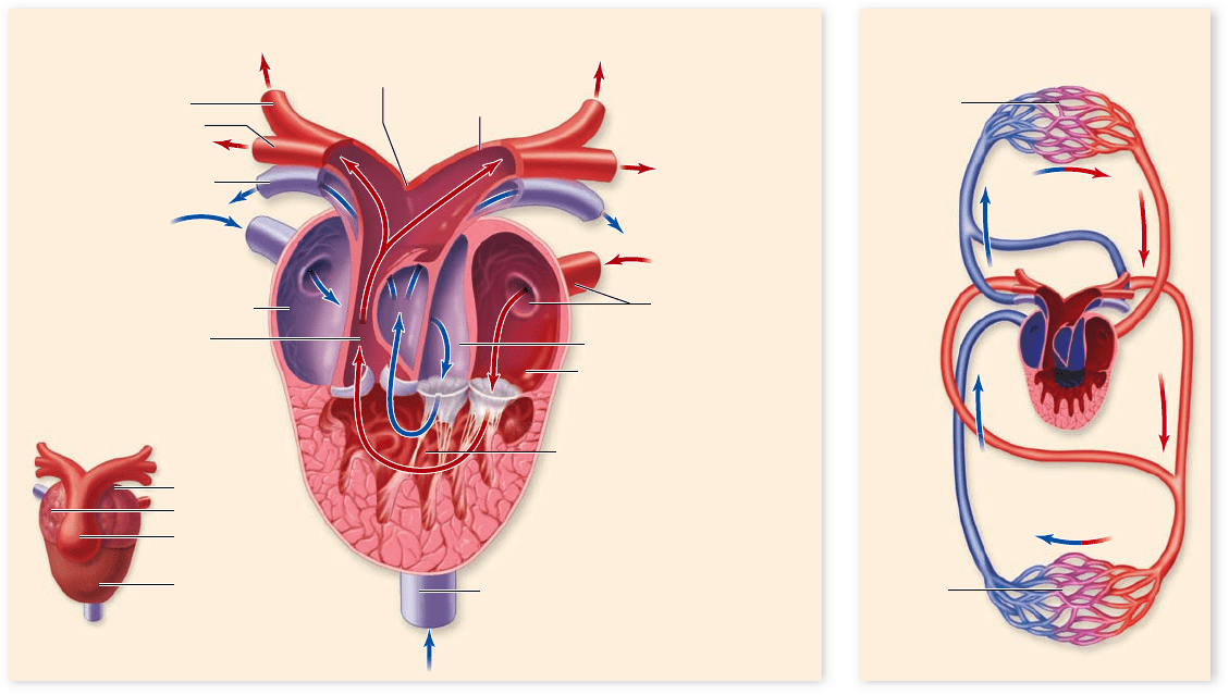
Mammals, birds, and crocodilians have two
completely separated circulatory systems
Mammals, birds, and crocodilians have a four-chambered heart
with two separate atria and two separate ventricles (figure 50.7) .
The hearts of birds and crocodiles exhibit some differences, but
overall are quite similar, which is not surprising given their
close evolutionary relationship (figure 50.8) . However, the ex-
treme similarity of the hearts of birds and mammals—so alike
that a single illustration can suffice for both (see figure 50.7)—
is a remarkable case of convergent evolution (see figure 50.8).
In a four-chambered heart, the right atrium receives de-
oxygenated blood from the body and delivers it to the right
ventricle, which pumps the blood to the lungs. The left atrium
receives oxygenated blood from the lungs and delivers it to the
left ventricle, which pumps the oxygenated blood to the rest of
the body (see figure 50.7).
The heart in these vertebrates is a two-cycle pump. Both
atria fill with blood and simultaneously contract, emptying
their blood into the ventricles. Both ventricles also contract at
the same time, pushing blood simultaneously into the pulmo-
nary and systemic circulations.
The increased efficiency of the double circulatory system
in mammals and birds is thought to have been important in the
evolution of endothermy. More efficient circulation is neces-
sary to support the high metabolic rate required for mainte-
nance of internal body temperature about a set point.
Throughout the evolutionary history of the vertebrate heart,
the sinus venosus has served as a pacemaker, the site where the
impulses that initiate the heartbeat originate. Although the sinus
venosus constitutes a major chamber in the fish heart, it is reduced
in size in amphibians and is further reduced in reptiles. In mam-
mals and birds, the sinus venosus is no longer present as a separate
chamber, although some of its tissue remains in the wall of the
right atrium. This tissue, the sinoatrial (SA) node, is still the site
where each heartbeat originates as detailed later in the chapter.
Learning Outcomes Review 50.3
The chordate heart has evolved from a muscular region of a vessel, to the
two-chambered heart of fi sh, the three-chambered heart of amphibians
and most reptiles, and the four-chambered heart of crocodilians, birds, and
mammals. Deoxygenated blood travels in the pulmonary circuit from the
right atrium into the right ventricle and then to the lungs; it returns to
the left atrium. Oxygenated blood travels in the systemic circuit from the
left atrium into the left ventricle and then to the body; it returns to the
right atrium.
■ What is the physiological advantage of having
separated ventricles?
Figure 50.7
The heart and circulation of mammals and birds. a. The path of blood through the four-chambered heart. b. The
right side of the heart receives deoxygenated blood and pumps it to the lungs; the left side of the heart receives oxygenated blood and pumps it
to the body. In this way, the pulmonary and systemic circulations are kept completely separate.
chapter
50
The Circulatory System
1025www.ravenbiology.com
rav32223_ch50_1018-1037.indd 1025rav32223_ch50_1018-1037.indd 1025 11/19/09 11:44:39 AM11/19/09 11:44:39 AM
Apago PDF Enhancer
2-chamber
heart
3-chamber
heart
4-chamber
heart
4-chamber
heart
Lancelets Fish Mammals TurtlesAmphibians CrocodiliansSquamates Birds
Figure 50.8
Evolution of the
heart in vertebrates.
Despite their similarity,
the four-chambered
hearts of mammals
and birds evolved
convergently.
50.4
The Four-Chambered Heart
and the Blood Vessels
Learning Outcomes
Explain the cardiac cycle.1.
Describe the role of autorhythmic cells of the SA node.2.
Define blood pressure and how it is measured3.
As mentioned earlier, the heart of mammals, birds, and croco-
dilians goes through two contraction cycles, one of atrial con-
traction to send blood to the ventricles, and one of ventricular
contraction to send blood to the pulmonary and systemic cir-
cuits. These two contractions plus the resting period between
these make up the complete cardiac cycle encompassed by
the heartbeat.
The cardiac cycle drives
the cardiovascular system
The heart has two pairs of valves. One pair, the atrioventricular
(AV) valves, maintains unidirectional blood flow between the
atria and ventricles. The AV valve on the right side is the
tricuspid valve, and the AV valve on the left is the bicuspid, or
mitral, valve. Another pair of valves, together called the
semilunar valves, ensure one-way flow out of the ventricles to
the arterial systems. The pulmonary valve is located at the exit
of the right ventricle, and the aortic valve is located at the exit
of the left ventricle. These valves open and close as the heart
goes through its cycle. The closing of these valves produces the
“lub-dub” sounds heard with a stethoscope.
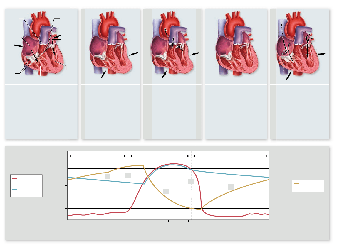
The cardiac cycle is portrayed in figure 50.9 . It begins as
blood returns to the resting heart through veins that empty
into the right and left atria. As the atria fill and the pressure in
them rises, the AV valves open and blood flows into the ven-
tricles. The ventricles become about 80% filled during this
time. Contraction of the atria tops up the final 20% of the
80 mL of blood the ventricles receive, on average, in a resting
person. These events occur while the ventricles are relaxing, a
period called ventricular diastole.
After a slight delay, the ventricles contract, a period called
ventricular systole. Contraction of each ventricle increases the
pressure within each chamber, causing the AV valves to force-
fully close (the “lub” sound), preventing blood from backing up
into the atria. Immediately after the AV valves close, the pres-
sure in the ventricles forces the semilunar valves open and blood
flows into the arterial systems. As the ventricles relax, closing of
the semilunar valves prevents backflow (the “dub” sound).
Contraction of heart muscle is
initiated by autorhythmic cells
As in other types of muscle, contraction of heart muscle is stimu-
lated by membrane depolarization (see chapters 44 and 47). In skel-
etal muscles, only nerve impulses from motor neurons can normally
initiate depolarization. The heart, by contrast, contains specialized
“self-excitable” muscle cells called autorhythmic fibers, which can
initiate periodic action potentials without neural activation.
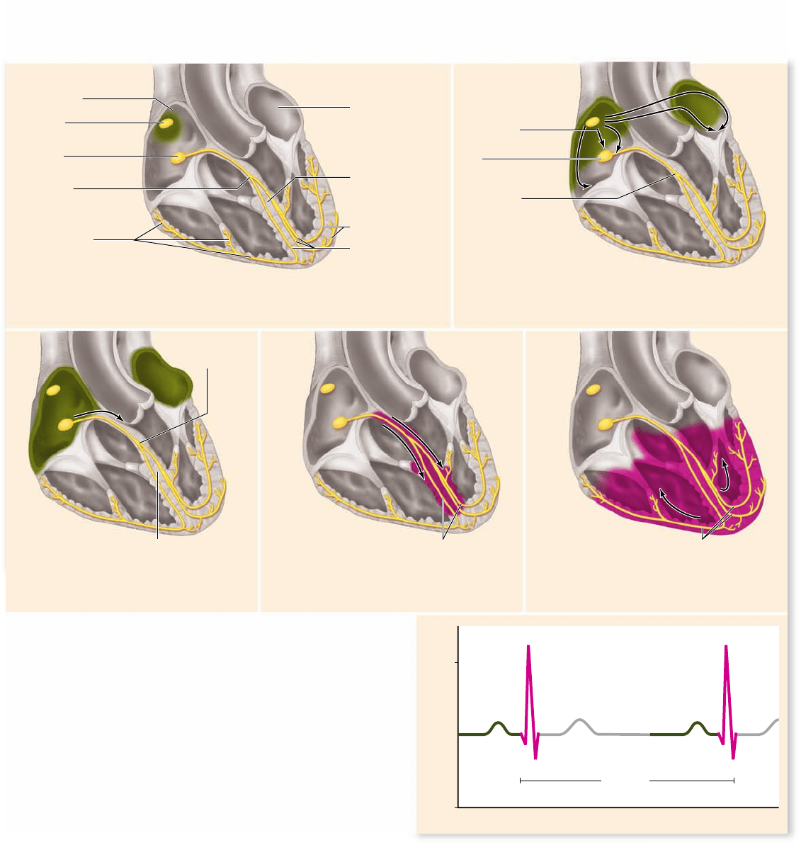
The most important group of autorhythmic cells is the
sinoatrial (SA) node, described earlier (figure 50.10) . Located
in the wall of the right atrium, the SA node acts as a pacemaker
for the rest of the heart by producing spontaneous action po-
tentials at a faster rate than other autorhythmic cells. These
spontaneous action potentials are due to a constant leakage of
Na
+
ions into the cell that depolarize the membrane. When the
1026
part
VII
Animal Form and Function
rav32223_ch50_1018-1037.indd 1026rav32223_ch50_1018-1037.indd 1026 11/19/09 11:44:44 AM11/19/09 11:44:44 AM
Apago PDF Enhancer
AV
valves
Right
atrium
Left
atrium
Aortic
valve
Pulmonary
valve
Right ventricle
Left
ventricle
4. “Dup”: The ventricles
relax, the pressure in
the ventricles falls at
the end of systole, and
since pressure is now
greater in the aorta and
pulmonary artery, the
aortic and pulmonary
valves slam shut.
2. “Lub”: The ventricles
contract, the
atrioventricular (AV)
valves close, and
pressure in the
ventricles builds
up until the aortic
and pulmonary
valves open.
3. Blood is pumped out
of ventricles and into
the aorta and
pulmonary artery.
5. The ventricles fill
with blood.
1. The atria contract.
Diastole
“Lub”
1.
2.
3.
4.
5.
“Dup”
DiastoleSystole
pressure in
left ventricle
pressure
in aorta
volume in
left ventricle
Pressure (mm Hg)
Time (seconds)
0
25
0.10 1.00.90.80.6 0.70.50.40.30.2
50
75
100
130 mL
65 mL
125
Figure 50.9
The cardiac cycle. a. Contraction and relaxation of the atria and ventricles moves blood through the heart. b. Blood
pressure and volume changes through the cardiac cycle, shown here for the left ventricle.
threshold is reached, an action potential occurs. At the end of
the action potential, the membrane is again below threshold
and the process begins again. The cells of the SA node generate
an action potential every 0.6 sec, equivalent to about 100 a min-
ute. As we will see later in the chapter, the autonomic nervous
system can modulate this rate.
Each depolarization initiated by this pacemaker is transmit-
ted through two pathways: one to the cardiac muscle fibers of the
left atrium, and the other to the right atrium and the atrioven-
tricular (AV) node. Once initiated, depolarizations spread quickly
from one muscle fiber to another in a wave that envelops the right
and left atria nearly simultaneously. The rapid spread of depolar-
ization is made possible because special conducting fibers are
present and because the cardiac muscle cells are coupled by groups
of gap junctions located within intercalated disks (see chapter 44 ).
A sheet of connective tissue separating the atria from the
ventricles blocks the spread of excitation through muscle fibers
from one chamber to the other. The AV node provides the only
pathway for conduction of the depolarization from the atria to
the ventricles. The fibers of the AV node slow down the con-
duction of the depolarizing signals, delaying the contraction of
the ventricle by about 0.1 sec. This delay permits the atria to
finish contracting and emptying their blood into the ventricles
before the ventricles contract.
From the AV node, the wave of depolarization is con-
ducted rapidly over both ventricles by a network of fibers called
the atrioventricular bundle, or bundle of His. These fibers relay
the depolarization to Purkinje fibers, which directly stimulate
the myocardial cells of the left and right ventricles, causing
their almost simultaneous contraction.
The stimulation of myocardial cells produces an action po-
tential that leads to contraction. Contraction is controlled by Ca
2+
and the troponin/tropomyosin system similar to skeletal muscle
(see chapter 47), but the shape of the action potential is different.
The initial rising phase due to an influx of Na
+
from voltage-gated
Na
+
channels is followed by a plateau phase that leads to more
sustained contraction. The plateau phase is due to the opening of
voltage-gated Ca
2+
channels. The resulting influx of Ca
2+
keeps
the membrane depolarized when the Na
+
channels inactivate.
This, in turn, leads to more voltage-gated Ca
2+
channels in the
sarcoplasmic reticulum opening. The additional Ca
2+
in the cyto-
plasm produces a more sustained contraction. The Ca
2+
is
chapter
50
The Circulatory System
1027www.ravenbiology.com
rav32223_ch50_1018-1037.indd 1027rav32223_ch50_1018-1037.indd 1027 11/19/09 11:44:45 AM11/19/09 11:44:45 AM
Apago PDF Enhancer
Millivolts
Seconds
R
P wave T wave
Q
S
1 sec
+1
-1
0
1. The impulse begins at the SA node and travels to the
AV node.
2. The impulse is delayed at the AV node. It
then travels to the AV bundle.
3. From the AV bundle, the impulse travels
down the interventricular septum.
4. The impulse spreads to branches from the
interventricular septum.
5. Finally reaching the Purkinje fibers, the impulse
is distributed throughout the ventricles.
SA node
(pacemaker)
AV node
AV bundle
Left and right
bundle branches
Left and right
bundle branches
Purkinje fibers
Purkinje fibers
Left atrium
Right atrium
Purkinje fibers
Interventricular
septum
AV bundle
Interventricular
septum
AV
AV bundle
Internodal
pathway
F igure 50.10
The path of electrical excitation in the
heart. The events occurring during contraction of the heart are
correlated with the measurement of electrical activity by an
electrocardiogram (ECG also called EKG). The depolarization/
contraction of the atrium is shown in green above and corresponds
to the P wave of the ECG (also in green). The depolarization/
contraction of the ventricle is shown in red above and corresponds
to the QRS wave of the ECG (also in red). The T wave on the ECG
corresponds to the repolarization of the ventricles. The atrial
repolarization is masked by the QRS wave.
removed from the cytoplasm by a pump in the sarcoplasmic re-
ticulum similar to skeletal muscle, and an additional carrier in the
plasma membrane pumps Ca
2+
into the interstitial space.
The electrical activity of the heart can be recorded from
the surface of the body with electrodes placed on the limbs and
chest. The recording, called an electrocardiogram (ECG or
EKG), shows how the cells of the heart depolarize and repolarize
during the cardiac cycle (see figure 50.10). Depolarization causes
contraction of the heart, and repolarization causes relaxation.
The first peak in the recording, P, is produced by the de-
polarization of the atria, and is associated with atrial systole.
The second, larger peak, QRS, is produced by ventricular de-
polarization; during this time, the ventricles contract (ventricu-
lar systole). The last peak, T, is produced by ventricular
repolarization; at this time, the ventricles begin diastole.
Arteries and veins branch to
and from all parts of the body
The right and left pulmonary arteries deliver oxygen-depleted
blood from the right ventricle to the right and left lungs. As
1028
part
VII
Animal Form and Function
rav32223_ch50_1018-1037.indd 1028rav32223_ch50_1018-1037.indd 1028 11/19/09 11:44:46 AM11/19/09 11:44:46 AM
Apago PDF Enhancer
150
120
75
0
150
120
75
0
150
120
75
0
cuff pressure
blood pressure
artery closed
artery open
1. Cuff pressure: 150 mm Hg
Artery closed
2. Cuff pressure: 120 mm Hg
Systolic pressure
3. Cuff pressure: 75 mm Hg
Diastolic pressure
Cuff
Blood pressure gauge
Artery always closed
“No sound”
Artery alternates closed/open
“Pulse sound”
C
C
O
CCCCOOOOO
Artery always open
“No sound”
Stethoscope
0
50
150100
200
250 0
50
150100
200
250
0
50
150100
200
250
C O
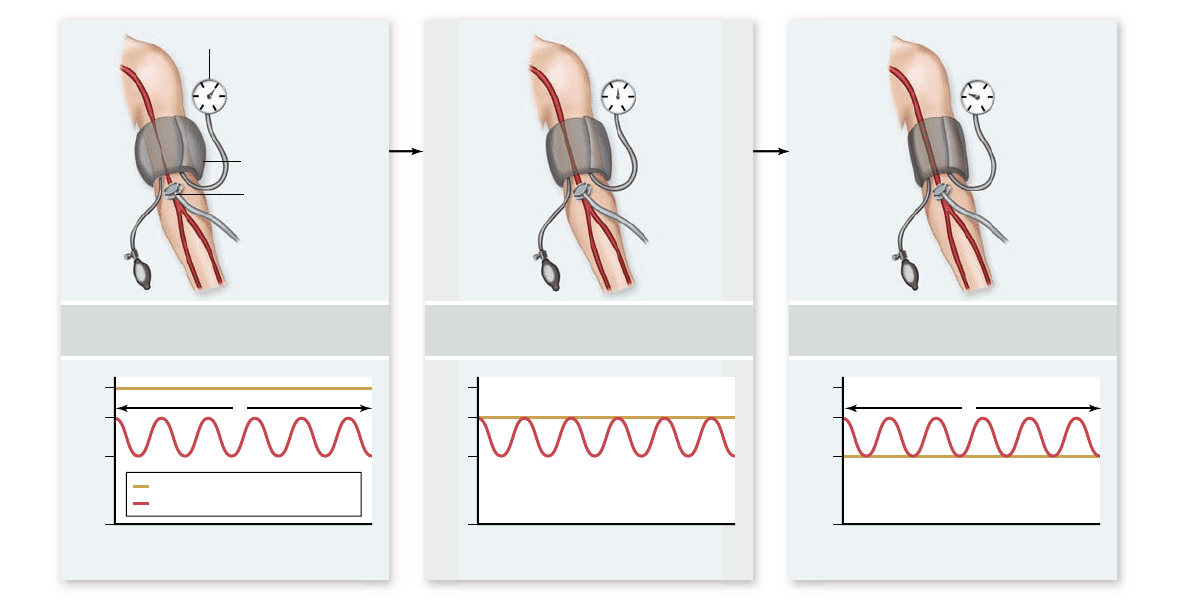
Figure 50.11
Measurement of blood pressure. The blood pressure cuff is tightened to stop the blood ow through the brachial
artery. As the cuff is loosened, the maximal (systolic) pressure becomes greater than the cuff pressure and blood can momentarily pass through,
producing a pulse that can be heard with a stethoscope. The pressure at this point is recorded as the systolic pressure. As the cuff pressure
continues to drop, blood pressure is greater than cuff pressure for larger portions of the cardiac cycle. Eventually, even the minimum pressure
during the cycle is greater than the cuff pressure, at which time the blood vessel is no longer distorted and silent laminar ow returns, replacing
the pulsing sound. The diastolic pressure is recorded as the pressure at which a sound is no longer heard.
previously mentioned, the pulmonary veins return oxygenated
blood from the lungs to the left atrium of the heart.
The aorta and all its branches are systemic arteries, car-
rying oxygen-rich blood from the left ventricle to all parts of
the body. The coronary arteries are the first branches off the
aorta; these supply oxygenated blood to the heart muscle itself
(see figure 50.7b ). Other systemic arteries branch from the
aorta as it makes an arch above the heart and as it descends and
traverses the thoracic and abdominal cavities.
The blood from the body’s organs, now lower in oxygen,
returns to the heart in the systemic veins. These eventually
empty into two major veins: the superior vena cava, which
drains the upper body, and the inferior vena cava, which drains
the lower body. These veins empty into the right atrium, com-
pleting the systemic circulation.
The flow of blood through the arteries, capillaries, and
veins is driven by the pressure generated by ventricular con-
traction. The ventricles must contract forcefully enough to
move the blood through the entire circulatory system.
Arterial blood pressure can be measured
As the ventricles contract, great pressure is generated within
them and transferred through the arteries once the aortic
valve opens. The pulse that you can detect in your wrist or
neck results from changes in pressure as elastic arteries ex-
pand and contract with the periodic blood flow. Doctors use
blood pressure as an general indicator of cardiovascular
health because a variety of conditions can cause increases or
decreases in pressure.
A sphygmomanometer measures the blood pressure in the
brachial artery found on the inside part of the arm, above the
elbow (figure 50.11) . A cuff wrapped around the upper part of
the arm is tightened enough to stop the flow of blood to the
lower part of the arm. As the cuff is slowly loosened, eventu-
ally the blood pressure produced by the heart is greater than
the constricting pressure of cuff and blood begins pulsating
through the artery, producing a sound that can be detected
using a stethoscope. The point at which this pulsing sound
begins marks the peak pressure, or systolic pressure, at
which ventricles are contracting. As the cuff is loosened fur-
ther, the point is reached where the pressure of the cuff is
lower than the blood pressure throughout the cardiac cycle, at
which time the blood vessel is no longer distorted and the
pulsing sound stops. This point marks the minimum pressure
between heartbeats or diastolic pressure, at which the ven-
tricles are relaxed.
The blood pressure is written as a ratio of systolic over
diastolic pressure, and for a healthy person in his or her twen-
ties, a typical blood pressure is 120/75 (measured in millimeters
chapter
50
The Circulatory System
1029www.ravenbiology.com
rav32223_ch50_1018-1037.indd 1029rav32223_ch50_1018-1037.indd 1029 11/19/09 11:44:47 AM11/19/09 11:44:47 AM
Apago PDF Enhancer
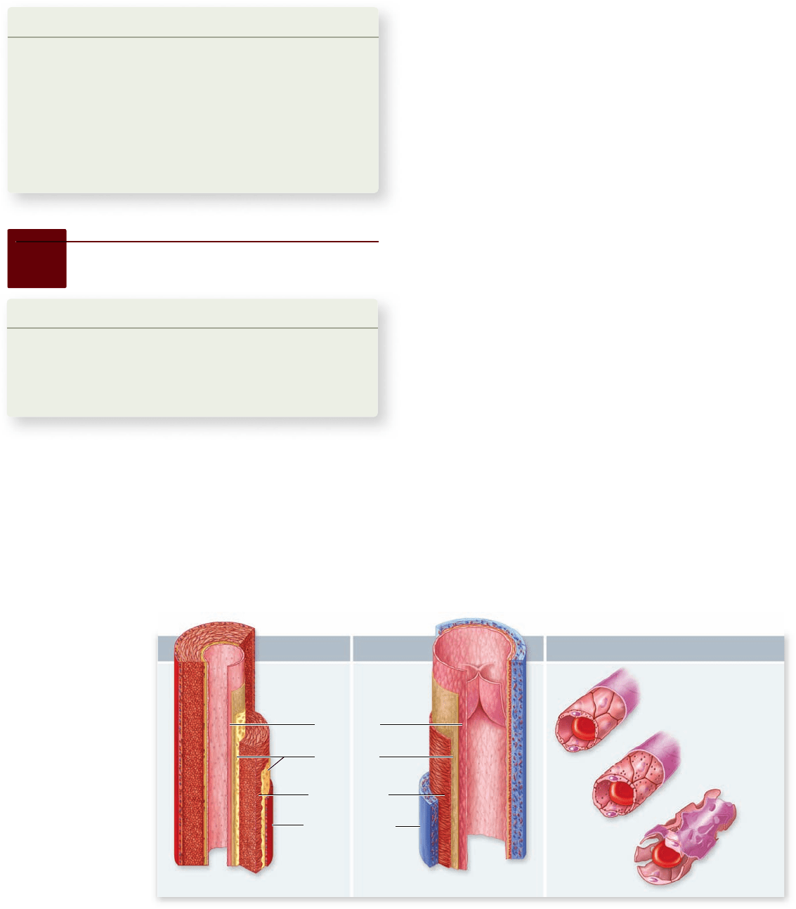
Capillary Vein Artery
Sinusoid
Fenestrated
capillary
Capillary
Elastic layer
Endothelium
Connective tissue
Smooth muscle
a. b. c.
Figure 50.12
The
structure of blood
vessels. Arteries (a)
and veins (b) have the
same tissue layers, but
the smooth muscle layer
in arteries is much
thicker and there are
two elastic layers.
c. Capillaries are
composed of only a
single layer of
endothelial cells.
(Not to scale.)
of mercury, or mm Hg). The medical condition called
hypertension (high blood pressure) is defined as either a sys-
tolic pressure greater than 150 mm Hg or a diastolic pressure
greater than 90 mm Hg.
Learning Outcomes Review 50.4
The cardiac cycle consists of systole and diastole; the ventricles contract
at systole and relax at diastole. The SA node in the right atrium initiates
waves of depolarization that stimulate fi rst the atria and then travel to the
AV node, which stimulates the ventricles. Blood pressure is expressed as
the ratio of systolic pressure over diastolic pressure and is measured with a
device called a sphygmomanometer.
■ What would happen without a delay between auricular
and ventricular contraction?
50.5
Characteristics of Blood Vessels
Learning Outcomes
Describe the four tissue layers in blood vessels.1.
Explain the distinctions among arteries, capillaries, 2.
and veins.
Describe how the lympathic system operates.3.
You already know that blood leaves the heart through vessels
known as arteries. These continually branch, forming a hollow
“tree” that enters each organ of the body. The finest, microscopic
branches of the arterial tree are the arterioles. Blood from the
arterioles enters the capillaries, an elaborate latticework of very
narrow, thin-walled tubes. After traversing the capillaries, the
blood is collected into microscopic venules, which lead to larger
vessels called veins, and these carry blood back to the heart.
Larger vessels are composed
of four tissue layers
Arteries, arterioles, veins, and venules all have the same basic
structure (figure 50.12 ). The innermost layer is an epithelial
sheet called the endothelium. Covering the endothelium is a thin
layer of elastic fibers, a smooth muscle layer, and a connective
tissue layer. The walls of these vessels, therefore, are thick
enough to significantly reduce exchange of materials between
the blood and the tissues outside the vessels.
The walls of capillaries, in contrast, are composed only of
endothelium, so molecules and ions can leave the blood plasma
by diffusion, by filtration through pores between the cells of
the capillary walls, and by transport through the endothelial
cells. Therefore, exchange of gases and metabolites between
the blood and the interstitial fluids and cells of the body takes
place through the capillaries.
Arteries and arterioles have
evolved to withstand pressure
The larger arteries contain more elastic fibers in their walls
than other blood vessels, allowing them to recoil each time they
receive a volume of blood pumped by the heart. Smaller arter-
ies and arterioles are less elastic, but their relatively thick
smooth muscle layer enables them to resist bursting.
The narrower the vessel, the greater the frictional resis-
tance to flow. In fact, a vessel that is half the diameter of an-
other has 16 times the frictional resistance. Resistance to blood
flow is inversely proportional to the fourth power of the radius
of the vessel. Therefore, within the arterial tree, the small arter-
ies and arterioles provide the greatest resistance to blood flow.
Contraction of the smooth muscle layer of the arterioles
results in vasoconstriction, which greatly increases resistance
and decreases flow. Relaxation of the smooth muscle layer re-
sults in vasodilation, decreasing resistance and increasing
blood flow to an organ. Chronic vasoconstriction of the arteri-
oles can result in hypertension, or high blood pressure.
1030
part
VII
Animal Form and Function
rav32223_ch50_1018-1037.indd 1030rav32223_ch50_1018-1037.indd 1030 11/19/09 11:44:48 AM11/19/09 11:44:48 AM
Apago PDF Enhancer
a.
b.
Increase in heat
loss across epidermis
Decrease in heat
loss across epidermis
Vasoconstriction
Epidermis
Venule
Arteriole
Capillary
Precapillary
sphincter (contracted)
Precapillary
sphincter (relaxed)
Air or water
Vasodilation
Open
valve
Contracting
skeletal
muscles
Vein
Valve
closed
Blood flows
toward heart
Figure 50.13
Regulation of heat exchange. The amount
of heat gained or lost at the body’s surface can be regulated by
controlling the ow of blood to the surface. a. Constriction of
surface blood vessels limits ow and heat loss when the animal is
warmer than the surrounding air; when the animal is cooler than
the surrounding air (not shown here), constriction minimizes heat
gain; (b) dilation of these vessels increases ow and heat exchange.
Vasoconstriction and vasodilation are important means
of regulating body heat in both ectotherms and endotherms
(figure 50.13) . By increasing blood flow to the skin, an animal
can increase the rate of heat exchange, which is beneficial for
gaining or losing heat. Conversely, shunting blood away from
the skin is effective when an animal needs to minimize heat
exchange, as might happen in cold weather.
Capillaries form a vast network
for exchange of materials
The huge number and extensive branching of the capillaries
ensure that every cell in the body is within 100 micrometers
( μm) of a capillary. On the average, capillaries are about 1 mm
long and 8 μm in diameter, this diameter is only slightly larger
than a red blood cell (5 to 7 μm in diameter). Despite the close
fit, normal red blood cells are flexible enough to squeeze
through capillaries without difficulty.
The rate of blood flow through vessels is governed by
hydrodynamics. The smaller the cross-sectional area of a ves-
sel, the faster fluid moves through it. Given this, flow in the
capillaries would be expected to be the fastest in the system.
This would not be ideal for diffusion, and is actually not the
case. Although each capillary is very narrow, so many of them
exist that the capillaries have the greatest total cross-sectional
area of any other type of vessel. Consequently, blood moving
through capillaries goes more slowly and has more time to ex-
change materials with the surrounding extracellular fluid. By
the time the blood reaches the end of a capillary, it has released
some of its oxygen and nutrients and picked up carbon dioxide
and other waste products. Blood loses pressure and velocity as
it moves through the arterioles and capillaries, but as cross-
sectional area decreases in the venous side, velocity increases.
Venules and veins have
less muscle in their walls
Venules and veins have the same tissue layers as arteries, but
they have a thinner layer of smooth muscle. Less muscle is
needed because the pressure in the veins is only about one-
tenth that in the arteries. Most of the blood in the cardiovascu-
lar system is contained within veins, which can expand to hold
additional amounts of blood. You can see the expanded veins in
your feet when you stand for a long time.
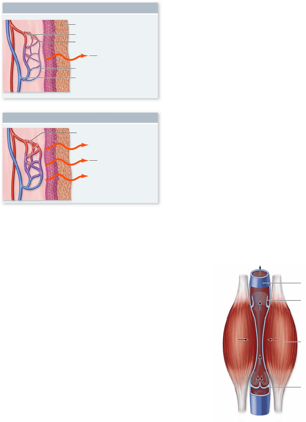
The venous pressure alone is not sufficient to return
blood to the heart from the feet and legs, but several other
sources of pressure provide help. Most significantly, skeletal
muscles surrounding the veins can contract to move blood by
squeezing the veins, a mechanism called the venous pump.
Blood moves in one direction through the veins back to the
heart with the help of venous valves (figure 50.14). When a
Figure 50.14
One-way ow of
blood through
veins. Venous valves
ensure that blood
moves through the
veins in only one
direction, back to
the heart.
chapter
50
The Circulatory System
1031www.ravenbiology.com
rav32223_ch50_1018-1037.indd 1031rav32223_ch50_1018-1037.indd 1031 11/19/09 11:44:49 AM11/19/09 11:44:49 AM