Raven P.H., Johnson G.B., Mason K.A. Biology (Ninth Edition)
Подождите немного. Документ загружается.

Apago PDF Enhancer
Blood pressure
Osmotic pressure
Lymphatic
capillary
Interstitial
fluid
Interstitial
fluid
Capillary bed Arteriole Venule
a.
b.
Lymphatic
capillary
Excess interstitial fluid
becomes lymph
Blood
flow
Capillary
Net filtration due to
blood pressure
Pressure
Direction of blood flow
Net absorption due to
osmotic pressure
Filtration
Absorption
Arteriole Venule
Arteriole Venule
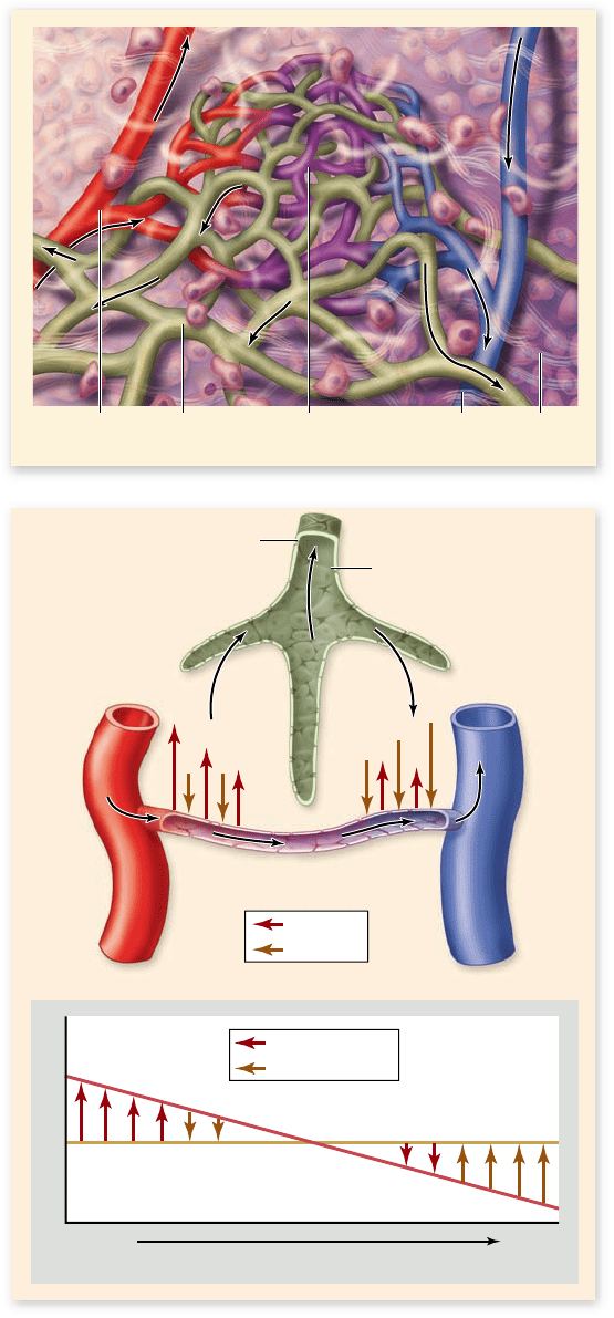
Figure 50.15
Relationship between blood, lymph, and
interstitial uid. a. Vessels of the circulatory and lymphatic
systems with arrows indicating the direction of ow of uid in the
vessels. b. Plasma uid, minus proteins, is ltered out of capillaries,
forming interstitial uid that bathes tissues. Much of this uid is
returned to the capillaries by osmosis due to the higher protein
concentration in plasma. Excess interstitial uid drains into
open-ended lymphatic capillaries, which ultimately return the uid
to the cardiovascular system.
person’s veins expand too much with blood, the venous valves
may no longer work and the blood may pool in the veins. Veins
in this condition are known as varicose veins.
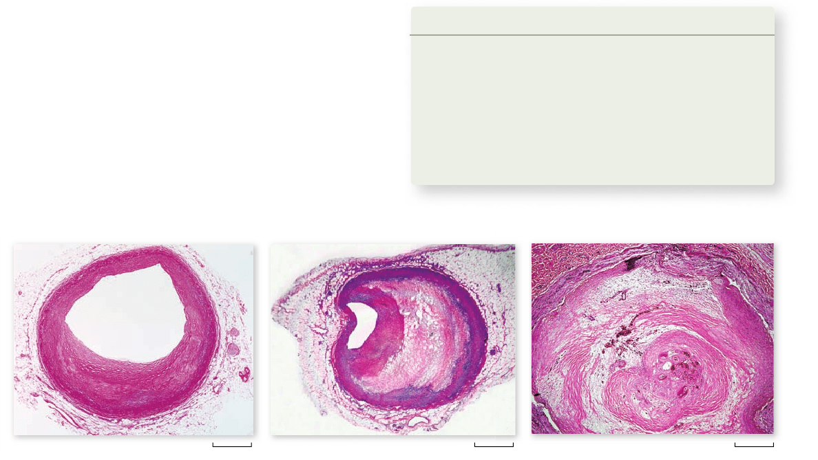
The lymphatic system handles uids
that leave the cardiovascular system
The cardiovascular system is considered a closed system be-
cause all its vessels are connected with one another—none are
simply open-ended. But a significant amount of water and sol-
utes in the blood plasma filter through the walls of the capillar-
ies to form the interstitial (tissue) fluid. Most of the fluid leaves
the capillaries near their arteriolar ends, where the blood pres-
sure is higher; it is returned to the capillaries near their venular
ends (figure 50.15).
Fluid returns by osmosis (see chapter 5). Most of the
plasma proteins cannot escape through the capillary pores be-
cause of their large size, and so the concentration of proteins
in the plasma is greater than the protein concentration in the
interstitial fluid. The difference in protein concentration pro-
duces an osmotic pressure gradient that causes water to move
into the capillaries from the interstitial space.
High capillary blood pressure can cause too much inter-
stitial fluid to accumulate. In pregnant women, for example, the
enlarged uterus, carrying the fetus, compresses veins in the ab-
dominal cavity, thereby adding to the capillary blood pressure
in the woman’s lower limbs. The increased interstitial fluid can
cause swelling of the tissues, or edema, of the feet.
Edema may also result if the plasma protein concentra-
tion is too low. Fluids do not return to the capillaries, but re-
main as interstitial fluid. Low protein concentration in the
plasma may be caused either by liver disease, because the liver
produces most of the plasma proteins, or by insufficient dietary
protein such as occurs in starvation.
Even under normal conditions, the amount of fluid fil-
tered out of the capillaries is greater than the amount that re-
turns to the capillaries by osmosis. The remainder does
eventually return to the cardiovascular system by way of an
open circulatory system called the lymphatic system.
The lymphatic system consists of lymphatic capillar-
ies, lymphatic vessels, lymph nodes, and lymphatic organs,
including the spleen and thymus. Excess fluid in the tissues
drains into blind-ended lymph capillaries with highly per-
meable walls. This fluid, now called lymph, passes into pro-
gressively larger lymphatic vessels, which resemble veins
and have one-way valves (similar to figure 50.14). The
lymph eventually enters two major lymphatic vessels, which
drain into the left and right subclavian veins located under
the collarbones.
Movement of lymph in mammals results as skeletal mus-
cles squeeze against the lymphatic vessels, a mechanism similar
to the venous pump that moves blood through veins. In some
cases, the lymphatic vessels also contract rhythmically. In many
fishes, all amphibians and reptiles, bird embryos, and some
adult birds, movement of lymph is propelled by lymph hearts.
As the lymph moves through lymph nodes and lymphatic
organs, it is modified by phagocytic cells (see chapter 4) that
line the channels of those organs. In addition, the lymph nodes
1032
part
VII
Animal Form and Function
rav32223_ch50_1018-1037.indd 1032rav32223_ch50_1018-1037.indd 1032 11/19/09 11:44:50 AM11/19/09 11:44:50 AM
Apago PDF Enhancer
a. b. c.
2000 µm 2500 µm 1000 µm
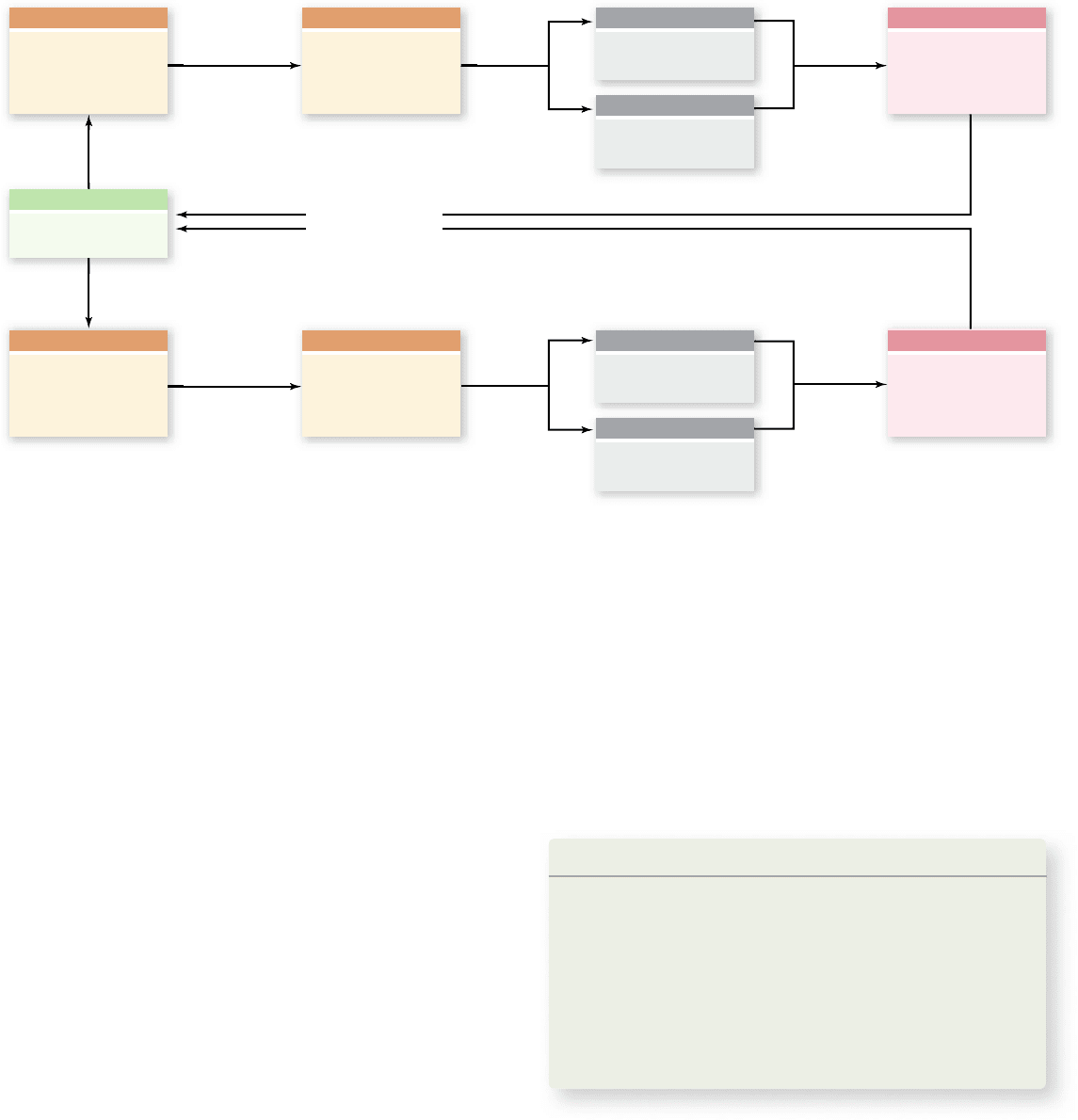
Figure 50.16
Atherosclerosis. a. The coronary artery shows only minor blockage. b. The artery exhibits severe atherosclerosis—
much of the passage is blocked by buildup on the interior walls of the artery. c. The coronary artery is essentially completely blocked.
and lymphatic organs contain germinal centers, where the acti-
vation and proliferation of lymphocytes occurs.
Cardiovascular diseases
a ect the delivery system
Cardiovascular diseases are the leading cause of death in the
United States; more than 80 million people have some form of
cardiovascular disease. Many disease conditions result from
problems in arteries, such as blockage or rupture.
Atherosclerosis, or hardening of the arteries, is an
accumulation within the arteries of fatty materials, abnormal
amounts of smooth muscle, deposits of cholesterol or fibrin,
or various kinds of cellular debris. These accumulations
cause an increase in vascular resistance, which impedes blood
flow (figure 50.16) . The lumen (interior) of the artery may
be further narrowed by a clot that forms as a result of the
atherosclerosis. In the severest cases, the artery becomes
completely blocked.
The accumulation of cholesterol in vessels is affected by
a number of factors including total serum cholesterol and the
levels of different cholesterol carrier proteins. Because cho-
lesterol is not very water-soluble, it is carried in blood in the
form of lipoprotein complexes. Two main forms are observed
that differ in density: low-density lipoproteins (LDL) and
high-density lipoproteins (HDL) —often called “bad choles-
terol” and “good cholesterol,” respectively. The reason for
this is that HDLs tend to take cholesterol out of circulation,
transporting it to the liver for elimination, and LDL is the
carrier that brings cholesterol to all cells in the body. The
problem arises when cells have enough cholesterol. This
causes a reduction in the amount of LDL receptors, leading to
high levels of circulating LDLs, which can end up being de-
posited in blood vessels.
Atherosclerosis is promoted by genetic factors, smok-
ing, hypertension (high blood pressure), and the effects of
cholesterol just discussed. Stopping smoking is the single
most effective action a smoker can take to reduce the risk of
atherosclerosis.
Arteriosclerosis occurs when calcium is deposited in
arterial walls. It tends to occur when atherosclerosis is severe.
Not only do such arteries have restricted blood flow, but they
also lack the ability to expand as normal arteries do. This de-
crease in flexibility forces the heart to work harder because
blood pressure increases to maintain flow.
Heart attacks (myocardial infarctions) are the main cause
of cardiovascular deaths in the United States, accounting for
about one-fifth of all deaths. Heart attacks result from an insuf-
ficient supply of blood to one or more parts of the heart muscle,
which causes myocardial cells in those parts to die. Heart at-
tacks may be caused by a blood clot forming somewhere in the
coronary arteries and may also result if an artery is blocked by
atherosclerosis. Recovery from a heart attack is possible if the
portion of the heart that was damaged is small enough that the
heart can still contract as a functional unit.
Angina pectoris, which literally means “chest pain,” oc-
curs for reasons similar to those that cause heart attacks, but it
is not as severe. The pain may occur in the heart and often also
in the left arm and shoulder. Angina pectoris is a warning sign
that the blood supply to the heart is inadequate but is still suf-
ficient to avoid myocardial cell death.
Strokes are caused by an interference with the blood sup-
ply to the brain. They may occur when a blood vessel bursts in
the brain (hemorrhagic stroke), when blood flow in a cerebral
artery is blocked by a blood clot or by atherosclerosis (ischemic
stroke). The effects of a stroke depend on the severity of the
damage and where in the brain the stroke occurs.
Learning Outcomes Review 50.5
The four layers of blood vessels are (1) endothelium, (2) an elastic layer,
(3) smooth muscle, and (4) connective tissue. In contrast, capillaries have
only endothelium. Arteries have more muscle in their walls than do veins
to help withstand greater pressure; large arteries also have more elastic
fi bers for recoil. Excess interstitial fl uid, called lymph, is returned to the
cardiovascular system via the lymphatic system, a one-way system.
■ What is the connection between the lymphatic and
circulatory systems?
chapter
50
The Circulatory System
1033www.ravenbiology.com
rav32223_ch50_1018-1037.indd 1033rav32223_ch50_1018-1037.indd 1033 11/19/09 11:44:51 AM11/19/09 11:44:51 AM

Apago PDF Enhancer
50.6
Regulation of Blood Flow
and Blood Pressure
Learning Outcomes
Describe how exertion affects cardiac output.1.
Explain how hormones regulate blood volume.2.
Although the autonomic nervous system does not initiate the
heartbeat, it does modulate its rhythm and force of contraction.
In addition, several mechanisms regulate characteristics of the
cardiovascular system, including cardiac output, blood pres-
sure, and blood volume.
The nervous system may speed
up or slow down heart rate
Heart rate is under the control of the autonomic nervous sys-
tem. The cardiac center of the medulla oblongata (a part of the
hindbrain; see chapter 44) consists of two neuronal centers that
modulate heart rate. The cardioacceleratory center sends sig-
nals by way of the sympathetic cardiac accelerator nerves to the
SA node, AV node, and myocardium. These nerves secrete nor-
epinephrine, which increases the heart rate. Sympathetic ner-
vous system stimulation can also increase contractility of the
heart muscle itself, thus ejecting more blood per contraction
(stroke volume).
The cardioinhibitory center sends signals via the para-
sympathetic fibers in the vagus nerve to the SA and AV nodes.
The vagus nerve secretes acetylcholine, which inhibits the de-
velopment of action potentials and so slows the heart down.
Cardiac output increases with exertion
Cardiac output is the volume of blood pumped by each ven-
tricle per minute. It is calculated by multiplying the heart rate
by the stroke volume, which is the volume of blood ejected by
each ventricle per beat. For example, if the heart rate is 72 beats
per minute and the stroke volume is 70 mL, the cardiac output
is 5 L/min, which is about average in a resting human.
Cardiac output increases during exertion because of an
increase in both heart rate and stroke volume. When exertion
begins, such as running, the heart rate increases up to about 100
beats per minute to provide more oxygen to cells in the body.
As movement becomes more intense, skeletal muscles squeeze
on veins more vigorously, returning blood to the heart more
rapidly. In addition, the ventricles contract more strongly, so
they empty more completely with each beat.
During exercise, the cardiac output increases to a maxi-
mum of about 25 L/min in an average young adult. Although
the cardiac output has increased fivefold, not all organs receive
five times the blood flow; some receive more, others less. Arte-
rioles in some organs, such as in the digestive system, constrict,
while the arterioles in the working muscles and heart dilate.
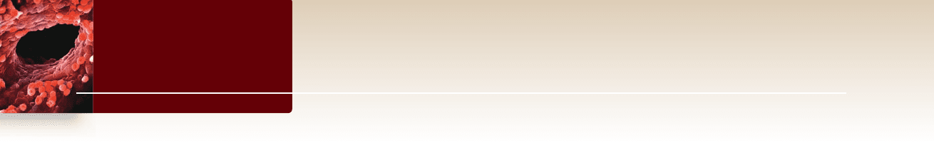
The baroreceptor re ex maintains
homeostasis in blood pressure
The arterial blood pressure (BP) depends on two factors: the
cardiac output (CO) and the resistance (R) to blood flow in the
vascular system. This relationship can be expressed as:
BP = CO × R
An increased blood pressure, therefore, could be pro-
duced by an increase in either heart rate or blood volume (be-
cause both increase the cardiac output), or by vasoconstriction,
which increases the resistance to blood flow. Conversely,
blood pressure falls if the heart rate slows or if the blood vol-
ume is reduced—for example, by dehydration or excessive
bleeding (hemorrhage).
Changes in arterial blood pressure are detected by
baroreceptors located in the arch of the aorta and in the ca-
rotid arteries (see chapter 46). These sensors are stretch re-
ceptors sensitive to expansion and contraction of arteries.
When the baroreceptors detect a fall in blood pressure, the
number of impulses to the cardiac center is decreased, result-
ing in increased sympathetic stimulation and decreased para-
sympathetic stimulation of the heart and other targets. This
increases heart rate and stroke volume to amplify cardiac out-
put. This also causes vasoconstriction of blood vessels in the
skin and viscera, raising resistance. These combine to increase
blood pressure, closing the feedback loop in this direction
(figure 50.17, top).
When baroreceptors detect a rise in blood pressure, the
number of impulses to the cardiac center is increased. This has
the opposite effect of decreasing sympathetic stimulation and
increasing parasympathetic simulation of the heart. This lowers
heart rate and stroke volume to reduce cardiac output. The car-
diac center also sends signals causing vasodilation of blood ves-
sels in the skin and viscera, lowering resistance. These combine
to decrease blood pressure closing the feedback loop in
this direction. Thus, the baroreceptor reflex forms a negative
feedback loop responding to changes in blood pressure
(figure 50.17, bottom).
Blood volume is regulated by hormones
Blood pressure depends in part on the total blood volume be-
cause this can affect the cardiac output. A decrease in blood
volume decreases blood pressure, if all else remains equal.
Blood volume regulation involves the effects of four hormones:
(1) antidiuretic hormone, (2) aldosterone, (3) atrial natriuretic
hormone, and (4) nitric oxide.
Antidiuretic hormone (ADH), also called vasopressin ,
is secreted by the posterior-pituitary gland in response to an
increase in the osmolarity of the blood plasma (see chapter 46 ).
Dehydration, for example, causes the blood volume to decrease.
Osmoreceptors in the hypothalamus promote thirst and stimu-
late ADH secretion from the posterior pituitary gland. ADH, in
turn, stimulates the kidneys to retain more water in the blood,
excreting less in the urine. A dehydrated person thus drinks
more and urinates less, helping to raise the blood volume and
restore homeostasis.
1034
part
VII
Animal Form and Function
rav32223_ch50_1018-1037.indd 1034rav32223_ch50_1018-1037.indd 1034 11/19/09 11:44:57 AM11/19/09 11:44:57 AM
Apago PDF Enhancer
Blood pressure
Stimulus
Baroreceptor senses
contraction of blood
vessels and low
blood pressure
Sensor
Baroreceptor senses
expansion of blood
vessels and high
blood pressure
Sensor
Cardiac center of
medulla sends
fewer impulses
Integrating Center
Cardiac center of
medulla increases
frequency of
impulses
Integrating Center
Decreases
sympathetic
Effector
Increases
parasympathetic
Increased heart rate,
stroke volume,
vasoconstriction
Response
Decreased heart rate,
stroke volume,
vasodilation
Response
Negative feedback
Raises blood pressure
Lowers blood pressure
( – )
( + )
( – )
( – )
( – )
( + )
Effector
Increases
sympathetic
Eff
ector
Decreases
parasympathetic
Effector
Figure 50.17
Baroreceptor negative feedback loops control blood pressure. Baroreceptors form the afferent portion of a
feedback loop controlling blood pressure. The frequency of nerve impulses from these stretch receptors correlates with blood pressure. This
information is processed in the cardiac center of the medulla. The efferent portion of the loop involves sympathetic and parasympathetic
nerves that innervate the heart. This control can raise or lower heart rate and stroke volume to raise and lower blood pressure in response to
baroreceptors signaling.
Whenever the kidneys experience a decreased blood flow,
a group of kidney cells initiate the release of an enzyme known
as renin into the blood. Renin activates a blood protein, angio-
tensin, which stimulates vasoconstriction throughout the body
while stimulating the adrenal cortex to secrete aldosterone.
This steroid hormone acts on the kidneys to promote the re-
tention of Na
+
and water in the blood (see chapter 46).
When excess Na
+
is present, less aldosterone is secreted
by the adrenals, so that less Na
+
is retained by the kidneys. Na
+
excretion in the urine is promoted by another hormone, atrial
natriuretic hormone. This hormone is secreted by the right
atrium of the heart in response to stretching caused by an in-
creased blood volume. The action of atrial natriuretic hormone
completes a negative feedback loop, lowering the blood volume
and pressure.
Nitric oxide (NO) is a gas produced by endothelial cells
of blood vessels. As described in chapter 46, it is one of a num-
ber of paracrine regulators of blood vessels. In solution, NO
passes outward through the cell layers of the vessel, causing the
smooth muscles that encase it to relax and the blood vessels to
dilate (become wider). For over a century, heart patients have
been prescribed nitroglycerin to relieve chest pain, but only
now has it become clear that nitroglycerin acts by releasing ni-
tric oxide.
Learning Outcomes Review 50.6
Cardiac output is the heart rate times the heart’s stroke volume. As exertion
increases, cardiac output increases to meet the body’s demands. Blood
pressure depends on cardiac output and the resistance to blood fl ow due to
constriction of the arteries. The blood volume is regulated by antidiuretic
hormone, aldosterone, and atrial natriuretic hormone; nitric oxide causes
vasodilation that lessens resistance.
■ What are the connections between regulation of heart
rate and breathing rate?
chapter
50
The Circulatory System
1035www.ravenbiology.com
rav32223_ch50_1018-1037.indd 1035rav32223_ch50_1018-1037.indd 1035 11/19/09 11:44:57 AM11/19/09 11:44:57 AM
Apago PDF Enhancer
Chapter Review
Arteries and veins branch to and from all parts of the body.
Arteries and arterioles carry oxygenated blood to the body; veins and
venules return deoxygenated blood to the heart (see gure 50.7).
Arterial blood pressure can be measured.
A sphygmomanometer measures the peak (systolic) and minimum
(diastolic) blood pressure. Blood pressure is expressed as the ratio of
systolic to diastolic.
50.5 Characteristics of Blood Vessels
Larger vessels are composed of four tissue layers.
Arteries and veins consist of endothelium, elastic bers, smooth
muscle, and connective tissues (see gure 50.12). Capillaries have
only one layer of endothelium.
Arteries and arterioles have evolved to withstand pressure.
Arteries and arterioles have thicker muscular layer and more
elastic bers to control blood ow and to recoil with changes in
blood pressure.
Capillaries form a vast network for exchange of materials.
Capillaries are the region of the circulatory system where exchange
takes place with the body’s tissues (see gure 50.13).
Venules and veins have less muscle in their walls.
The return of blood to the heart through veins is facilitated by
skeletal muscle contractions and one-way valves (see gure 50.14).
The lymphatic system handles uids that leave the
cardiovascular system.
Fluid from plasma lters out of capillaries, then returns
via the separate, one-way lymphatic system (see gure 50.15).
The lymphatic system connects with the blood circulation at the
subclavian veins.
Cardiovascular diseases a ect the delivery system.
Atherosclerosis is an accumulation of fatty materials in arteries; it is
one cause of a heart attack, which results from an insuf cient supply
of blood to heart muscle. Strokes are caused by blockage of the blood
supply to the brain.
50.6 Regulation of Blood Flow
and Blood Pressure
The nervous system may speed up or slow down heart rate.
Norepinephrine from sympathetic neurons increases heart rate;
acetylcholine from parasympathetic neurons decreases the rate.
Cardiac output increases with exertion.
Both heart rate and stroke volume increase with exertion.
The baroreceptor re ex maintains homeostasis in blood pressure.
Arterial blood pressure is monitored by baroreceptors
in the aortic arch and carotid arteries, which relay impulses to the
cardiac center (see gure 50.17).
Blood volume is regulated by hormones.
Blood volume regulation and arterial resistance involves the effects
of four hormones: (1) antidiuretic hormone, (2) aldosterone, (3) atrial
natriuretic hormone, and (4) nitric oxide.
50.1 The Components of Blood
Blood plasma is a uid matrix.
Plasma is 92% water plus nutrients, hormones, ions, plasma proteins,
and wastes (see gure 50.1).
Formed elements include circulating cells and platelets.
Blood cells include erythrocytes (red cells), leukocytes (white cells),
and platelets. Erythrocytes contain hemoglobin for oxygen transport,
and leukocytes are part of the immune system. Platelets help initiate
blood clotting (see gure 50.3).
Formed elements arise from stem cells.
Blood cells are derived from pluripotent stem cells in bone marrow
by hematopoiesis (see gure 50.2).
Blood clotting is an example of an enzyme cascade.
Upon initiation of clotting, brinogen, normally dissolved in the
plasma, is turned into brin, an insoluble protein, via an enzyme
cascade. As a wound heals, the clot must be dissolved.
50.2 Invertebrate Circulatory Systems
Open circulatory systems move uids in a one-way path.
Sponges pass water through channels, and cnidarians circulate water
through a gastrovascular cavity. Small animals can use body cavity
uids for circulation.
Closed circulatory systems move uids in a loop.
Closed systems have a distinct circulatory uid, such as blood,
enclosed in vessels and transported in a loop.
50.3 Vertebrate Circulatory Systems
In shes, more e cient circulation developed concurrently with gills.
Fishes have a linear heart with two pumping chambers to increase
ef ciency of blood ow through the gills; from the gills, the blood
moves into the rest of the body (see gure 50.5).
In amphibians and most reptiles, lungs required a separate circulation.
Pulmonary circulation pumps blood to the lungs, and systemic
circulation pumps blood to the body.
Amphibian hearts have two atria that separate blood ow to the
lungs and body, and a single ventricle ( gure 50.6). The heart of most
reptiles has a septum that partially divides the ventricle, reducing
mixing of blood from the atria.
Mammals, birds, and crocodilians have two completely separated
circulatory systems.
The four-chambered heart has two ventricles (see gure 50.7). The
extreme similarity between the heart of mammals and birds is an
example of convergent evolution.
50.4 The Four-Chambered Heart
and the Blood Vessels
The cardiac cycle drives the cardiovascular system.
The unidirectional ow of blood through the heart is maintained
by two atrioventricular valves (see gure 50.9). During diastole
ventricles relax and atria contract; during systole ventricles contract.
Contraction of heart muscle is initiated by autorhythmic cells.
Contraction is initiated by the SA node, a natural pacemaker, and
impulses then travel to the AV node (see gure 50.10).
1036
part
VII
Animal Form and Function
rav32223_ch50_1018-1037.indd 1036rav32223_ch50_1018-1037.indd 1036 11/19/09 11:44:58 AM11/19/09 11:44:58 AM
Apago PDF Enhancer
Review Questions
to the bottom of the ventricles before it emanates over
ventricular tissue.
b. The depolarization from the SA node is initiated
from motor neurons coming down from our brain;
depolarization from the AV node is initiated from motor
neurons coming up from our spinal cord.
c. Gravity carries the depolarization from the SA node down
from the top of the heart; contraction of the diaphragm
forces depolarization from the AV node from the bottom up.
d. This statement is false; both contract from the bottom.
2. A molecule of CO
2
that is generated in the cardiac muscle
of the left ventricle would not pass through which of the
following structures before leaving the body?
a. Right atrium c. Right ventricle
b. Left atrium d. Left ventricle
3. Blood clots are made of
a. brin. c. prothrombin.
b. brinogen. d. all of these.
4. The difference between the amphibian and mammal hearts
is that
a. in the amphibian heart, oxygenated and deoxygenated
blood mix completely in the single ventricle.
b. in the amphibian heart, there are two SA nodes so that
contractions occur simultaneously throughout the heart.
c. in the ventricle in the amphibian heart, internal channels
reduce mixing of blood.
d. in the amphibian heart, only the left aorta pumps oxygen
obtained by diffusion through the skin.
5. Contraction of the smooth muscle layers of the arterioles
a. increases the frictional resistance to blood ow.
b. may be a way of increasing heat exchange through the skin.
c. can increase blood ow to an organ.
d. includes all of the above.
SYNTHESIZE
1. Humans have a number of mechanisms that help to maintain
blood pressure, particularly when it falls too low. Explain how the
kidney and the endocrine systems help to maintain blood pressure.
2. What is the difference among blood, lymph, and hemolymph?
3. Is the evolution of the four-chambered heart related to the
evolution of endothermy?
4. What do you think are the clinical symptoms indicating that a
person requires the surgical implantation of a mechanical
pacemaker?
UNDERSTAND
1. An ECG measures
a. changes in electrical potential during the cardiac cycle.
b. Ca
2+
concentration of the ventricles in diastole.
c. the force of contraction of the atria during systole.
d. the volume of blood being pumped during the contraction cycle.
2. Systole is vitally important to heart function and begins in the
heart with the
a. activation of the AV node.
b. activation of the SA node.
c. opening of the voltage-gated potassium gates.
d. opening of the semilunar valves.
3. Which of the following is the correct sequence of events in the
circulation of blood?
a. Heart
arteries arterioles capillaries venules
lymph heart
b. Heart
arteries arterioles capillaries veins
venules heart
c. Heart
arteries arterioles capillaries venules
veins heart
d. Heart
arterioles arteries capillaries venules
veins heart
4. Which of the following statements is not true?
a. Only arteries carry oxygenated blood.
b. Both arteries and veins have a layer of smooth muscle.
c. Both arteries and veins branch out into capillary beds.
d. Precapillary sphincters regulate blood ow through capillaries.
5. The lymphatic system is like the circulatory system in that
they both
a. have nodes that lter out pathogens.
b. have a network of arteries.
c. have capillaries.
d. are closed systems.
6. Which pairing of structure and function is incorrect?
a. Erythrocytes: oxygen transport
b. Platelets: blood clotting
c. Plasma: waste transport
d. All of these are correct.
7. When a sphygmomanometer is used,
a. blood pulses through the vein when systolic pressure is
greater than the pressure caused by the cuff.
b. pulsing ceases when blood pressure falls below the
systolic pressure.
c. blood does not move through the vein when cuff pressure
is greater than maximal blood pressure.
d. cuff pressure stops decreasing when it equals
systolic pressure.
APPLY
1. In vertebrate hearts, atria contract from the top, and ventricles
contract from the bottom. How is this accomplished?
a. Depolarization from the SA node proceeds across the atria
from the top; depolarization from the AV node is carried
ONLINE RESOURCE
www.ravenbiology.com
Understand, Apply, and Synthesize—enhance your study with
animations that bring concepts to life and practice tests to assess
your understanding. Your instructor may also recommend the
interactive eBook, individualized learning tools, and more.
chapter
50
The Circulatory System
1037www.ravenbiology.com
rav32223_ch50_1018-1037.indd 1037rav32223_ch50_1018-1037.indd 1037 11/19/09 11:44:58 AM11/19/09 11:44:58 AM
Apago PDF Enhancer
T
Chapter
51
Osmotic Regulation
and the
Urinary System
Introduction
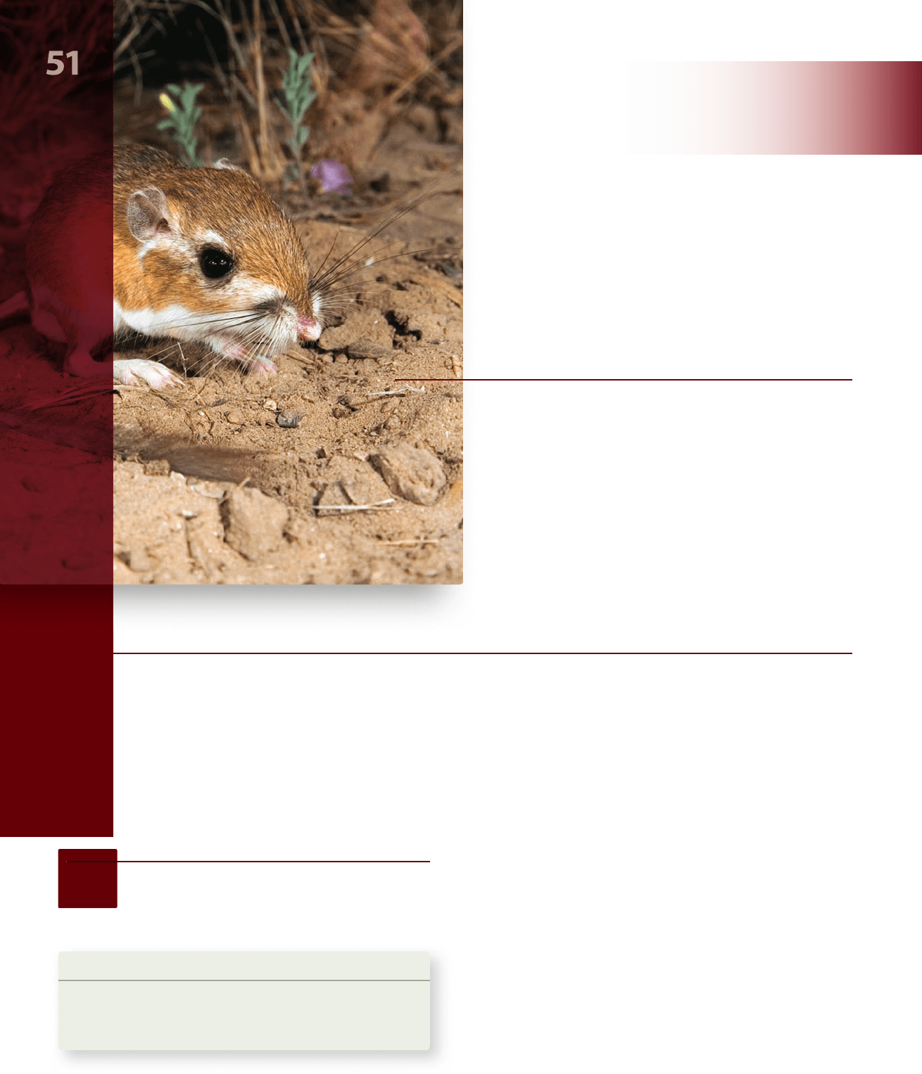
The majority of your body weight is actually water, but you exist in a very dehydrating environment. The kangaroo rat pictured
lives in a desert environment that is even more dehydrating and yet is so parsimonious with water that it never needs to drink;
it generates sufficient water as a by-product of oxidizing its food. Fish can exist in both freshwater and marine environments,
facing the challenge of either gaining or losing water, respectively. Life in these different environments is possible because
elaborate mechanisms enable organisms to control the osmotic strength of their blood and extracellular fluids. The regulation
of internal fluid and its composition is an example of homeostasis —the ability of living organisms to maintain internal
conditions within an optimal range. In this chapter, we describe the osmoregulatory systems of a number of animals, including
the mammalian urinary system. These organ systems maintain the water and ionic balance of fluids in the body.
Chapter Outline
51.1 Osmolarity and Osmotic Balance
51.2 Osmoregulatory Organs
51.3 Evolution of the Vertebrate Kidney
51.4 Nitrogenous Wastes: Ammonia, Urea,
and Uric Acid
51.5 The Mammalian Kidney
51.6 Hormonal Control of Osmoregulatory Functions
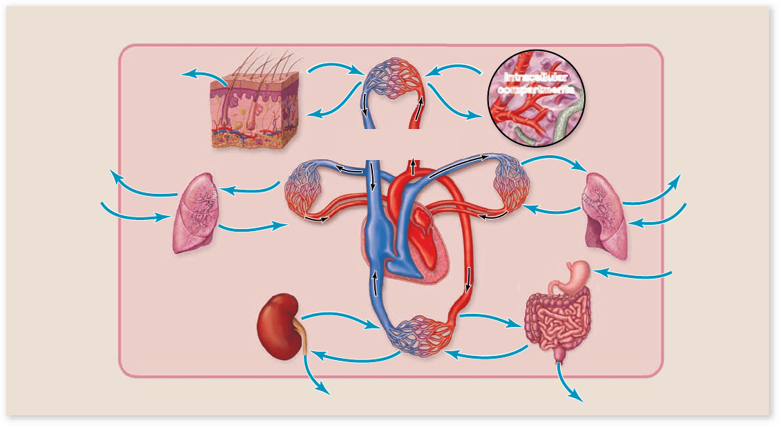
Water in a multicellular animal’s body is distributed between
the intracellular and extracellular compartments (figure 51.1) .
To maintain osmotic balance, the extracellular compartment of
an animal’s body (including its blood plasma) must be able to
take water from the environment and to excrete excess water
into the environment. Inorganic ions must also be exchanged
between the extracellular body fluids and the external envi-
ronment to maintain homeostasis. Exchanges of water and
electrolytes between the body and the external environment
occur across specialized epithelial cells and, in most vertebrates,
through a filtration process in the kidneys.
51.1
Osmolarity and Osmotic
Balance
Learning Outcomes
Explain the importance of osmotic balance.1.
Describe how organisms are classified based on their 2.
method of osmotic regulation.
CHAPTER
rav32223_ch51_1038-1054.indd 1038rav32223_ch51_1038-1054.indd 1038 11/19/09 3:13:51 PM11/19/09 3:13:51 PM
Apago PDF Enhancer
Extracellular compartment
(including blood)
Solutes
and H
2
O
Solutes
and H
2
O
Urine (excess H
2
O)
Solutes
and H
2
O
Solutes
and H
2
O
Food
and
H
2
O
Waste
Animal body
External environment
H
2
O
(Sweat)
Intracellular
compartments
CO
2
and H
2
O
O
2
CO
2
and H
2
O
O
2
Solutes
and H
2
O
Solutes
and H
2
O
Figure 51.1
The interaction between intracellular and extracellular compartments of the body and the external
environment. Water can be taken in from the environment or lost to the environment. Exchanges of water and solutes between the
extracellular uids of the body and the environment occur across transport epithelia, and water and solutes can be ltered out of the blood by
the kidneys. Overall, the amount of water and solutes that enters and leaves the body must be balanced in order to maintain homeostasis.
placed in a hypertonic solution loses water to the surrounding so-
lution and shrinks. In contrast, an animal cell placed in a hypotonic
solution gains water and expands. A cell in an isotonic solution
shows no net water movement. In medical care, isotonic solu-
tions such as normal saline and 5% dextrose are used to bathe
exposed tissues and are given as intravenous fluids.
Osmoconformers live in marine environments
The osmolarity of body fluids in most marine invertebrates is
the same as that of seawater (although the concentrations of
particular solutes, such as Mg
2+
, are not equal). Because the ex-
tracellular fluids are isotonic to seawater, no osmotic gradient
exists, and there is no tendency for water to leave or enter the
body. Such organisms are termed osmoconformers—they are
in osmotic equilibrium with their environment.
Among the vertebrates, only the primitive hagfish are
strict osmoconformers. The sharks and their relatives in the
class Chondrichthyes (cartilaginous fish) are also isotonic to
seawater, even though their blood level of NaCl is lower than
that of seawater; the difference in total osmolarity is made up
by retaining urea, as described later on.
Osmoregulators control
their osmolarity internally
All other vertebrates are osmoregulators—that is, animals
that maintain a relatively constant blood osmolarity despite
Most vertebrates maintain homeostasis for both the total
solute concentration of their extracellular fluids and the concen-
tration of specific inorganic ions. Sodium (Na
+
) is the major cat-
ion in extracellular fluids, and chloride (Cl
–
) is the major anion.
The divalent cations, calcium (Ca
2+
) and magnesium (Mg
2+
), the
monovalent cation K
+
, as well as other ions, also have important
functions and are maintained at constant levels.
Osmotic pressure is a measure
of concentration di erence
You learned in chapter 5 that osmosis is the diffusion of water
across a semipermeable membrane. Osmosis always occurs from
a more dilute solution (with a lower solute concentration) to a
less dilute solution (with a higher solute concentration). The os-
motic pressure of a solution is a measure of its tendency to take
in water by osmosis. This is the amount of pressure needed to
balance the pressure created by the movement of water.
A solution with a higher concentration of solute exerts
more osmotic pressure. This is measured as the osmolarity of
a solution, the number of osmotically active moles of solute per
liter of solution. Notice that osmolarity can differ from molar
concentration if a substance dissociates in solution into more
than one osmotically active particle. For example, a 1 molar (M)
solution of sucrose is also 1 osmolar (Osm), but a 1 M solution of
NaCl is 2 Osm as it dissociates into two osmotically active ions.
The tonicity of a solution is a measure of the ability of the
solution to change the volume of a cell by osmosis. An animal cell
chapter
51
Osmotic Regulation and the Urinary System
1039www.ravenbiology.com
rav32223_ch51_1038-1054.indd 1039rav32223_ch51_1038-1054.indd 1039 11/19/09 1:22:45 PM11/19/09 1:22:45 PM
Apago PDF Enhancer
Collecting
tubule
Excretory
pore
Cilia
Fluid
Flame cell
the different concentration in the surrounding environment.
The maintenance of a relatively constant body fluid osmo-
larity has permitted vertebrates to exploit a wide variety of
ecological niches. Achieving this constancy, however, requires
continuous regulation.
Freshwater vertebrates have a much higher solute con-
centration in their body fluids than that of the surrounding
water. In other words, they are hypertonic to their environ-
ment. Because of their cells’ higher osmotic pressure, water
tends to enter their bodies. Consequently, they have adapted to
prevent water from entering their bodies as much as possible
and to eliminate the excess water that does enter. In addition,
freshwater vertebrates tend to lose inorganic ions to their en-
vironment and so must actively transport these ions back into
their bodies.
In contrast, most marine vertebrates are hypotonic to
their environment; their body fluids have only about one-third
the osmolarity of the surrounding seawater. These animals are
therefore in danger of losing water by osmosis, and adaptations
have evolved to help them retain water to prevent dehydration.
They do this by drinking seawater and eliminating the excess
ions through kidneys and gills.
The body fluids of terrestrial vertebrates clearly have a
higher concentration of water than does the air surrounding
them. Therefore, they tend to lose water to the air by evapo-
ration from the skin and lungs. All reptiles, birds, and mam-
mals, as well as amphibians during the time when they live on
land, face this problem. Urinary/osmoregulatory systems have
evolved in these vertebrates that help them retain water.
Learning Outcomes Review 51.1
Osmotic balance must be maintained so that tissues can carry out metabolic
functions. Physiological mechanisms help most vertebrates keep blood
osmolarity and ion concentrations relatively constant. Marine invertebrates
are osmoconformers; their body fl uids are isotonic to their environment.
Most vertebrates are osmoregulators; their body fl uids are either hypertonic
or hypotonic compared to their environment.
■ During osmosis, does water move toward regions of
higher or lower osmolarity?
tubules (little tubes) that expel fluid and wastes from the body.
In addition, more elaborate systems can be found in inverte-
brates. In vertebrates, the urinary system is highly complex.
Invertebrates make use of
specialized cells and tubules
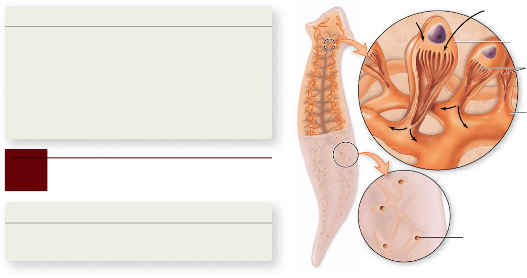
In flatworms, tubules called protonephridia branch through-
out the body into bulblike flame cells (figure 51.2) . Although
these simple excretory structures open to the outside of the
body, they do not open to the inside; rather, the movement of
cilia within the flame cells must draw in fluid from the body.
Water and metabolites are then reabsorbed, and the substances
to be excreted are expelled through excretory pores.
Other invertebrates have a system of tubules that open
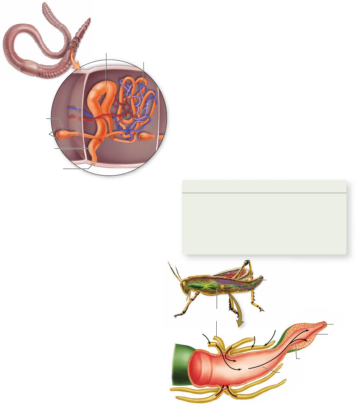
both to the inside and to the outside of the body. In the earth-
worm, these tubules are known as nephridia (orange structures
in figure 51.3) . The nephridia obtain fluid from the body cav-
ity through a process of filtration into funnel-shaped structures
called nephrostomes. The term filtration is used because the fluid
is formed under pressure and passes through small openings,
so that molecules larger than a certain size are excluded. This
filtered fluid is isotonic to the fluid in the coelom, but as it
passes through the tubules of the nephridia, NaCl is removed
by active transport processes.
The general term for transport out of the tubule and into
the surrounding body fluids is reabsorption. Because salt is
Figure 51.2
The protonephridia of atworms. A
branching system of tubules, bulblike ame cells, and excretory
pores make up the protonephridia of atworms. Cilia inside the
ame cells draw in uids from the body by their beating action.
Substances are then expelled through pores that open to the
outside of the body.
51.2
Osmoregulatory Organs
Learning Outcomes
Describe invertebrate osmoregulatory organs.1.
Define reabsorption and secretion.2.
A variety of mechanisms have evolved in animals to cope with
problems of water balance. In many animals, the removal of
water or salts from the body is coupled with the removal of
metabolic wastes through the excretory system. Single-celled
protists employ contractile vacuoles for this purpose, as do
sponges. Other multicellular animals have a system of excretory
1040
part
VII
Animal Form and Function
rav32223_ch51_1038-1054.indd 1040rav32223_ch51_1038-1054.indd 1040 11/19/09 1:22:46 PM11/19/09 1:22:46 PM
Apago PDF Enhancer
Waste
Malpighian
tubules
Rectum
(active transport)
(active
transport)
(osmosis)
Midgut
Hindgut
Rectum
Ions and H
2
O
K
+
H
2
O
Anus
Coelomic fluid
Nephrostome
Pore for
urine excretion
Capillary
network
Bladder
Nephridium
Figure 51.3
The nephridia of annelids. Most
invertebrates, such as the annelid shown here, have nephridia
(orange). These consist of tubules that receive a ltrate of coelomic
uid, which enters the funnel-like nephrostomes. Salt can be
reabsorbed from these tubules, and the uid that remains, urine, is
released from pores into the external environment.
Figure 51.4
The Malpighian
tubules of insects.
The Malpighian tubules of
insects are extensions of the digestive tract that collect water and
wastes from the body’s circulatory system. K
+
is secreted into these
tubules, drawing water with it osmotically. Much of this water
(arrows) is reabsorbed across the wall of the hindgut.
The vertebrate kidney lters
and then reabsorbs
The kidneys of vertebrates, unlike the Malpighian tubules
of insects, create a tubular fluid by filtering the blood under
pressure. In addition to waste products and water, the filtrate
contains many small molecules, including glucose, amino acids,
and vitamins, that are of value to the animal. These molecules
and most of the water are reabsorbed from the tubules into
the blood, while wastes remain in the filtrate. Additional wastes
may be secreted by the tubules and added to the filtrate, and the
final waste product, urine, is eliminated from the body.
It may seem odd that the vertebrate kidney should fil-
ter out almost everything from blood plasma (except proteins,
which are too large to be filtered) and then spend energy to
take back or reabsorb what the body needs. But selective re-
absorption provides great flexibility. Various vertebrate groups
have evolved the ability to reabsorb molecules that are espe-
cially valuable in particular habitats. This flexibility is a key
factor underlying the successful colonization of many diverse
environments by the vertebrates. In the rest of this chapter, we
focus on the vertebrate kidney and its elimination of waste ma-
terials, notably nitrogen compounds.
Learning Outcomes Review 51.2
Many invertebrates fi lter fl uid into a system of tubules and then reabsorb
ions and water, leaving waste products for excretion. Insects create an
excretory fl uid by secreting K
+
and waste products into tubules, which draw
water osmotically. The vertebrate kidney produces a fi ltrate that enters
tubules and is modifi ed to become urine.
■ How are the function of Malpighian tubules and
kidneys similar?
reabsorbed from the filtrate, the urine excreted is more dilute
than the body fluids—that is, the urine is hypotonic. The kid-
neys of mollusks and the excretory organs of crustaceans (called
antennal glands) also produce urine by filtration and reclaim
certain ions by reabsorption.
Insects have a unique
osmoregulatory system
The excretory organs in insects are the Malpighian tubules
(figure 51.4) , extensions of the digestive tract that branch off
anterior to the hindgut. Urine is not formed by filtration in
these tubules because there is no pressure difference between
the blood in the body cavity and the tubule. Instead, waste mol-
ecules and potassium (K
+
) ions are secreted into the tubules by
active transport.
Secretion is the opposite of reabsorption—ions or mol-
ecules are transported from the body fluid into the tubule. The
secretion of K
+
creates an osmotic gradient that causes water
to enter the tubules by osmosis from the body’s open circula-
tory system. Most of the water and K
+
is then reabsorbed into
the circulatory system through the epithelium of the hindgut,
leaving only small molecules and waste products to be excreted
from the rectum along with feces. Malpighian tubules thus pro-
vide a very efficient means of water conservation.
chapter
51
Osmotic Regulation and the Urinary System
1041www.ravenbiology.com
rav32223_ch51_1038-1054.indd 1041rav32223_ch51_1038-1054.indd 1041 11/19/09 1:22:47 PM11/19/09 1:22:47 PM