Raven P.H., Johnson G.B., Mason K.A. Biology (Ninth Edition)
Подождите немного. Документ загружается.

Apago PDF Enhancer
a.
b.
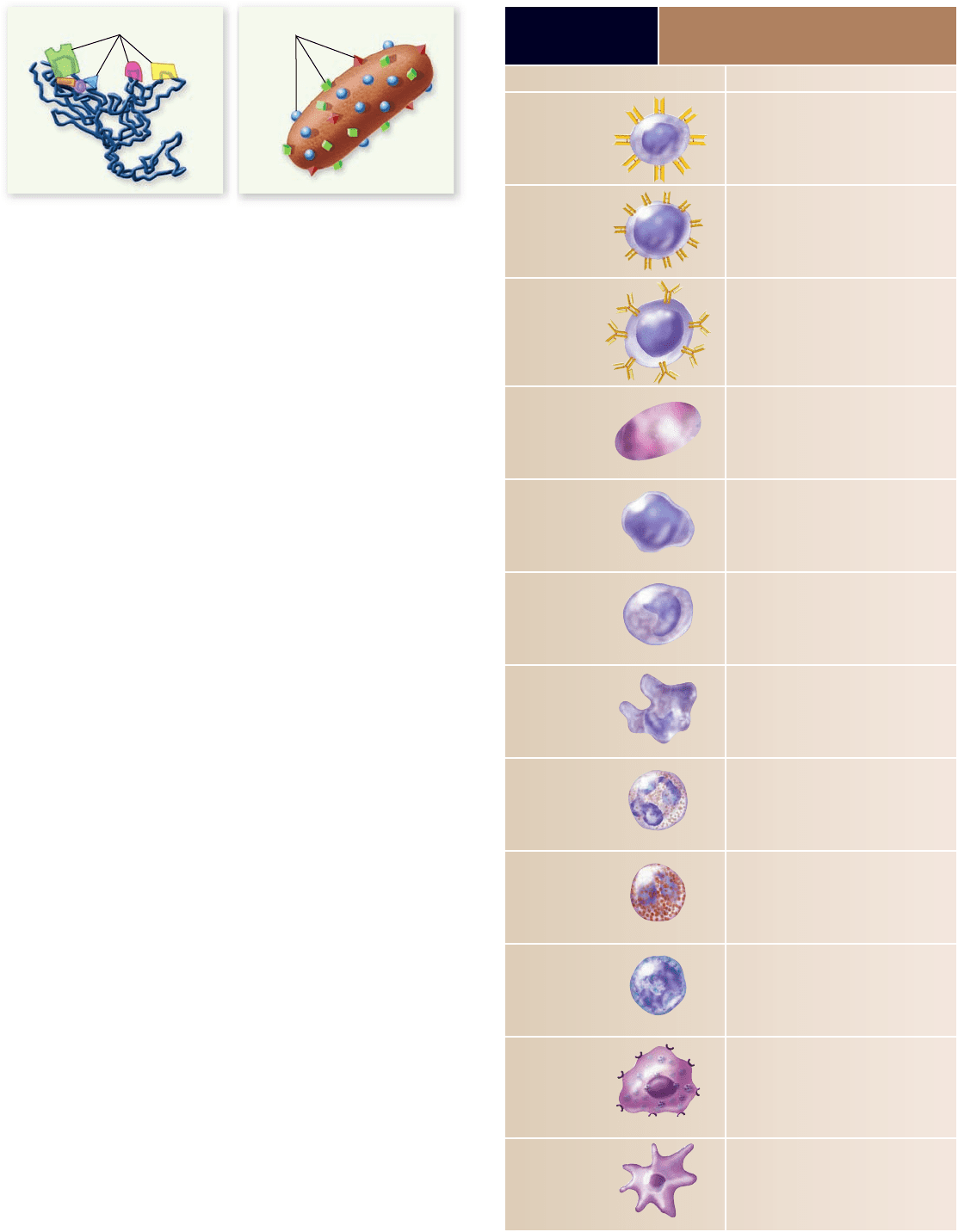
EpitopesEpitopes
Protein Bacterium
1062
part
VII
Animal Form and Function
They may also be components of foods or pollens. A large an-
tigen is likely to have many different parts, known as antigenic
determinants, or epitopes (figure 52.7) , each of which can stimu-
late a distinct immune response.
Hematopoiesis gives rise to the cells
of the immune system
All the cells that are found in the blood are derived from the di-
vision and differentiation of hematopoietic stem cells, a process
called hematopoiesis (see chapter 50). Embryologically, these
stem cells are initially found in the yolk sac, then migrate to
the fetal liver and spleen and finally to the bone marrow. Stem
cells give rise to lymphoid progenitors and myeloid progeni-
tors. A lymphoid progenitor, in turn, gives rise to both the B
and T lymphocytes as well as to natural killer cells. A myeloid
progenitor gives rise to all the other cells of the immune system
as well as to erythrocytes and platelets (see figure 50.2) .
Although the lymphocytes are responsible for adaptive
immunity, all the other leukocytes illustrated in figure 50.2
play supporting roles in this specific response or are part of
innate immunity. Monocytes give rise to the macrophages,
and these along with the neutrophils are phagocytic cells.
Eosinophils are important in the elimination of helminths
(flatworms; see chapter 33), either via secretion of digestive
enzymes through perforin pores inserted in the plasma mem-
brane of the helminths or occasionally by phagocytosis. They
also play a role in exacerbating chronic inflammatory diseases
such as asthma or inflammatory bowel disease.
Basophils and mast cells are not phagocytic but rather
secrete inflammatory mediators such as histamine and pros-
taglandin in response to the binding of complement proteins
during the elimination of pathogens. These cells, and mast cells
in particular, are also activated during an allergic response, and
the inflammatory mediators they release cause the symptoms of
allergy.
Dendritic cells are important in the activation of T cells,
as will be described further. Dendritic cells also form a link
Figure 52.7
Many di erent epitopes are exhibited
by any one antigen. a. A single protein, with associated
carbohydrate, may have many different antigenic determinants
called epitopes, each of which can stimulate a distinct immune
response. b. A pathogen such as a bacterium has many proteins on
its surface, and there are likely to be multiple copies of each. Note
that the protein and bacterium are not drawn to scale with respect
to each other.
TABLE 52.1
Cells of the
Immune System
Cell Type Function
Helper T cell Speci cally recognizes foreign peptides
on antigen-presenting cells, inducing the
release of cytokines that activate B cells
or macrophages
Cytotoxic T cell Speci cally recognizes and kills “altered-
self” cells: virally infected, or tumor cells
B cell Binds speci c soluble antigens with its
membrane-bound antibody; serves as
an antigen-presenting cell to T
H
cells; on
activation di erentiates into plasma and
memory B cells
Plasma cell Derived from activated B cell; is a
biochemical factory devoted to the secretion
of antibodies directed against speci c
antigens
Natural killer cell Rapidly recognizes and kills virally infected
or tumor cells
Monocyte Precursor of macrophage; located in blood
Macrophage Phagocytic tissue cell that is a component
of the body’s rst cellular line of defense;
also serves as an antigen-presenting cell to
T
H
cells
Neutrophil A phagocytic cell that is a component of the
body’s rst cellular line of defense; found in
the blood in large numbers until attracted to
tissues during in ammation
Eosinophil Important to the elimination of parasites and
involved in chronic in ammatory diseases
Basophil Circulating cell that releases mediators such
as histamine that promote in ammation
Mast cell Located primarily under mucosal surfaces
and releases mediators such as histamine
that promote in ammation; triggered both
during in ammatory and allergic responses
Dendritic cell Important antigen-presenting cell to naive
T
H
cells; also helps in the activation of naive
T
C
cells
rav32223_ch52_1055-1083.indd 1062rav32223_ch52_1055-1083.indd 1062 11/19/09 1:45:14 PM11/19/09 1:45:14 PM
Apago PDF Enhancer
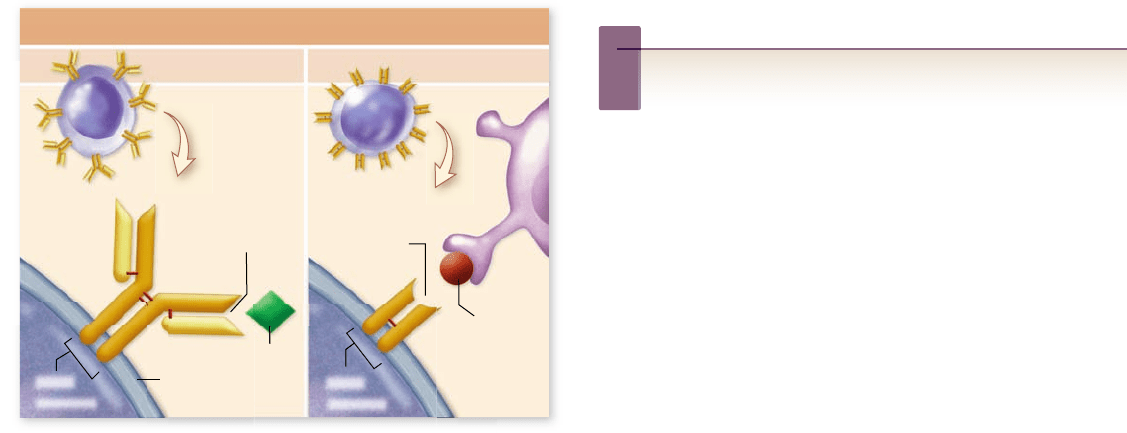
Lymphocyte Receptor Proteins
B Cell T Cell
Antigen
Antigen
Antigen-binding
site
Plasma membraneB-cell
receptor
Antigen-binding
site
T-cell
receptor
chapter
52
The Immune System
1063www.ravenbiology.com
between innate and adaptive immunity. Dendritic cells have
a variety of TLRs that recognize pathogens and stimulate
secretion of cy to kines and the inflammatory response. Thus
they can present antigens and recognize pathogen patterns
via innate receptors as well. The roles of these cells are sum-
marized in table 52.1 .
Lymphocytes carry out the
adaptive immune responses
The adaptive immune system is characterized by
Speci city of recognition of antigen1.
Wide diversity of antigens can be speci cally recognized2.
Memory, whereby the immune system responds more 3.
quickly and more intensely to an antigen it encountered
previously than to one it is meeting for the rst time
Ability to distinguish self-antigens from nonself 4.
The cells in the blood involved in the adaptive immune re-
sponse are leukocytes derived from a stem cell line called lym-
phoid progenitor cells (see gure 50.2). These lymphocytes
have receptor proteins on their surfaces that recognize spe-
cific epitopes on an antigen and direct an immune response
against either the antigen in solution or on the cell surface
(figure 52.8) . This response is also affected by signals derived
from the innate system described earlier. The innate system
dominates early in infection by a new pathogen, and the adap-
tive response dominates in later stages of infection.
Lymphocytes and antigen recognition
Although all the receptor proteins on any one lymphocyte have
the same epitope specificity, it is rare that any two lymphocytes
Figure 52.8
B- and T-cell receptors bind antigens.
B-cell receptors are immunoglobulin (Ig) molecules with a
characteristic Y-shaped structure. Every B cell has a single kind of
Ig on its surface that binds to a single antigenic determinant. T-cell
receptors are simpler than Ig molecules, but also bind to speci c
antigenic determinants. T cells only bind to antigens bound to
another cell.
have identical specificities. This feature produces the diversity
of immune responses that ensures that at least some epitopes of
any antigen that might be encountered are recognized.
A lymphocyte that has never before encountered anti-
gen is referred to as a naive lymphocyte. When a naive lympho-
cyte binds to a foreign antigen, the lymphocyte is activated,
causing it to divide producing a clone of cells with identical
antigen specificity, a process called clonal selection. Some of
these cells respond immediately to the antigen, and others be-
come memory cells, which can remain in our bodies for years
and perhaps for the remainder of our lives. Memory cells are
easily and rapidly activated on subsequent encounters with
the same antigen.
B cells
Lymphocytes called B lymphocytes, or B cells, respond
to antigens by secreting proteins called antibodies, or
immunoglobulins (Ig). Antigen recognition occurs when an
antigen binds to immunoglobulins on the B cell’s membrane.
Binding to antigen, in conjunction with other signals to be de-
scribed later, initiates a signaling pathway that leads to the pro-
duction of plasma cell’s that secrete antibodies specific for the
epitope recognized by the antibody in the B-cell membrane. This
B-cell–mediated response producing secreted antibodies is
called humoral immunity.
T cells
Other lymphocytes, called T lymphocytes, or T cells, do
not secrete antibodies but instead regulate the immune re-
sponses of other cells or directly attack the cells that carry
the specific antigens. These cells participate in the other arm
of adaptive immunity called cell-mediated immunity. Both
cell-mediated and humoral immunity processes are described
in detail in later sections.
Inquiry question
?
Jenner used cowpox virus to elicit an immune response
against smallpox. What does this tell us about the antigenic
properties of the two viruses?
Adaptive immunity can
be active or passive
Immunity can be acquired in different ways. First, an individual
can gain immunity when infected by a pathogen and perhaps
developing the disease it causes. Alternatively, an individual can
be immunized with portions of a pathogen or with a less viru-
lent form of the pathogen. Both of these situations result in
active immunity, associated with the activation of specific lym-
phocytes and the generation of memory cells by the individual.
Second, an individual can gain immunity by obtaining anti-
bodies from another individual. This happened to you before
you were born, as some antibodies made by your mother were
transferred to your body across the placenta. Immunity gained
in this way is called passive immunity, and it does not result in
the generation of memory cells. The immunity is only effective
as long as the antibodies remain in your body. Like any other
proteins, they will degrade in time.
rav32223_ch52_1055-1083.indd 1063rav32223_ch52_1055-1083.indd 1063 11/19/09 1:45:15 PM11/19/09 1:45:15 PM
Apago PDF Enhancer
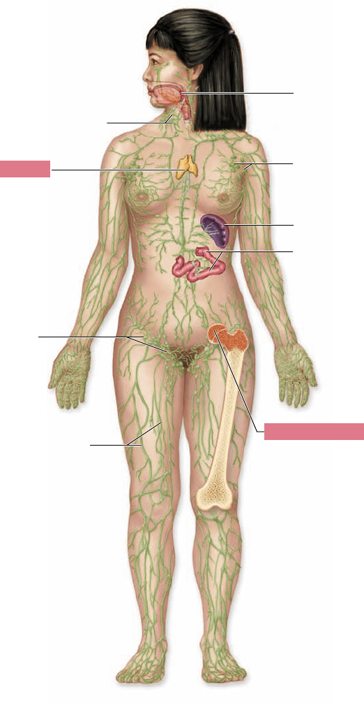
Cervical lymph nodes
Thymus
Inguinal
lymph
nodes
Lymphatic vessels
Axillary
lymph nodes
Spleen
Red bone marrow
Tonsils
Mucosa-
associated
lymphatic
tissue
(MALT) (in
small intestine)
1064
part
VII
Animal Form and Function
Figure 52.9
Organs of the speci c immune system.
There are two types of immune system organs: primary lymphoid
organs (red boxes), in which B and T lymphocytes mature and
acquire their speci c receptors, and secondary lymphoid organs
(labeled in black) in which antigen is collected and through which
the mature naive lymphocytes circulate in order to meet and be
stimulated by antigen.
The immune system is supported
by two classes of organs
The organs of the immune system consist of the primary
lymphoid organs—the bone marrow and the thymus—
as well as the secondary lymphoid organs—the lymph nodes,
spleen, and mucosa-associated lymphoid tissue, or MALT
(figure 52.9) .
The primary lymphoid organs
The bone marrow is not only the source of stem cells, it is where
B cells mature. After hematopoiesis gives rise to the most imma-
ture B cells, progenitor B cells, these cells complete their matura-
tion in the bone marrow. It is here that DNA rearrangements of
the immunoglobulin genes, to be discussed later, dictate the speci-
ficity of each B cell. Every B cell has about 10
5
Ig molecules on
its surface, all with identical specificity of epitope binding and all
different from cell to cell.
Any lymphocytes that are likely to bind to self-antigens
undergo apoptosis (figure 52.10a) . The remainder are released
to circulate in the blood and lymph and pass through the sec-
ondary lymphoid organs, where they may encounter antigen.
After their origin in the bone marrow, progenitor T cells mi-
grate to the thymus, a primary lymphoid organ located just above
the heart. The thymus is very large in infants; it starts to shrink in
the teenage years, which it continues to do throughout life.
The antigen receptor on T cells is designated the T-cell
receptor, or TCR. The TCR is produced by gene rearrange-
ments as T cells mature in the thymus, similar to those that oc-
cur for Ig genes of progenitor B cells. Thus, T cells may express
about 10
5
identical TCRs per T cell, all likely to be different
from one T cell to the next.
B cells recognize an epitope of an intact antigen that may
or may not be a protein. In contrast, T cells recognize only a
peptide fragment of a protein antigen, and this peptide frag-
ment must be bound to one of a series of self-proteins that are
present on the surface of almost all of the body’s cells. These
proteins are encoded by genes in the major histocompatibility
complex, or MHC. The MHC is discussed in detail in subse-
quent sections.
During selection in the thymus, T cells are exposed to
many thymic cells, all expressing self-MHC proteins with
bound self-peptides on their surfaces. If a T cell’s TCRs
bind too strongly to these self-MHC protein complexes,
that T cell becomes “self-reactive” and undergoes apoptosis
(figure 52.10b). Conversely, if the T cell’s TCR does not bind
MHC complexes at all, it is also eliminated. Only about 5% of
the progenitor T cells that enter the thymus pass this rigorous
two-step selection and avoid apoptosis.
The secondary lymphoid organs
The locations of the secondary lymphoid organs promote the
filtering of antigens that enter any part of an individual’s body.
Bacteria attached to a thorn stuck in the skin, for example, enter
the lymph that bathes the tissues. Lymph is eventually returned
to the blood circulation through a series of vessels referred to
as the lymphatics (see chapter 50). On its way, the lymph is fil-
tered in the thousands of lymph nodes, which are located at the
junction of lymphatic vessels (see figure 52.9).
The many mature but naive B and T lymphocytes that
have entered a lymph node on exiting the primary lymphoid
organs, or the memory cells that are located here, become
activated on meeting with antigen. Antibodies secreted on acti-
vation of B cells in the lymph nodes, as well as the clonal prog-
eny of the activated B and T cells, then leave the lymph node
and enter the blood circulation when the lymph is returned to
blood vessels near the heart.
rav32223_ch52_1055-1083.indd 1064rav32223_ch52_1055-1083.indd 1064 11/19/09 1:45:16 PM11/19/09 1:45:16 PM
Apago PDF Enhancer
a. b.
Ig on B cell
surface binds,
inducing
apoptosis
Ig does not
bind; B cell
leaves bone
marrow
Bone marrow
stromal cell
Dendritic cell
Igs
TCRs
TCR binds
tightly, inducing
apoptosis
TCR binds
weakly;
T cell leaves
thymus
MHC–
peptide
complex
Cells that cannot
bind MHC are
also eliminated
chapter
52
The Immune System
1065www.ravenbiology.com
Lymphocytes responding to antigens in a lymph node
may pass out of capillaries supplying blood to the lymph node
and enter the node’s tissues. This is the cause of the “swollen
glands” that sometimes accompany infection. The local lymph
nodes enlarge due to the vast influx of lymphocytes.
Some antigens are found primarily in the blood, or in the
blood as well as in the tissues. One example is the bacterium
Neisseria meningitidis, a cause of a potentially fatal meningitis
(infection of the meninges, layers of membranes covering the
brain). Immune responses to such antigens occur in the spleen.
The splenic artery carries blood to the spleen where it
then subdivides into arterioles. Antigens released into the
ground tissue of the spleen are recognized by B and T cells
present in the white pulp, regions of the spleen immediately
surrounding the arterioles. Lymphocytes in the white pulp may
be activated, as in the lymph node. Antibodies along with some
of the activated lymphocytes exit via the splenic vein.
The final important secondary lymphoid organ is the
mucosa-associated lymphoid tissue (MALT), which includes
the tonsils, the appendix, and a large number of follicles located
in the connective tissue under mucosal surfaces. These follicles
are composed of lymphocytes, primarily B cells but also some T
cells, and some macrophages. Any antigens that pass through the
mucosa immediately encounter lymphocytes in these follicles and
their entry further into the body may be stopped at this point.
If invading organisms manage to escape or evade innate
defenses of mucosal surfaces as well as the specific responses
of the lymphocytes in the MALT, then they still face a further
Figure 52.10
Selection against
self-reactive lymphocytes in
primary lymphoid organs. After
B and T lymphocytes acquire their
speci c receptors, self-reactive cells are
eliminated by apoptosis. a. If Igs on
the surface of a maturing B cell bind to
an epitope on a bone marrow stromal
cell, that cell will undergo apoptosis.
The small percentage (10%) of B cells
whose Igs do not recognize stromal cell
epitopes will be released from the bone
marrow. b. If TCRs on a maturing
T cell bind too tightly to self-MHC/
self-peptide complexes on dendritic cells
in the thymus, that T cell will undergo
apoptosis. Cells that don’t bind MHC
complexes at all are also eliminated.
The very small percentage (2–5%)
of maturing T cells that bind MHC
peptide complexes with intermediate
af nity are released from the thymus.
These cells bind self-MHC/foreign
peptide complexes with high af nity.
chance of being stopped by responses in the other secondary
lymphoid organs.
Two forms of adaptive immunity have evolved
Adaptive immunity, involving the ability to distinguish be-
tween self and nonself, was long thought to have evolved once
in vertebrates. The type of adaptive immunity described in this
chapter first arose in the cartilaginous fish that evolved some
450 mya (see chapter 35).
Sharks and rays possess a thymus and a spleen, as well
as a rather diffuse MALT. These animals mount cell-mediated
responses with T cells bearing TCRs, and humoral responses
with B cells that secrete Ig. Bone marrow in which hemato-
poiesis occurs appeared first in amphibians, although its exact
role appears to vary in different species. Lymph nodes appeared
first in birds, and their immune system differs little from that
of mammals.
Recently, a second form of adaptive immunity has been
described in jawless fish. This system does not involve B and
T cells with their characteristic receptors. Instead, lymphocytes
have receptor proteins composed of variable repeats rich in the
amino acid leucine. These proteins appear to function much
like Ig, but with a completely different protein architecture.
The number of different receptor proteins produced by this
system appears similar to the number of potential Ig. The gen-
eration of diversity in the two systems appears to have some
similarity as the different lymphocyte receptors in jawless fish
rav32223_ch52_1055-1083.indd 1065rav32223_ch52_1055-1083.indd 1065 11/19/09 1:45:17 PM11/19/09 1:45:17 PM

Apago PDF Enhancer
1066
part
VII
Animal Form and Function
TABLE 52.2
Lymphocyte Recognition of Antigen
Recognize epitopes
of soluble or
particulate antigen
Recognize peptides
bound to
self-MHC proteins
Class of MHC
proteins recognized
Cell types on
which recognized
MHC is expressed
B cells Yes No None NA
T
H
(CD4
+
) cells No Yes Class II Antigen-presenting cells: dendritic
cells, B cells, and macrophages
T
C
(CD8
+
) cells No Yes Class I All nucleated cells
The MHC carries self and nonself information
As discussed earlier, the surfaces of most vertebrate cells ex-
hibit glycoproteins encoded by the MHC. In humans, the name
given to the proteins encoded by the MHC complex is human
leukocyte antigens (HLAs). The genes encoding the MHC
proteins are highly polymorphic (have many alleles). For ex-
ample, the HLA proteins are specified by genes that are the
most polymorphic known, with nearly 500 alleles detected for
some of the proteins. Only rarely will two individuals have the
same combination of alleles, and the HLAs are thus different
for each individual, much as fingerprints are.
MHC proteins on the tissue cells serve as self markers
that enable an individual’s immune system, specifically its T
cells, to distinguish its own cells from foreign cells, an ability
called self versus nonself recognition.
There are two classes of MHC proteins. MHC class
I proteins are present on every nucleated cell of the body.
MHC class II proteins, however, are found only on antigen-
presenting cells (in addition to MHC class I); these cells in-
clude macrophages, B cells, and dendritic cells (table 52.2) . T
C
cells respond to peptides bound to MHC class I proteins, and
T
H
cells respond to peptides bound to MHC class II proteins.
Most of the time, the peptides bound to MHC proteins
are derived from self-proteins from the individual’s own cells.
For this reason, it is important that T cells undergo selection in
the thymus so that those that bind too strongly to peptides of
self-proteins on self-MHC are eliminated. In this way, T cells
normally are activated only outside the primary lymphoid or-
gans in which they mature, when they encounter peptides of
foreign proteins on self-MHC—for example, in the case of vi-
ral infection or cancer.
Cytotoxic T cells eliminate virally
infected cells and tumor cells
Activated cytotoxic T cells recognize “altered-self” cells, par-
ticularly those that are virally infected or tumor cells. The
TCRs of cytotoxic T lymphocytes recognize peptides of en-
dogenous antigens bound to MHC class I proteins. Peptides of
endogenous antigens are generated in a cell’s cytosol and then
are pumped by special transport proteins into the rough en-
doplasmic reticulum where they become bound to MHC class
I proteins. These proteins continue on their way through the
endomembrane system to the cell surface.
are also assembled by DNA rearrangements. The makeup of
the genes involved and the mechanism of these rearrangements
is currently unknown.
It is unclear whether this newly described form of adap-
tive immunity was present in the ancestor to all chordates, or if
it evolved in the lineage that gave rise to jawless fish. Given the
differences in the two systems, it is likely that they represent
independent events. If this other form of adaptive immunity
was present in the ancestor to all chordates, some vestige may
remain in modern vertebrates, including humans.
Learning Outcomes Review 52.2
Adaptive immunity is able to recognize individual pathogens and mount
a specifi c response. Lymphocytes, produced in bone marrow, must acquire
their specifi c receptors and undergo selection for self-reactivity in primary
lymphoid organs. These mature but naive lymphocytes circulate to
secondary lymphoid organs, where they may encounter foreign antigens.
B cells produce circulating antibodies (humoral immunity); T cells kill
pathogens or help other cells respond to them (cell-mediated immunity).
■ What type of adult stem cells are found in the
immune system?
52.3
Cell-Mediated Immunity
Learning Outcomes
Describe the function of cytotoxic T cells.1.
Explain the role of helper T cells.2.
T cells may be characterized as either cytotoxic T cells (T
C
)
or helper T cells ( T
H
) . These cells can also be identified based on
cell surface markers. T
C
cells have CD8 protein on their cell
surface, making them CD8
+
cells. T
H
cells have CD4 protein
on their cell surface, making them CD4
+
cells.
To be activated, both of these T cell types must rec-
ognize peptide fragments bound to MHC proteins, but the
two cell types may be distinguished by (1) recognition of
different classes of MHC proteins, which have distinct cell
distributions, and (2) differing roles of the T cells after they
are activated.
rav32223_ch52_1055-1083.indd 1066rav32223_ch52_1055-1083.indd 1066 11/19/09 1:45:18 PM11/19/09 1:45:18 PM
Apago PDF Enhancer
Virus
Dendritic cell
MHC class I
Viral peptide
TCR specific for this
MHC–peptide complex
Naive cytotoxic T cell
CD8
Cytokines
Cytokines
Clonal
expansion
Memory cytotoxic T cells
Activated cytotoxic T cells
Destroys
altered cell
Persists after
altered cells
are destroyed
Infected cell
Apoptosis of infected cell
a. b.
chapter
52
The Immune System
1067www.ravenbiology.com
An endogenous antigen may be a self-protein, or it
may be a viral protein produced within a virally infected cell
or an unusual protein produced by a cancerous cell. T
C
cells
respond only to the peptides of these unusual proteins bound
to self-MHC class I. T-cell activation occurs in a secondary
lymphoid organ, as described earlier. In a lymph node, for ex-
ample, T cells encounter antigen-presenting cells. Dendritic
cells in particular often present antigens that activate T
C
cells.
Because not all viruses can infect dendritic cells, the den-
dritic cells must ingest viruses or tumor cells and then, through
a mechanism referred to as cross-presentation, place the viral
or tumor peptides on MHC class I proteins. Binding of the
T
C
cell through its TCR and its CD8 site to the dendritic cell
induces clonal expansion of the T
C
cell, generating many acti-
vated T
C
cells as well as memory T
C
cells (figure 52.11) . The
activated T
C
cells then circulate around the body where they
bind to “target” host cells that express the same combination of
foreign peptide on self-MHC class I (figure 52.12) .
Apoptosis of the target cell is induced in a very similar
fashion to that used by NK cells; a T
C
cell secretes perforin
monomers that create pores in the target’s membrane; gran-
zymes enter and activate caspases, which in turn cause apopto-
sis of the target.
Helper T cells secrete proteins
that direct immune responses
Activated helper T cells, T
H
cells, secrete low-molecular-
weight proteins known as cytokines. A vast array of cytokines
is known, many but not all of which are secreted by T
H
cells.
These cytokines bind to specific receptors on the membranes
of many other cells, particularly but not exclusively those of the
immune system. On binding, they initiate signaling cascades in
these cells that promote their activation or differentiation.
Figure 52.11
Cytotoxic T cells induce apoptosis
of “altered-self” cells. Naive cytotoxic T cells are initially
activated on TCR recognition of foreign peptide displayed on self-
MHC class I proteins on dendritic cells in a secondary lymphoid
organ. Activation results in clonal expansion and differentiation
into memory cells and activated cells. Activated progeny of the
T
C
cell can induce apoptosis of any cell in the periphery (outside
the secondary lymphoid organ) that displays the same self-MHC
class I–peptide combination on its surface. This will most likely be
a virally infected cell or a tumor cell.
Figure 52.12
Cytotoxic T cells destroy tumor cells.
a. The cytotoxic T cell (orange) comes into contact with a tumor
cell (purple). b. The T cell recognizes that the tumor cell is
“altered-self” and induces the apoptosis of the tumor cell.
rav32223_ch52_1055-1083.indd 1067rav32223_ch52_1055-1083.indd 1067 11/19/09 1:45:19 PM11/19/09 1:45:19 PM

Apago PDF Enhancer
1068
part
VII
Animal Form and Function
Because cytokines are quite potent, they are generally se-
creted at very low concentrations so that, with a few exceptions,
they bind only to nearby cells. IL-1 is an exception in that it
travels to the hypothalamus to induce the fever response. Dif-
ferent subsets of T
H
cells secrete cytokines specific for different
cell receptors, so it is largely the T
H
cells and the cytokines they
secrete that determine whether an immune response will be
humoral or cell-mediated in nature.
T
H
cells respond to exogenous antigen that has been
brought into an antigen-presenting cell. Macrophages or
dendritic cells acquire these antigens by phagocytosis or en-
docytosis, and B cells gain them through receptor-mediated
endocytosis. Once inside these cells, the antigen is gradually
degraded in increasingly acidic endosomes or lysosomes. Pep-
tides of the antigen join with MHC class II proteins in certain
of these endosomes, and the MHC class II–peptide complexes
are then transported to and displayed on the cell surface of the
antigen-presenting cell. T
H
cells encounter these cells within
the secondary lymphoid organs and bind to the complexes.
The CD4 protein of the T
H
cells additionally bind to con-
served regions of MHC class II.
A naive T
H
cell expresses a protein called CD28 that must
bind to a protein called B7 if that T cell is to be activated. B7 is
found only on antigen-presenting cells and is at highest levels
on dendritic cells. This requirement ensures that T
H
cells are
activated only when needed; this careful regulation is necessary
due to the potency of the cytokines these cells release.
As with T
C
cells, an activated T
H
cell gives rise to a
clone of T
H
cells including both effector T
H
cells and mem-
ory T
H
cells, with identical TCR specificity. Most of the
effector cells will leave the lymphoid organ and circulate
around the body.
T cells are the primary cells that
mediate transplant rejection
When T cells encounter the nonself MHC–peptide complexes
present on transplanted tissue, such as a kidney, the TCRs on
many of the T cells can weakly bind to these complexes. This
is simply a case of cross-reactivity: The structure of a nonself
MHC–peptide complex sufficiently resembles that of the self-
MHC–foreign-peptide complex. The result is that the T cell
binds to the foreign tissue cell.
Although the interactions between TCRs and nonself
MHC–peptide complexes are relatively weak, many interac-
tions occur between any one T cell and any one transplanted
cell because a high density of MHC proteins is present on the
surface of all cells. This activates the T cells and initiates the
attack on the foreign tissues.
Because of the genetic basis of MHC proteins, the
more closely two individuals are related, the less their MHC
proteins vary, and thus the more likely they will be to toler-
ate each other’s tissues. As a result, relatives are often sought
as donors for patients in need of an organ transplant, and
HLA typing is done to find matching alleles.
A variety of drugs are used to suppress immune system
rejection of a transplant; most individuals with a non-MHC-
matched transplant continue to take some of these drugs for the
remainder of their lives. One very effective drug is cyclosporin,
which blocks the activation of lymphocytes.
Cells of the innate immune
system release cytokines
Many cells in addition to T
H
cells release cytokines, always in a
carefully regulated fashion. For example, macrophages that have
been activated by phagocytosis of antigen, or by the binding of
PAMP molecules to TLRs on their surface, release cytokines
such as interleukin-12 (IL-12) that can, in turn, bind to T
H
cells
to increase their level of activation. Macrophages with TLRs
bound to PAMP also release other cytokines, such as tumor ne-
crosis factor-α (TNF-α). These cytokines bind to blood vessels
to induce a local or even systemic increase in vascular permeabil-
ity. This links the innate response to the adaptive response.
Learning Outcomes Review 52.3
T cells respond to peptides of foreign antigens displayed on self-MHC
proteins. Activated T
C
cells induce apoptosis of altered self cells—those that
are virally infected or are tumor cells. T
H
cells secrete cytokines that promote
either cell-mediated or humoral immune responses.
■ How are T-cell receptors different from Toll-
like receptors?
52.4
Humoral Immunity
and Antibody Production
Learning Outcomes
Explain how antibody diversity is generated.1.
List the five classes of immunoglobulins.2.
Explain how vaccination prevents disease.3.
The B-cell receptors for antigen are the immunoglobulin mol-
ecules present as integral proteins in the plasma membrane. As
noted earlier, each B cell exhibits about 10
5
immunoglobulin
molecules of identical specificity for a particular epitope of an
antigen. Naive B cells in secondary lymph organs encounter
antigens. When immunoglobulin molecules on a B cell bind
to a specific epitope on an antigen, and the B cell receives ad-
ditional required signals, particularly cytokines secreted by
T
H
cells, then that B cell becomes activated, proliferating into
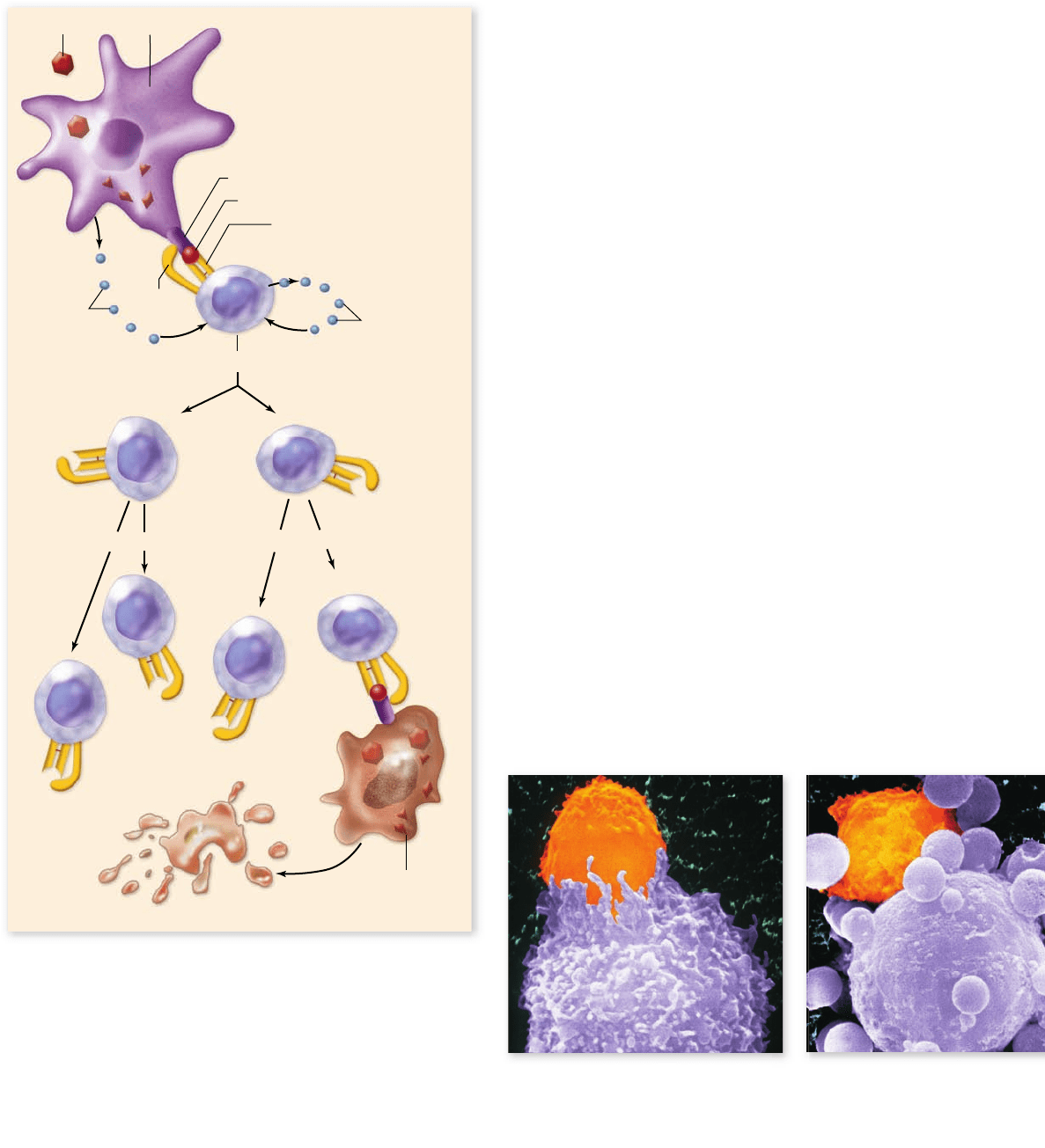
plasma cells and memory cells (figure 52.13) .
Each plasma B cell is a miniature factory producing solu-
ble antibodies of the same specificity as the membrane-bound
antibodies of the parent B cell. These antibodies enter the
lymph and blood circulation as well as the extracellular fluid,
and they bind to the appropriate epitopes of antigen encoun-
tered anywhere in the body. Any one antigen may present a
variety of epitopes, so that different B cells might recognize
different epitopes of a single antigen.
rav32223_ch52_1055-1083.indd 1068rav32223_ch52_1055-1083.indd 1068 11/19/09 1:45:25 PM11/19/09 1:45:25 PM
Apago PDF Enhancer
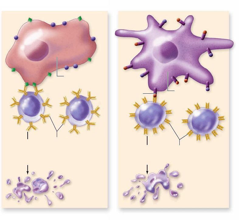
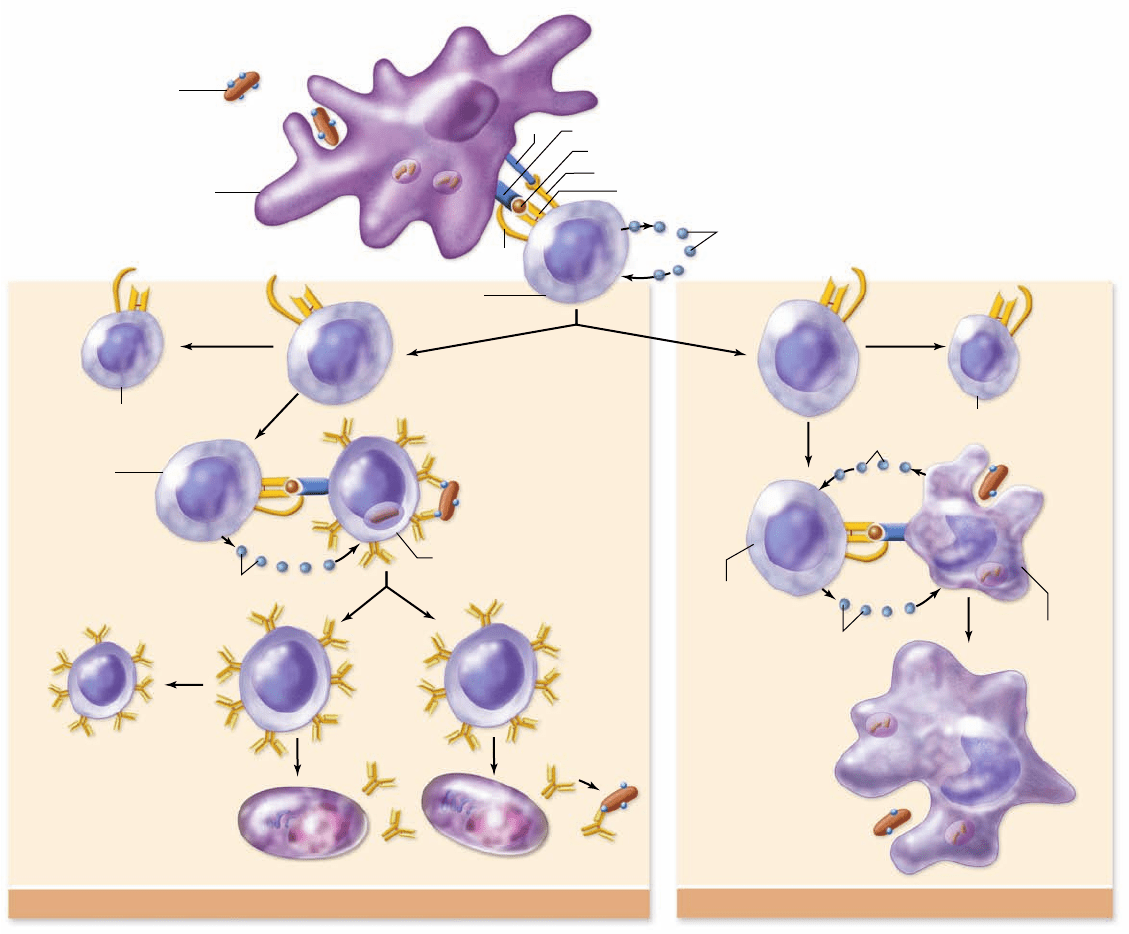
Humoral Response Cellular Response
Antigen
(such as a
bacterium)
Dendritic cell
CD4
B7
CD28
MHC class II
Bacterial peptide
TCR specific for this MHC–peptide complex
Cytokines (IL-2)
Cytokines (IL-12)
Naive
helper T cell
Clonal
expansion
Memory helper T cell
Activated
helper T cell
Naive B cell
Cytokines (IL-4)
Memory B cell
Clonal
expansion
Plasma cells secreting antibody specific to this antigen
Memory helper T cell
Activated
helper T cell
Cytokines (IFN-γ)
Macrophage
Macrophage that can
better destroy invading
antigens
chapter
52
The Immune System
1069www.ravenbiology.com
Once immunoglobulins coat an antigen, many other cells
and processes may be activated to eliminate the antigen. The
immunity to avian cholera that Pasteur observed in his chickens
resulted from such antibodies and from the continued presence
of the progeny of the B cells that produced them.
Immunoglobulin structure reveals
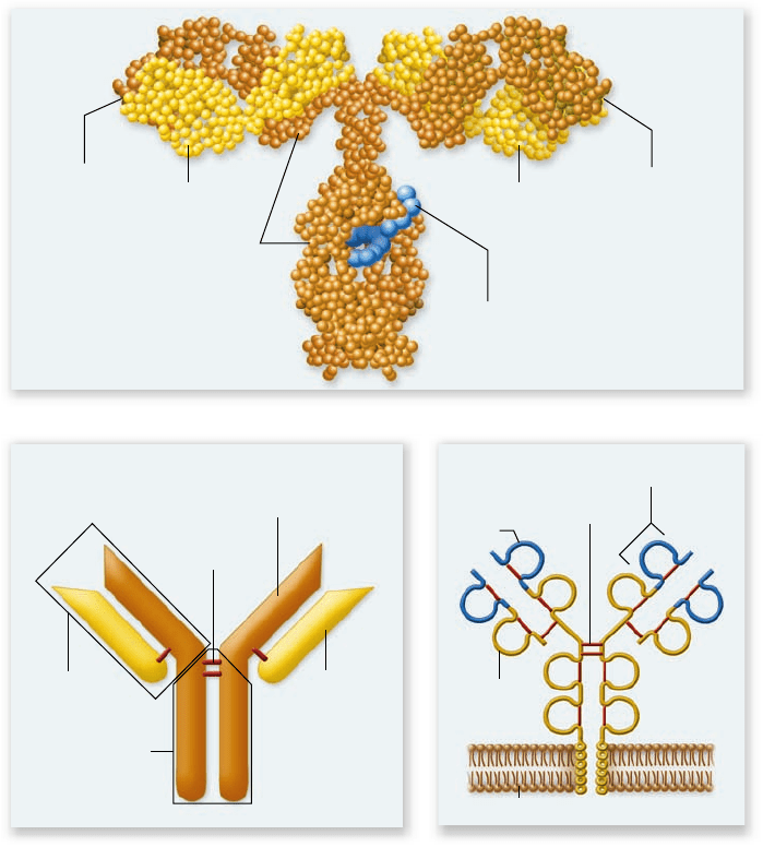
variable and constant regions
Each immunoglobulin molecule consists of two identical short
polypeptides called light chains and two identical longer poly-
peptides called heavy chains (figure 52.14) . The four chains
in an immunoglobulin molecule are held together by disulfide
bonds, forming a Y-shaped molecule (figure 52.14a). Each
“arm” of the molecule is referred to as an Fab region, and the
“stem” is the Fc region (figure 52.14b).
Antibody specificity: The variable region
Comparison of the amino acid sequences of many differ-
ent immunoglobulin molecules has demonstrated that the
specificity of immunoglobulins for antigen epitopes resides
in the amino-terminal half of each Fab region. This half of
the Fab has an amino acid sequence that varies from one im-
munoglobulin to the next and is thus designated the variable
region. Both the light chain and the heavy chain have a vari-
able region.
Figure 52.13
Helper T cells secrete cytokines promoting either cell-mediated or humoral immune responses. Naive
helper T cells are initially activated by TCR bound to a foreign peptide displayed on self-MHC class II proteins on dendritic cells. Activation
results in clonal expansion and differentiation into memory cells and activated cells. T
H
cells promote the humoral response when they
recognize the same antigen displayed by a B cell. Cytokines such as interleukin-4 (IL-4) released from the T
H
cell then activate the B cell ,
producing memory cells and plasma that secrete antibodies against the antigen. T
H
cells also secrete interferon-γ (IFN-γ), which stimulates
cells involved in the cellular response such as the macrophage shown here. Macrophages secrete other cytokines that stimulate T
H
cells.
rav32223_ch52_1055-1083.indd 1069rav32223_ch52_1055-1083.indd 1069 11/19/09 1:45:25 PM11/19/09 1:45:25 PM
Apago PDF Enhancer
a.
b. c.
Light chainLight chain
Antigen-
binding site
Antigen-
binding site
Heavy chains
Carbohydrate chain
Heavy chain
Light chain
S–S bridge
One Fab
F
C
Constant
region
Variable
region
Cell membrane
An immunoglobulin fold
S–S bridge
1070
part
VII
Animal Form and Function
Figure 52.14
The structure of an
immunoglobulin molecule.
a. In this model
of an immunoglobulin (Ig) molecule, amino acids
in the peptide chains are represented by small
spheres. The molecule consists of two heavy
chains (brown) and two light chains (yellow). The
four chains form a Y shape, with two identical
antigen-binding sites at the arms of the Y, the Fab
regions, and a stem, or Fc region. The two Fab
regions are joined to the Fc region by a exible
hinge. b. A more schematic depiction showing
heavy chains (brown) and light chains (yellow) as
rods. The two identical halves of the molecule are
joined by disul de bonds (red) as are the heavy and
light chains of each half. c. Ig molecule shown as a
membrane protein. This depiction highlights the
domain structure of heavy and light chains. Each
chain contains a series of domains, each about
110 amino acids, which include an immunoglobulin
fold motif. These are represented as loops with
globular structure maintained by disul de bonds
(red). The amino-terminal half of each Fab is a
variable region (blue) that binds to an epitope
and the remainder of the molecule is the
constant region.
although not generally on the same antigen because of steric
(shape) constraints. This ability to bind with two epitopes al-
lows the formation of antigen–antibody complexes containing
multiple antibody and antigen molecules (figure 52.15a) .
Function of antibody classes: The constant region
Although the specificity of each immunoglobulin is deter-
mined by its variable region, the function of the immunoglob-
ulin depends on its class, as determined by the heavy-chain
constant region, and particularly the Fc portion of the con-
stant region.
Many cells have Fc receptors that can bind the Fc re-
gion of a particular class of immunoglobulin. Therefore,
when an immunoglobulin binds to an antigen through its
antigen-binding site, another cell, such as a phagocytic cell,
may be brought close to the antigen by binding to the Fc re-
gion of the immunoglobulin (figure 52.15c). This binding of
antigen–antibody complex to Fc receptors can also activate
these cells. In this way, specific immunoglobulins can promote
the interaction of nonspecific cells with the antigen, generally
resulting in the elimination of the antigen.
The amino acid sequence of the remainder of the im-
munoglobulin is relatively constant from one immuno-
globulin to the next and is thus designated the constant region
(figure 52.14c). Both light and heavy chains also exhibit con-
stant regions. Careful analysis shows that light-chain constant
regions of mammalian immunoglobulins consist of two different
sequences, designated κ (kappa) and λ (lambda), which have ap-
parently equivalent function. The heavy-chain constant regions
consist of five different sequences: μ (mu), δ (delta), γ (gamma),
α (alpha), and ε (epsilon). When each of these heavy chains is
bound to either type of light chain, they give rise to a particular
class of immunoglobulin: IgM, IgD, IgG, IgA, and IgE.
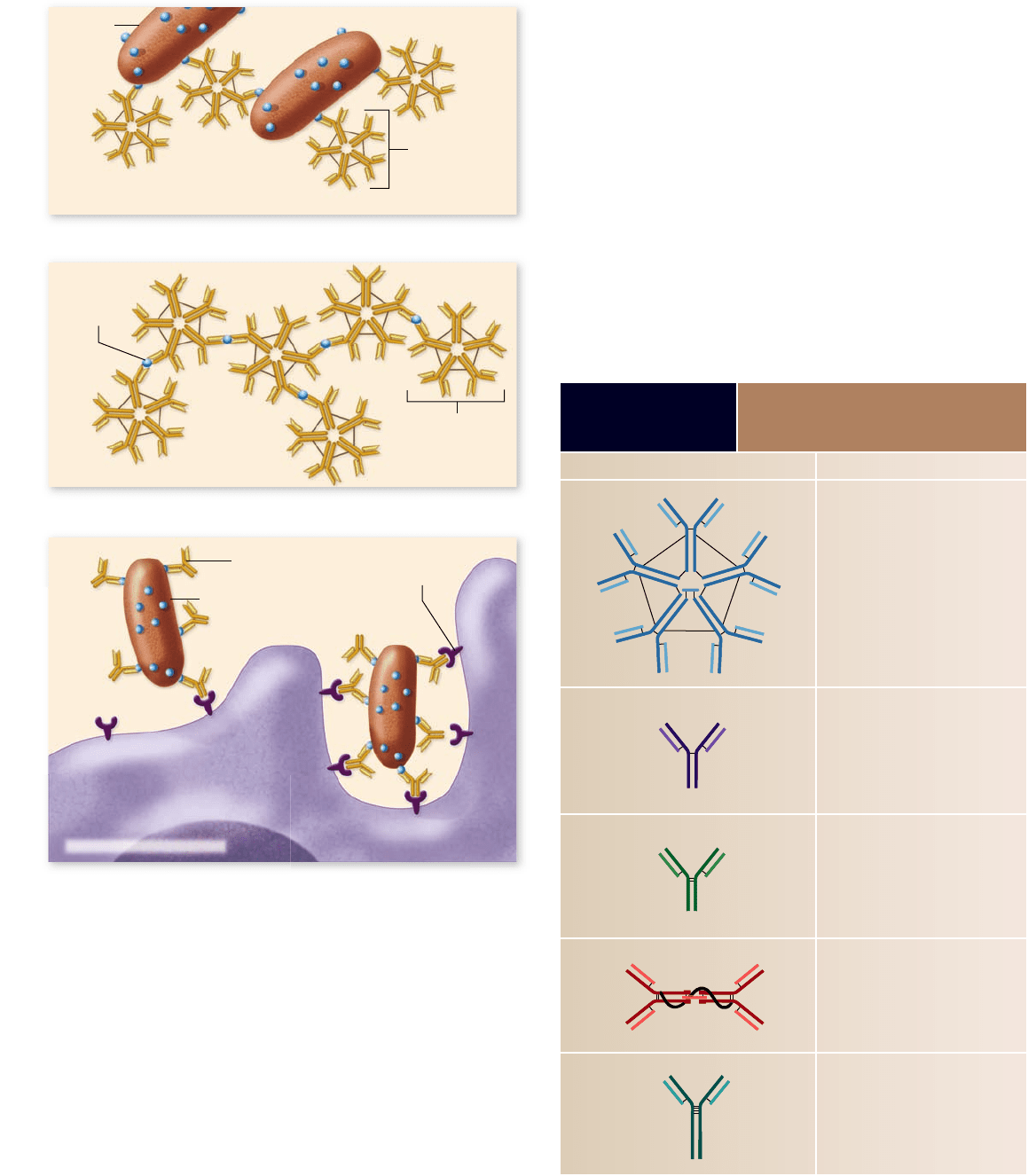
Binding of antibody with antigen
The variable regions of the heavy and light chains fold together
to form a sort of cleft, the antigen-binding site (see figure 52.14).
The size and shape of the antigen-binding site, as well as which
amino acids line its surface, determine the specificity of each im-
munoglobulin for an antigen epitope.
Because each immunoglobulin is composed of two iden-
tical halves, they can each bind with two identical epitopes,
rav32223_ch52_1055-1083.indd 1070rav32223_ch52_1055-1083.indd 1070 11/19/09 1:45:26 PM11/19/09 1:45:26 PM
Apago PDF Enhancer
a.
b.
c.
FcR for IgG
Macrophage or neutrophil
Bacterium
Small,
soluble
antigen
Antigen
Secreted IgM
Secreted IgM
Secreted IgG
chapter
52
The Immune System
1071www.ravenbiology.com
The ve classes of immunoglobulins
have di erent functions
The five classes of antibodies are based on the sequence and
structure of the constant regions of their heavy chains. These
five classes have different functions in the protection of an in-
dividual. Characteristics of the different classes are summarized
in table 52.3 and are described in the following sections.
Keep in mind that antibodies don’t kill invading patho-
gens directly; rather, they cause destruction of pathogens by
targeting them for attack by other, nonspecific cells or by acti-
vating the complement system.
IgM is a receptor on the surface of all mature, naive B cells
and is the first type of antibody to be secreted during an im-
mune response. Although IgM in the membrane of a B cell is
monomeric in form, it is secreted as a pentamer (five units) of
about 900,000 kDa. Its large size restricts it to the circulation,
but its pentameric form means that it very efficiently promotes
agglutination of larger antigens (figure 52.15a) and precipitation
Figure 52.15
Binding of antibody to antigens can
cause agglutination, precipitation, or neutralization of
the antigens. a. Binding of secreted IgM to larger particulate
antigens leads to the clumping, or agglutination, of the antigens.
b. Binding of secreted IgM to small soluble antigens can lead to
their precipitation. Secreted IgG, due to its high concentration
(75% of plasma Ig), can also agglutinate and precipitate antigens.
IgG does not precipitate as ef ciently as IgM because of the
pentameric nature of secreted IgM. c. Secreted IgG can coat or
neutralize an antigen by blocking its ability to bind to a host.
Macrophages and neutrophils that have Fc receptors for IgG can
thus attach to an antigen–antibody complex, which they will then
phagocytose and destroy.
TABLE 52.3
Five Classes of
Immunoglobulins
Class Function
IgM First antibody secreted during the
primary immune response; promotes
agglutination and precipitation
reactions and activates complement
IgD Present only on surfaces of B cells;
serves as antigen receptor
IgG Major antibody secreted during the
secondary response; neutralizes
antigens and promotes their
phagocytosis and activates
complement
IgA Most abundant form of antibody in
body secretions; high density of IgA-
secreting plasma cells in the MALT
IgE Fc binds to mast cells and basophils;
allergen binding to V regions promotes
the release of mediators, which
triggers allergic reactions
Pentamer
Monomer
Dimer
Monomer
Monomer
rav32223_ch52_1055-1083.indd 1071rav32223_ch52_1055-1083.indd 1071 11/19/09 1:45:27 PM11/19/09 1:45:27 PM