Raven P.H., Johnson G.B., Mason K.A. Biology (Ninth Edition)
Подождите немного. Документ загружается.

Apago PDF Enhancer
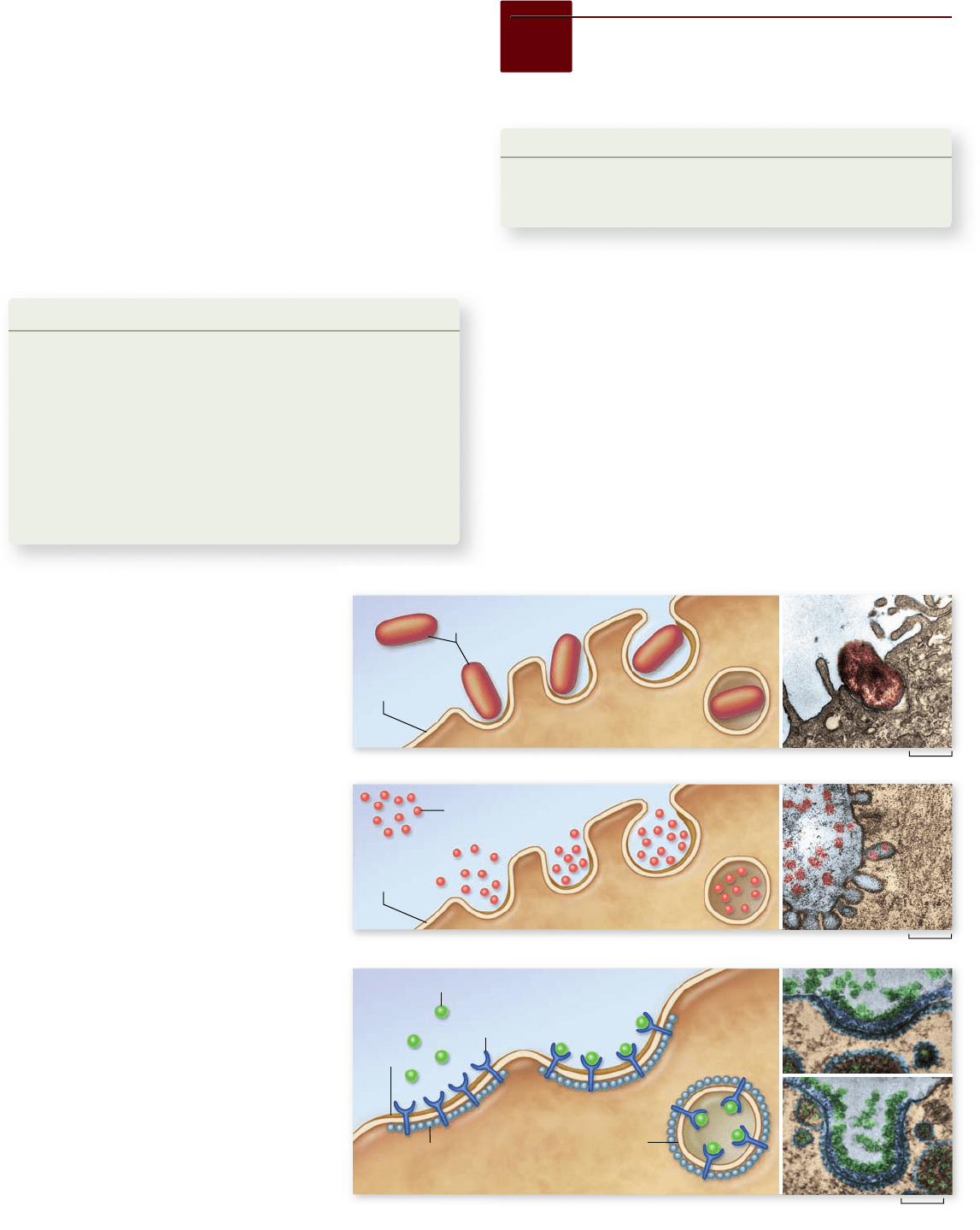
a. Phagocytosis
b. Pinocytosis
c. Receptor-mediated endocytosis
Cytoplasm
Plasma
membrane
Plasma
membrane
Cytoplasm
Target molecule
Receptor protein
Coated pit
Clathrin
Coated vesicle
0.1 µm
1 µm
0.093 µm
Bacterial
cells
Solute
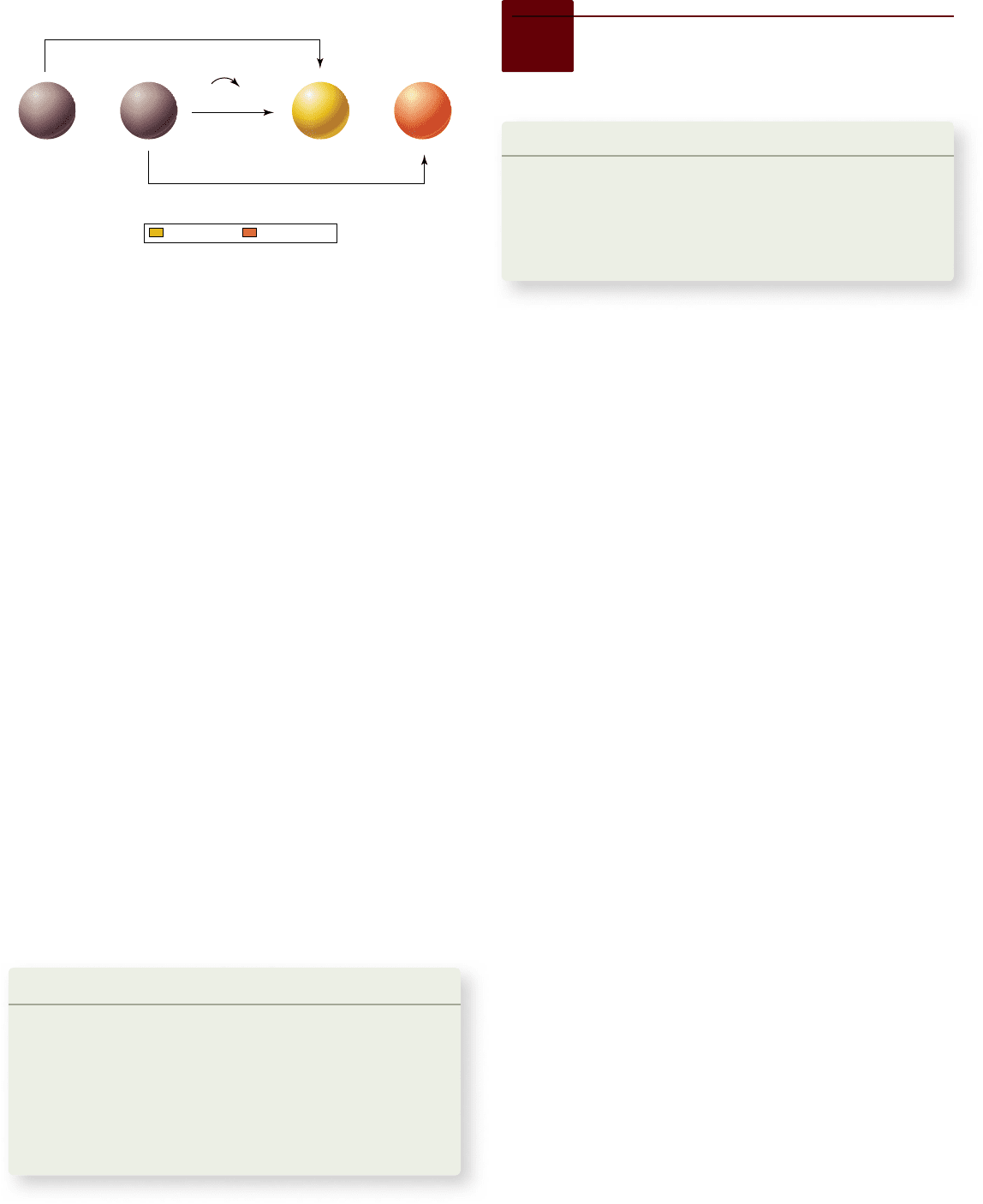
Figure 5.15
Endocytosis.
Both
(a) phagocytosis and (b) pinocytosis are forms of
endocytosis. c. In receptor-mediated endocytosis,
cells have pits coated with the protein clathrin
that initiate endocytosis when target molecules
bind to receptor proteins in the plasma
membrane. Photo inserts (false color has been
added to enhance distinction of structures):
(a) A TEM of phagocytosis of a bacterium,
Rickettsia tsutsugamushi, by a mouse peritoneal
mesothelial cell. The bacterium enters the
host cell by phagocytosis and replicates in
the cytoplasm. (b) A TEM of pinocytosis in
a smooth muscle cell. (c) A coated pit appears
in the plasma membrane of a developing egg
cell, covered with a layer of proteins. When
an appropriate collection of molecules gathers
in the coated pit, the pit deepens and will
eventually seal off to form a vesicle.
In a related process, called countertransport, the inward
movement of Na
+
is coupled with the outward movement of an-
other substance, such as Ca
2+
or H
+
. As in cotransport, both Na
+
and the other substance bind to the same transport protein, which
in this case is an antiporter, as the substances bind on opposite
sides of the membrane and are moved in opposite directions. In
countertransport, the cell uses the energy released as Na
+
moves
down its concentration gradient into the cell to eject a substance
against its concentration gradient. In both cotransport and coun-
tertransport, the potential energy in the concentration gradient of
one molecule is used to transport another molecule against its
concentration gradient. They differ only in the direction that the
second molecule moves relative to the first.
Learning Outcomes Review 5.5
Active transport requires both a carrier protein and energy, usually in
the form of ATP, to move molecules against a concentration gradient. The
sodium–potassium pump uses ATP to moved Na
+
in one direction and K
+
in
the other to create and maintain concentration diff erences of these ions.
In coupled transport, a favorable concentration gradient of one molecule
is used to move a diff erent molecule against its gradient, such as in the
transport of glucose by Na
+
.
■ Can active transport involve a channel protein. Why or
why not?
5.6
Bulk Transport by Endocytosis
and Exocytosis
Learning Outcomes
1. Distinguish between endocytosis and exocytosis.
2. Explain how endocytosis can be specific.
The lipid nature of cell plasma membranes raises a second prob-
lem. The substances cells require for growth are mostly large,
polar molecules that cannot cross the hydrophobic barrier a
lipid bilayer creates. How do these substances get into cells?
Two processes are involved in this bulk transport: endocytosis
and exocytosis.
Bulk material enters the cell in vesicles
In endocytosis, the plasma membrane envelops food particles
and fluids. Cells use three major types of endocytosis: phago-
cytosis, pinocytosis, and receptor-mediated endocytosis
(figure 5.15). Like active transport, these processes also re-
quire energy expenditure.
102
part
II
Biology of the Cell
rav32223_ch05_088-106.indd 102rav32223_ch05_088-106.indd 102 11/6/09 12:02:23 PM11/6/09 12:02:23 PM
Apago PDF Enhancer
Plasma membrane
a. b.
Secretory vesicle
Secretory product
Cytoplasm
0.069 µm
Phagocytosis and pinocytosis
If the material the cell takes in is particulate (made up of dis-
crete particles), such as an organism or some other fragment of
organic matter (figure 5.15a), the process is called phagocytosis
(Greek phagein, “to eat,” + cytos, “cell”). If the material the cell
takes in is liquid (figure 5.15b), the process is called pinocytosis
(Greek pinein, “to drink”). Pinocytosis is common among ani-
mal cells. Mammalian egg cells, for example, “nurse” from sur-
rounding cells; the nearby cells secrete nutrients that the
maturing egg cell takes up by pinocytosis.
Virtually all eukaryotic cells constantly carry out these
kinds of endocytotic processes, trapping particles and extracel-
lular fluid in vesicles and ingesting them. Endocytosis rates vary
from one cell type to another. They can be surprisingly high;
some types of white blood cells ingest up to 25% of their cell
volume each hour.
Receptor-mediated endocytosis
Molecules are often transported into eukaryotic cells through
receptor-mediated endocytosis. These molecules first bind
to specific receptors in the plasma membrane—they have a
conformation that fits snugly into the receptor. Different cell
types contain a characteristic battery of receptor types, each for
a different kind of molecule in their membranes.
The portion of the receptor molecule that lies inside the
membrane is trapped in an indented pit coated on the cyto-
plasmic side with the protein clathrin. Each pit acts like a mo-
lecular mousetrap, closing over to form an internal vesicle
when the right molecule enters the pit (figure 5.15c). The trig-
ger that releases the trap is the binding of the properly fitted
target molecule to the embedded receptor. When binding oc-
curs, the cell reacts by initiating endocytosis; the process is
highly specific and very fast. The vesicle is now inside the cell
carrying its cargo.
One type of molecule that is taken up by receptor-
mediated endocytosis is low-density lipoprotein (LDL). LDL
molecules bring cholesterol into the cell where it can be in-
corporated into membranes. Cholesterol plays a key role in
determining the stiffness of the body’s membranes. In the hu-
man genetic disease familial hypercholesterolemia, the LDL
receptors lack tails, so they are never fastened in the clathrin-
coated pits and as a result, do not trigger vesicle formation.
The cholesterol stays in the bloodstream of affected individu-
als, accumulating as plaques inside arteries and leading to
heart attacks.
It is important to understand that endocytosis in itself
does not bring substances directly into the cytoplasm of a cell.
The material taken in is still separated from the cytoplasm by
the membrane of the vesicle.
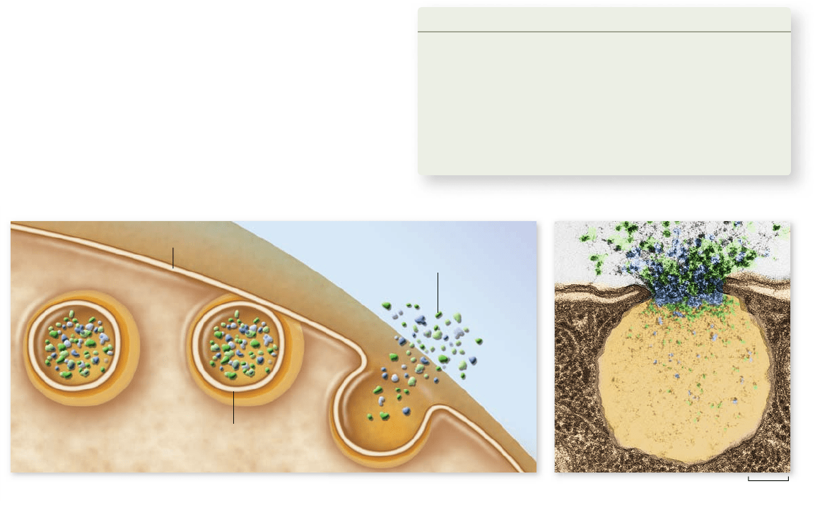
Material can leave the cell by exocytosis
The reverse of endocytosis is exocytosis, the discharge of ma-
terial from vesicles at the cell surface (figure 5.16). In plant
cells, exocytosis is an important means of exporting the materi-
als needed to construct the cell wall through the plasma mem-
brane. Among protists, contractile vacuole discharge is
considered a form of exocytosis. In animal cells, exocytosis pro-
vides a mechanism for secreting many hormones, neurotrans-
mitters, digestive enzymes, and other substances.
The mechanisms for transport across cell membranes are
summarized in table 5.2.
Learning Outcomes Review 5.6
Large molecules and other bulky materials can enter a cell by endocytosis
and leave the cell by exocytosis. These processes require energy. Endocytosis
may be mediated by specifi c receptor proteins in the membrane that trigger
the formation of vesicles.
■ What feature unites transport by receptor-mediated
endocytosis, transport by a carrier, and catalysis by
an enzyme?
Figure 5.16
Exocytosis. a. Proteins and other molecules are secreted from cells in small packets called vesicles, whose membranes fuse
with the plasma membrane, releasing their contents outside the cell. b. A false-colored transmission electron micrograph showing exocytosis.
chapter
5
Membranes
103www.ravenbiology.com
rav32223_ch05_088-106.indd 103rav32223_ch05_088-106.indd 103 11/9/09 10:29:51 AM11/9/09 10:29:51 AM
Apago PDF Enhancer
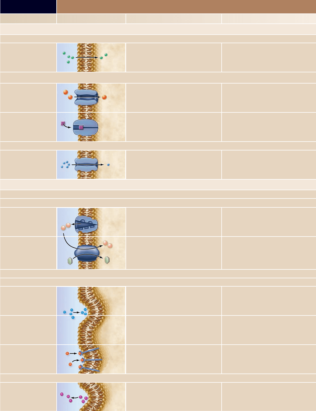
TABLE 5.2
Mechanisms for Transport Across Cell Membranes
Process How It Works Example
PASSIVE PROCESSES
Diff usion
Direct Random molecular motion produces net migration
of nonpolar molecules toward region of lower
concentration
Movement of oxygen into cells
Facilitated Diff usion
Protein channel Polar molecules or ions move through a protein
channel; net movement is toward region of lower
concentration
Movement of ions in or out of cell
Protein carrier Molecule binds to carrier protein in membrane and is
transported across; net movement is toward region
of lower concentration
Movement of glucose into cells
Osmosis
Aquaporins Di usion of water across the membrane via osmosis;
requires osmotic gradient
Movement of water into cells placed in a hypotonic
solution
ACTIVE PROCESSES
Active Transport
Protein carrier
Na
+
/K
+
pump
Carrier uses energy to move a substance across a
membrane against its concentration gradient
Na
+
and K
+
against their concentration gradients
Coupled transport
Molecules are transported across a membrane
against their concentration gradients by the
cotransport of sodium ions or protons down their
concentration gradients
Coupled uptake of glucose into cells against its
concentration gradient using a Na
+
gradient
Endocytosis
Membrane vesicle
Phagocytosis Particle is engulfed by membrane, which folds
around it and forms a vesicle
Ingestion of bacteria by white blood cells
Pinocytosis Fluid droplets are engulfed by membrane, which
forms vesicles around them
“Nursing” of human egg cells
Receptor-mediated
endocytosis
Endocytosis triggered by a speci c receptor, forming
clathrin-coated vesicles
Cholesterol uptake
Exocytosis
Membrane vesicle
Vesicles fuse with plasma membrane and
eject contents
Secretion of mucus; release of neurotransmitters
104
part
II
Biology of the Cell
rav32223_ch05_088-106.indd 104rav32223_ch05_088-106.indd 104 11/6/09 12:02:37 PM11/6/09 12:02:37 PM

Apago PDF Enhancer
Chapter Review
5.1 The Structure of Membranes
The uid mosaic model shows proteins embedded
in a uid lipid bilayer.
Membranes are sheets of phospholipid bilayers with associated
proteins ( gure 5.2). Hydrophobic regions of a membrane are
oriented inward and hydrophilic regions oriented outward. In the
uid mosaic model, proteins oat on or in the lipid bilayer.
Cellular membranes consist of four component groups.
In eukaryotic cells, membranes have four components: a phosopholipid
bilayer, transmembrane proteins (integral membrane proteins), an
interior protein network, and cell-surface markers. The interior
protein network is composed of cytoskeletal laments and peripheral
membrane proteins, which are associated with the membrane but are
not an integral part. Membranes contain glycoproteins and glycolipids
on the surface that act as cell identity markers.
Electron microscopy has provided structural evidence.
Transmission electron microscopy (TEM) and scanning electron
microscopy (SEM) have con rmed the structure predicted by the
uid mosaic model.
5.2 Phospholipids: The Membrane’s Foundation
Phospholipids are composed of two fatty acids and a phosphate
group linked to a three-carbon glycerol molecule.
Phospholipids spontaneously form bilayers.
The phosphate group of a phospholipid is polar and hydrophilic; the
fatty acids are nonpolar and hydrophobic, and they orient away from
the polar head of the phospholipids. The nonpolar interior of the lipid
bilayer impedes the passage of water and water-soluble substances.
The phospholipid bilayer is uid.
Hydrogen bonding of water keeps the membrane in its bilayer
con guration; however, phospholipids and unanchored proteins in
the membrane are loosely associated and can diffuse laterally.
Membrane uidity can change.
Membrane uidity depends on the fatty acid composition of the
membrane. Unsaturated fats tend to make the membrane more
uid because of the “kinks” of double bonds in the fatty acid tails.
Temperature also affects uidity. Some bacteria have enzymes
that alter the fatty acids of the membrane to compensate for
temperature changes.
5.3 Proteins: Multifunctional Components
Proteins and protein complexes perform key functions.
Transporters are integral membrane proteins that carry speci c
substances through the membrane. Enzymes often occur on the
interior surface of the membrane. Cell-surface receptors respond to
external chemical messages and change conditions inside the cell; cell
identity markers on the surface allow recognition of the body’s cells
as “self.” Cell-to-cell adhesion proteins glue cells together; surface
proteins that interact with other cells anchor to the cytoskeleton.
Structural features of membrane proteins relate to function.
Surface proteins are attached to the surface by nonpolar regions
that associate with polar regions of phospholipids. Transmembrane
proteins may cross the bilayer a number of times, and each
membrane-spanning region is called a transmembrane domain. Such
a domain is composed of hydrophobic amino acids usually arranged
in α-helices. In certain proteins, β-pleated sheets in the nonpolar
region form a pipelike passageway having a polar environment. An
example is the porin class of proteins.
5.4 Passive Transport Across Membranes
Transport can occur by simple di usion.
Simple diffusion is the passive movement of a substance along
a chemical or electrical gradient. Biological membranes pose a
barrier to hydrophilic polar molecules, while they allow hydrophobic
substances to diffuse freely.
Proteins allow membrane di usion to be selective.
Ions and large hydrophilic molecules cannot cross the phospholipid
bilayer. Diffusion can still occur with the help of proteins, thus
we call this facilitated diffusion. These proteins can be either
channels, or carriers. Channels allow the diffusion of ions based on
concentration and charge across the membrane. They are speci c for
different ions, but form an aqueous pore in the membrane. Carrier
proteins bind to the molecules they transport, much like an enzyme.
The rate of transport by a carrier is limited by the number of carriers
in the membrane.
Osmosis is the movement of water across membranes.
The direction of movement due to osmosis depends on the solute
concentration on either side of the membrane ( gure 5.12). Solutions
can be isotonic, hypotonic, or hypertonic. Cells in an isotonic
solution are in osmotic balance; cells in a hypotonic solution will gain
water; and cells in a hypertonic solution will lose water. Aquaporins
are water channels that facilitate the diffusion of water.
5.5 Active Transport Across Membranes
Active transport uses energy to move materials against a
concentration gradient.
Active transport uses specialized protein carriers that couple a
source of energy to transport. They are classi ed based on the
number of molecules and direction of transport. Uniporters
transport a speci c molecule in one direction; symporters transport
two molecules in the same direction; and antiporters transport two
molecules in opposite directions.
The sodium–potassium pump runs directly on ATP.
The sodium–potassium pump moves Na
+
out of the cell and K
+
into
the cell against their concentration gradients using ATP. In every
cycle of the pump, three Na
+
leave the cell and two K
+
enter it. This
pump appears to be almost universal in animal cells.
Coupled transport uses ATP indirectly.
Coupled transport occurs when the energy released by a diffusing
molecule is used to transport a different molecule against its
concentration gradient in the same direction. Countertransport is
similar to coupled transport, but the two molecules move in
opposite directions.
5.6 Bulk Transport by Endocytosis
and Exocytosis
Bulk transport moves large quantities of substances that cannot pass
through the cell membrane.
Bulk material enters the cell in vesicles.
In endocytosis, the cell membrane surrounds material and pinches
off to form a vesicle. In receptor-mediated endocytosis, speci c
molecules bind to receptors on the cell membrane.
Material can leave the cell by exocytosis.
In exocytosis, material in a vesicle is discharged when the vesicle
fuses with the membrane.
chapter
5
Membranes
105www.ravenbiology.com
rav32223_ch05_088-106.indd 105rav32223_ch05_088-106.indd 105 11/6/09 12:02:40 PM11/6/09 12:02:40 PM

Apago PDF Enhancer
Review Questions
UNDERSTAND
1. The uid mosaic model of the membrane describes the
membrane as
a. containing a signi cant quantity of water in the interior.
b. composed of uid phospholipids on the outside and protein
on the inside.
c. composed of protein on the outside and uid phospholipids
on the inside.
d. made of proteins and lipids that can freely move.
2. What chemical property characterizes the interior
of the phospholipid bilayer?
a. It is hydrophobic. c. It is polar.
b. It is hydrophilic. d. It is saturated.
3. The transmembrane domain of an integral membrane protein
a. is composed of hydrophobic amino acids.
b. often forms an α-helical structure.
c. can cross the membrane multiple times.
d. is all of the above.
4. The speci c function of a membrane within a cell is determined
by the
a. degree of saturation of the fatty acids within
the phospholipid bilayer.
b. location of the membrane within the cell.
c. presence of lipid rafts and cholesterol.
d. type and number of membrane proteins.
5. The movement of water across a membrane is dependent on
a. the solvent concentration.
b. the solute concentration.
c. the presence of carrier proteins.
d. membrane potential.
6. If a cell is in an isotonic environment, then
a. the cell will gain water and burst.
b. no water will move across the membrane.
c. the cell will lose water and shrink.
d. osmosis still occurs, but there is no net gain or loss
of cell volume.
7. Which of the following is NOT a mechanism for bringing
material into a cell?
a. Exocytosis c. Pinocytosis
b. Endocytosis d. Phagocytosis
APPLY
1. A bacterial cell that can alter the composition of saturated and
unsaturated fatty acids in its membrane lipids is adapted to a
cold environment. If this cell is shifted to a warmer
environment, it will react by
a. increasing the amount of cholesterol in its membrane.
b. altering the amount of protein present in the membrane.
c. increasing the degree of saturated fatty acids in its membrane.
d. increasing the percentage of unsaturated fatty acids in its
membrane.
2. What variable(s) in uence(s) whether a nonpolar molecule
can move across a membrane by passive diffusion?
a. The structure of the phospholipids bilayer
b. The difference in concentration of the molecule
across the membrane
c. The presence of transport proteins in the membrane
d. All of the above
3. Which of the following does NOT contribute to the selective
permeability of a biological membrane?
a. Speci city of the carrier proteins in the membrane
b. Selectivity of channel proteins in the membrane
c. Hydrophobic barrier of the phospholipid bilayer
d. Hydrogen bond formation between water
and phosphate groups
4. How are active transport and coupled transport related?
a. They both use ATP to move molecules.
b. Active transport establishes a concentration gradient,
but coupled transport doesn’t.
c. Coupled transport uses the concentration gradient
established by active transport.
d. Active transport moves one molecule, but coupled
transport moves two.
5. A cell can use the process of facilitated diffusion to
a. concentrate a molecule such as glucose inside a cell.
b. remove all of a toxic molecule from a cell.
c. move ions or large polar molecules across the membrane
regardless of concentration.
d. move ions or large polar molecules from a region of high
concentration to a region of low concentration.
SYNTHESIZE
1. Figure 5.4 describes a classic experiment demonstrating the
ability of proteins to move within the plane of the cell’s plasma
membrane. The following table outlines three different
experiments using the fusion of labeled mouse and human cells.
Experiment Conditions Temperature (°C) Result
1 Fuse human and mouse cells 37
Intermixed membrane
proteins
2
Fuse human and mouse cells
in presence of ATP inhibitors
37
Intermixed membrane
proteins
3
Fuse human and mouse cells
4
No intermixing of membrane
proteins
What conclusions can you reach about the movement
of these proteins?
2. Each compartment of the endomembrane system of a cell is
connected to the plasma membrane. Create a simple diagram of
a cell including the RER, Golgi apparatus, vesicle, and the
plasma membrane. Starting with the RER, use two different
colors to represent the inner and outer halves of the bilayer for
each of these membranes. What do you observe?
3. The distribution of lipids in the ER membrane is symmetric, that
is, it is the same in both lea ets of the membrane. The Golgi
apparatus and plasma membrane do not have symmetric
distribution of membrane lipids. What kinds of processes could
achieve this outcome?
ONLINE RESOURCE
www.ravenbiology.com
Understand, Apply, and Synthesize—enhance your study with
animations that bring concepts to life and practice tests to assess
your understanding. Your instructor may also recommend the
interactive eBook, individualized learning tools, and more.
106
part
II
Biology of the Cell
rav32223_ch05_088-106.indd 106rav32223_ch05_088-106.indd 106 11/6/09 12:02:40 PM11/6/09 12:02:40 PM
Apago PDF Enhancer
L
Chapter Outline
6.1 The Flow of Energy in Living Systems
6.2 The Laws of Thermodynamics and Free Energy
6.3 ATP: The Energy Currency of Cells
6.4 Enzymes: Biological Catalysts
6.5 Metabolism: The Chemical Description
of Cell Function
Chapter
6
Energy and
Metabolism
Introduction
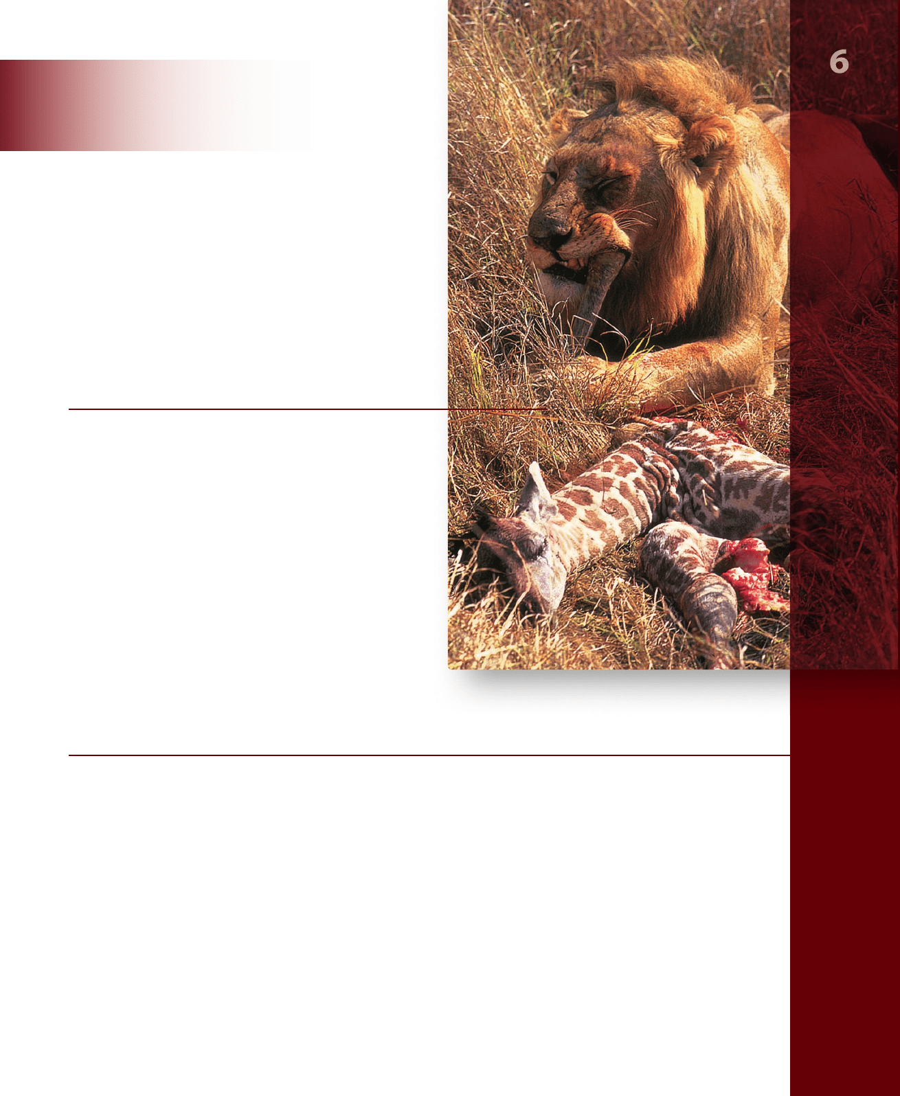
Life can be viewed as a constant flow of energy, channeled by organisms to do the work of living. Each of the significant
properties by which we define life—order, growth, reproduction, responsiveness, and internal regulation—requires a
constant supply of energy. Both the lion and the giraffe need to eat to provide energy for a wide variety of cellular functions.
Deprived of a source of energy, life stops. Therefore, a comprehensive study of life would be impossible without discussing
bioenergetics, the analysis of how energy powers the activities of living systems. In this chapter, we focus on energy—what
it is and how it changes during chemical reactions.
CHAPTER
rav32223_ch06_107-121.indd 107rav32223_ch06_107-121.indd 107 11/6/09 12:28:53 PM11/6/09 12:28:53 PM

Apago PDF Enhancer
a. Potential energy
b. Kinetic energy
Figure 6.1
Potential and kinetic energy. a. Objects that have the capacity to move
but are not moving have potential energy. The energy required for the girl to climb to the top
of the slide is stored as potential energy. b. Objects that are in motion have kinetic energy.
The stored potential energy is released as kinetic energy as the girl slides down.
6.1
The Flow of Energy
in Living Systems
Learning Outcomes
Explain what energy is and describe its different forms. 1.
Identify the source of energy for the biosphere.2.
Contrast oxidation and reduction reactions.3.
Thermodynamics is the branch of chemistry concerned with
energy changes. Cells are governed by the laws of physics and
chemistry, so we must understand these laws in order to under-
stand how cells function.
Energy can take many forms
Energy is defined as the capacity to do work. We think of energy
as existing in two states: kinetic energy and potential energy
(figure 6.1). Kinetic energy is the energy of motion. Moving ob-
jects perform work by causing other matter to move. Potential
energy is stored energy. Objects that are not actively moving but
have the capacity to do so possess potential energy. A boulder
perched on a hilltop has gravitational potential energy. As it be-
gins to roll downhill, some of its potential energy is converted
into kinetic energy. Much of the work that living organisms carry
out involves transforming potential energy into kinetic energy.
Energy can take many forms: mechanical
energy, heat, sound, electric current, light,
or radioactivity. Because it can exist in so
many forms, energy can be measured in many ways. Heat is the
most convenient way of measuring energy because all other
forms of energy can be converted into heat. In fact, the term
thermodynamics means “heat changes.”
The unit of heat most commonly employed in biology is
the kilocalorie (kcal). One kilocalorie is equal to 1000 calories
(cal). One calorie is the heat required to raise the temperature
of one gram of water one degree Celsius (°C). (You are probably
more used to seeing the term Calorie with a capital C. This is
used on food labels and is actually the same as kilocalorie. ) An-
other energy unit, often used in physics, is the joule; one joule
equals 0.239 cal.
The sun provides energy for living systems
Energy flows into the biological world from the Sun. It is esti-
mated that the Sun provides the Earth with more than
13 × 10
23
calories per year, or 40 million billion calories per
second! Plants, algae, and certain kinds of bacteria capture a
fraction of this energy through photosynthesis.
In photosynthesis, energy absorbed from sunlight is used
to combine small molecules (water and carbon dioxide) into
more complex ones (sugars). This process converts carbon from
an inorganic to an organic form. In the process, energy from
the Sun is stored as potential energy in the covalent bonds be-
tween atoms in the sugar molecules.
Breaking the bonds between atoms requires energy. In
fact, the strength of a covalent bond is measured by the amount
of energy required to break it. For example, it takes 98.8 kcal to
break one mole (6.023 × 10
23
) of the carbon–hydrogen (C
–
H)
bonds found in organic molecules. Fat molecules have many
C
–
H bonds, and breaking those bonds provides lots of energy.
108
part
II
Biology of the Cell
rav32223_ch06_107-121.indd 108rav32223_ch06_107-121.indd 108 11/6/09 12:28:58 PM11/6/09 12:28:58 PM
Apago PDF Enhancer
lower energy higher energy
Gain of electron (reduction)
e
:
A B
Loss of electron (oxidation)
A
+
B
+
A
;
B
:
Figure 6.2
Redox reactions. Oxidation is the loss of an
electron; reduction is the gain of an electron. In this example, the
charges of molecules A and B appear as superscripts in each
molecule. Molecule A loses energy as it loses an electron, and
molecule B gains that energy as it gains an electron.
This is one reason animals store fat. The oxidation of one mole
of a 16- carbon fatty acid that is completely saturated with hy-
drogens yields 2340 kcal.
Oxidation–reduction reactions transfer
electrons while bonds are made or broken
During a chemical reaction, the energy stored in chemical bonds
may be used to make new bonds. In some of these reactions,
electrons actually pass from one atom or molecule to another. An
atom or molecule that loses an electron is said to be oxidized, and
the process by which this occurs is called oxidation. The name
comes from the fact that oxygen is the most common electron
acceptor in biological systems. Conversely, an atom or molecule
that gains an electron is said to be reduced, and the process is
called reduction. The reduced form of a molecule has a higher
level of energy than the oxidized form (figure 6.2).
Oxidation and reduction always take place together, be-
cause every electron that is lost by one atom through oxidation
is gained by another atom through reduction. Therefore,
chemical reactions of this sort are called oxidation –reduction,
or redox, reactions. Oxidation– reduction reactions play a key
role in the flow of energy through biological systems.
In the next two chapters, you will learn the details of
how organisms derive energy from the oxidation of organic
compounds via respiration, as well as from the energy in sun-
light via photosynthesis.
Learning Outcomes Review 6.1
Energy is defi ned as the capacity to do work. The two forms of energy are
kinetic energy, or energy of motion, and potential energy, or stored energy.
The ultimate source of energy for living systems is the Sun. Organisms derive
their energy from oxidation–reduction reactions. In oxidation, a molecule
loses an electron; in reduction, a molecule gains an electron.
■ What energy source might ecosystems at the bottom of
the ocean use?
6.2
The Laws of Thermodynamics
and Free Energy
Learning Outcomes
Explain the laws of thermodynamics.1.
Recognize how free energy can be used to predict the 2.
outcome of chemical reactions.
Contrast the course of a reaction with and without an 3.
enzyme catalyst.
All activities of living organisms—growing, running, thinking,
singing, reading these words—involve changes in energy. A set
of two universal laws we call the laws of thermodynamics gov-
ern all energy changes in the universe, from nuclear reactions
to a bird flying through the air.
The First Law states that energy
cannot be created or destroyed
The First Law of Thermodynamics concerns the amount of
energy in the universe. Energy cannot be created or destroyed;
it can only change from one form to another (from potential to
kinetic, for example). The total amount of energy in the uni-
verse remains constant.
The lion eating a giraffe at the beginning of this chap-
ter is acquiring energy. Rather than creating new energy or
capturing the energy in sunlight, the lion is merely transfer-
ring some of the potential energy stored in the giraffe’s tis-
sues to its own body, just as the giraffe obtained the potential
energy stored in the plants it ate while it was alive.
Within any living organism, chemical potential energy
stored in some molecules can be shifted to other molecules
and stored in different chemical bonds. It can also be con-
verted into other forms, such as kinetic energy, light, or elec-
tricity. During each conversion, some of the energy dissipates
into the environment as heat, which is a measure of the ran-
dom motion of molecules (and therefore a measure of one
form of kinetic energy). Energy continuously flows through
the biological world in one direction, with new energy from
the Sun constantly entering the system to replace the energy
dissipated as heat.
Heat can be harnessed to do work only when there is a
heat gradient—that is, a temperature difference between two
areas. Cells are too small to maintain significant internal tem-
perature differences, so heat energy is incapable of doing the
work of cells. Instead, cells must rely on chemical reactions
for energy.
Although the total amount of energy in the universe re-
mains constant, the energy available to do work decreases as
more of it is progressively lost as heat.
chapter
6
Energy and Metabolism
109www.ravenbiology.com
rav32223_ch06_107-121.indd 109rav32223_ch06_107-121.indd 109 11/6/09 12:29:02 PM11/6/09 12:29:02 PM
Apago PDF Enhancer
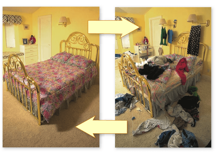
Disorder happens
spontaneously
Organization
requires energy
The Second Law states that some energy
is lost as disorder increases
The Second Law of Thermodynamics concerns the transfor-
mation of potential energy into heat, or random molecular mo-
tion. It states that the disorder in the universe, more formally
called entropy, is continuously increasing. Put simply, disorder is
more likely than order. For example, it is much more likely that
a column of bricks will tumble over than that a pile of bricks will
arrange themselves spontaneously to form a column.
In general, energy transformations proceed spontaneously
to convert matter from a more ordered, less stable form to a less
ordered, but more stable form. For this reason, the second law
is sometimes called “time’s arrow.” Looking at the photographs
in figure 6.3, you could put the pictures into correct sequence
using the information that time had elapsed with only natural
processes occurring. Although it might be great if our rooms
would straighten themselves up, we know from experience how
much work it takes to do so.
The Second Law of Thermodynamics can also be stated
simply as “entropy increases.” When the universe formed, it
held all the potential energy it will ever have. It has become
progressively more disordered ever since, with every energy ex-
change increasing the amount of entropy.
Chemical reactions can be predicted
based on changes in free energy
It takes energy to break the chemical bonds that hold the atoms
in a molecule together. Heat energy, because it increases atomic
motion, makes it easier for the atoms to pull apart. Both chemi-
cal bonding and heat have a significant influence on a molecule.
Chemical bonding reduces disorder; heat increases it. The net
effect, the amount of energy actually available to break and sub-
sequently form other chemical bonds, is called the free energy of
that molecule. In a more general sense, free energy is defined
as the energy available to do work in any system.
For a molecule within a cell, where pressure and volume
usually do not change, the free energy is denoted by the symbol
G (for “ Gibbs free energy”). G is equal to the energy contained
in a molecule’s chemical bonds (called enthalpy and designated
H) together with the energy term (TS) related to the degree of
disorder in the system, where S is the symbol for entropy and T
is the absolute temperature expressed in the Kelvin scale
(K = °C + 273):
G = H – TS
Chemical reactions break some bonds in the reactants and
form new ones in the products. Consequently, reactions can pro-
duce changes in free energy. When a chemical reaction occurs un-
der conditions of constant temperature, pressure, and volume—as
do most biological reactions—the change symbolized by the
Greek capital letter delta, Δ, in free energy (ΔG) is simply:
ΔG = ΔH – TΔS
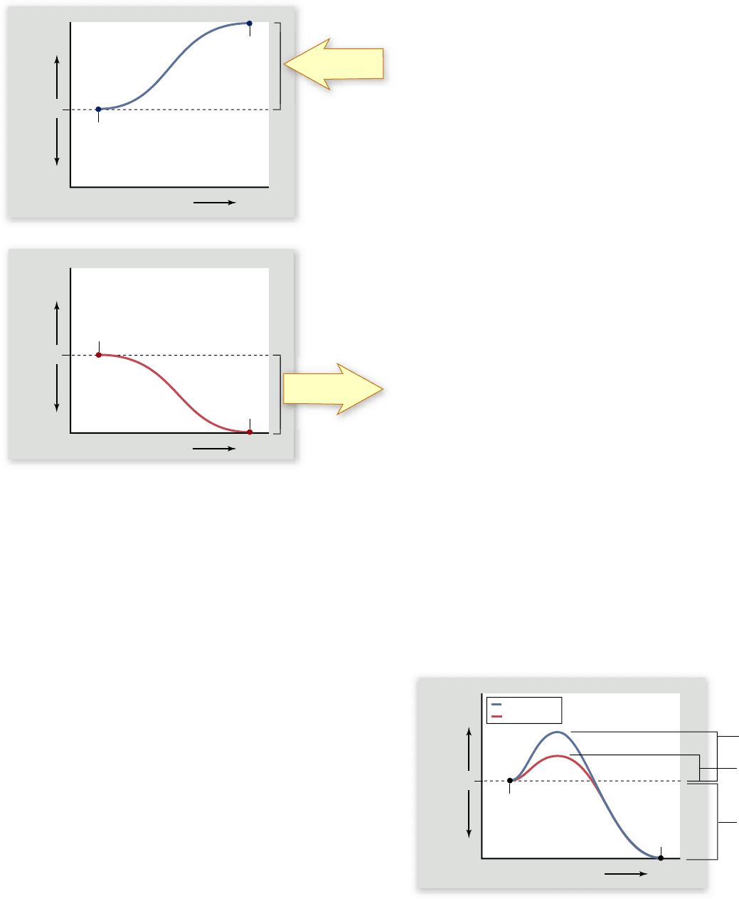
We can use the change in free energy, or ΔG, to predict
whether a chemical reaction is spontaneous or not. For some
reactions, the ΔG is positive, which means that the products of
the reaction contain more free energy than the reactants; the
bond energy (H) is higher, or the disorder (S) in the system is
lower. Such reactions do not proceed spontaneously because
they require an input of energy. Any reaction that requires an
input of energy is said to be endergonic (“inward energy”).
For other reactions, the ΔG is negative. In this case, the
products of the reaction contain less free energy than the reac-
tants; either the bond energy is lower, or the disorder is higher,
or both. Such reactions tend to proceed spontaneously. These
reactions release the excess free energy as heat and are thus said
to be exergonic (“outward energy”). Any chemical reaction
tends to proceed spontaneously if the difference in disorder
(TΔS) is greater than the difference in bond energies between
reactants and products (ΔH).
Note that spontaneous does not mean the same thing as
instantaneous. A spontaneous reaction may proceed very slowly.
Figure 6.4 sums up endergonic and exergonic reactions.
Figure 6.3
Entropy in action. As time
elapses, the room shown at right becomes more
disorganized. Entropy has increased in this
room. It takes energy to restore it to the ordered
state shown at left.
110
part
II
Biology of the Cell
rav32223_ch06_107-121.indd 110rav32223_ch06_107-121.indd 110 11/6/09 12:29:03 PM11/6/09 12:29:03 PM
Apago PDF Enhancer
a.
b.
Energy Released Energy Supplied
Course of Reaction
Free Energy (G)
Reactants
Products
Energy Released Energy Supplied
Course of Reaction
Free Energy (G)
Reactants
Products
Energy must
be supplied
DG>0
Energy is
released
DG<0
0
0
DG
Energy Released Energy Supplied
Course of Reaction
Free Energy (G)
Reactant
Product
uncatalyzed
catalyzed
Activation
energy
Activation
energy
0
Because chemical reactions are reversible, a reaction that
is exergonic in the forward direction will be endergonic in the
reverse direction. For each reaction, an equilibrium exists at
some point between the relative amounts of reactants and prod-
ucts. This equilibrium has a numeric value and is called the
equilibrium constant. This characteristic of reactions provides us
with another way to think about free energy changes: an exer-
gonic reaction has an equilibrium favoring the products, and an
endergonic reaction has an equilibrium favoring the reactants.
Spontaneous chemical reactions
require activation energy
If all chemical reactions that release free energy tend to occur
spontaneously, why haven’t all such reactions already occurred?
Consider the gasoline tank of your car: The oxidation of the
hydrocarbons in gasoline is an exergonic reaction, but your gas
tank does not spontaneously explode. One reason is that most
reactions require an input of energy to get started. In the case
of your car, this input consists of the electrical sparks in the
engine’s cylinders, producing a controlled explosion.
Activation energy
Before new chemical bonds can form, even bonds that contain
less energy, existing bonds must first be broken, and that re-
quires energy input. The extra energy needed to destabilize ex-
isting chemical bonds and initiate a chemical reaction is called
activation energy (figure 6.5).
The rate of an exergonic reaction depends on the activa-
tion energy required for the reaction to begin. Reactions with
larger activation energies tend to proceed more slowly because
fewer molecules succeed in getting over the initial energy hur-
dle. The rate of reactions can be increased in two ways: (1) by
increasing the energy of reacting molecules or (2) by lowering
activation energy. Chemists often drive important industrial re-
actions by increasing the energy of the reacting molecules,
which is frequently accomplished simply by heating up the re-
actants. The other strategy is to use a catalyst to lower the acti-
vation energy.
How catalysts work
Activation energies are not constant. Stressing particular chemi-
cal bonds can make them easier to break. The process of influ-
encing chemical bonds in a way that lowers the activation energy
needed to initiate a reaction is called catalysis, and substances
that accomplish this are known as catalysts (see figure 6.5).
Catalysts cannot violate the basic laws of thermodynam-
ics; they cannot, for example, make an endergonic reaction pro-
ceed spontaneously. By reducing the activation energy, a catalyst
accelerates both the forward and the reverse reactions by exactly
the same amount. Therefore, a catalyst does not alter the pro-
portion of reactant that is ultimately converted into product.
To understand this, imagine a bowling ball resting in a
shallow depression on the side of a hill. Only a narrow rim of
dirt below the ball prevents it from rolling down the hill. Now
imagine digging away that rim of dirt. If you remove enough
dirt from below the ball, it will start to roll down the hill—but
Figure 6.4
Energy in chemical reactions. a. In an
endergonic reaction, the products of the reaction contain more
energy than the reactants, and the extra energy must be supplied
for the reaction to proceed. b. In an exergonic reaction, the
products contain less energy than the reactants, and the excess
energy is released.
Figure 6.5
Activation energy and catalysis. Exergonic
reactions do not necessarily proceed rapidly because activation
energy must be supplied to destabilize existing chemical bonds.
Catalysts accelerate particular reactions by lowering the amount of
activation energy required to initiate the reaction. Catalysts do not
alter the free-energy change produced by the reaction.
chapter
6
Energy and Metabolism
111www.ravenbiology.com
rav32223_ch06_107-121.indd 111rav32223_ch06_107-121.indd 111 11/6/09 12:29:06 PM11/6/09 12:29:06 PM