Raven P.H., Johnson G.B., Mason K.A. Biology (Ninth Edition)
Подождите немного. Документ загружается.

Apago PDF Enhancer
Yolk
Cleaving embryonic cells
25 μm
ICM
Blastocoel
Blastodisc
Trophoblast
Yolk
cleavage (figure 54.9) . The resulting embryo is not spherical,
but rather has the form of a thin cap perched on the yolk.
Cleavage in mammals
Mammalian eggs contain very little yolk; however, mammalian
embryogenesis has many similarities to development of their
reptilian and avian relatives.
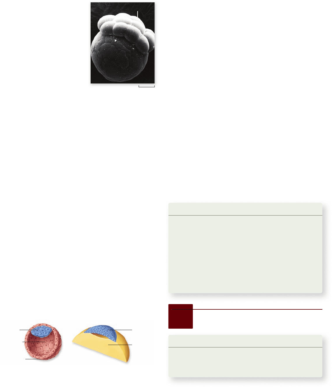
Because cleavage is not impeded by yolk in mammalian eggs,
it is holoblastic, forming a structure called a blastocyst, in which a
single layer of cells surrounds a central fluid-filled blastocoel. In ad-
dition, an inner cell mass (ICM) is located at one pole of the blas-
tocoel cavity (figure 54.10) . The ICM is similar to the blastodisc of
reptiles and birds, and it goes on to form the developing embryo.
The outer layer of cells, called the trophoblast, is similar to
the cells that form the membranes underlying the tough outer shell
of the reptilian egg. These cells have changed during the course of
mammalian evolution to carry out a very different function: Part
of the trophoblast enters the maternal endometrium (the epithelial
lining of the uterus) and contributes to the placenta , the organ that
permits exchanges between the fetal and maternal blood supplies.
The placenta will be discussed in more detail in a later section.
The major cleavage patterns of animal embryos are sum-
marized in table 54.2.
Blastomeres may or may not be
committed to developmental paths
Viewed from the outside, cleavage-stage embryos often look
like a simple ball or disc of similar cells. In many animals, this
appearance is misleading; for example, the unequal segregation
of cytoplasmic determinants into specific blastomeres of tuni-
cate embryos (described in chapter 19) commits those cells to
different developmental paths. The experimental destruction or
removal of these committed cells results in embryos deficient
in the tissues that would have developed from those cells.
In contrast, mammals exhibit highly regulative development ,
in which early blastomeres do not appear to be committed to a
particular fate. For example, if a blastomere is removed from an
early eight-cell stage human embryo (as is done in the process
of preimplantation genetic diagnosis), the remaining seven cells
of the embryo will “regulate” and develop into a complete indi-
vidual if implanted into the uterus of a woman. Similarly, em-
bryos that are split into two (either naturally or experimentally)
form identical twins. It therefore appears that inheritance of
maternally encoded determinants is not an important mecha-
nism in mammalian development, and body form is determined
primarily by cell–cell interactions.
The earliest patterning events in mammalian embryos
occur during the preimplantation stages that lead to formation
of the blastocyst. At the eight-cell stage, the outer surfaces of
many mammalian blastomeres flatten against each other in a
process called compaction, which serves to polarize the blasto-
meres. The polarized blastomeres then undergo asymmetrical
cell divisions. Cell lineage studies have shown that cells that are
in the interior of the embryo most often become ICM cells of
the mammalian blastocyst, whereas cells on the exterior of the
embryo usually become trophoblast cells.
Learning Outcomes Review 54.2
Cleavage is a series of rapid cell divisions that transforms the zygote into
the blastula—a hollow ball of cells. The amount of yolk is the major
determinant of cleavage pattern. Eggs with little yolk cleave completely
(holoblastic cleavage); eggs with a large yolk cannot cleave completely
(meroblastic cleavage). In many animals, each blastomere is committed to a
developmental path; in mammals, blastomeres are not committed but can
regulate as needed to produce a complete individual.
■ If the cells of a mammalian embryo were separated at
the four-cell stage, would they develop normally? What
about a frog embryo at the four-cell stage?
Figure 54.9
Meroblastic
cleavage. Only a portion of the
egg actively divides to form a mass
of cells in this type of cleavage,
which occurs in eggs with relatively
large amounts of yolk.
Figure 54.10
The embryos of mammals and birds are
more similar than they seem. A mammalian blastula (left),
called a blastocyst, is composed of a sphere of cells, the trophoblast,
surrounding a cavity, the blastocoel, and an inner cell mass (ICM).
An avian (bird) blastula consists of a cap of cells, the blastodisc,
resting atop a large yolk mass (right). The blastodisc will form an
upper and a lower layer with a compressed blastocoel in between.
54.3
Gastrulation
Learning Outcomes
Define gastrulation.1.
Compare gastrulation in different animals.2.
Name the extraembryonic membranes in amniotes.3.
In a complex series of cell shape changes and cell movements,
the cells of the blastula rearrange themselves to form the ba-
sic body plan of the embryo. This process, called gastrulation ,
forms the three primary germ layers and converts the blastula
1112
part
VII
Animal Form and Function
rav32223_ch54_1105-1131.indd 1112rav32223_ch54_1105-1131.indd 1112 11/19/09 4:17:49 PM11/19/09 4:17:49 PM
Apago PDF Enhancer
a. b. c.
Animal pole
Animal pole
Vegetal pole
Vegetal pole Blastopore
Blastocoel
Future
ectoderm
Future
endoderm
Blastocoel
Primary
mesenchyme
cells (PMC’s)
Ectoderm
Ectoderm
Filopodia
Anus
Ectoderm
Ectoderm
PMC
PMC
Archenteron
Archenteron
tightly attached to one another via desmosomes or adherens
junctions will move as cell sheets.
In embryos with little yolk and a hollow blastula, the cell
sheet at the vegetal pole of the blastula invaginates (dents in-
ward) to form the primitive gut tube. In embryos with large
yolky cells that are hard to move, sheets of smaller cells involute
(roll inward) from the surface of the blastula and move over the
basal surfaces of the outer cells. Other cells break away from cell
sheets and migrate as individual cells during ingression.
Avian and mammalian gastrulation begins with delami-
nation, in which one sheet of cells splits into two sheets. Each
migrating cell possesses particular cell-surface glycoproteins,
which adhere to specific molecules on the surfaces of other cells
or in the extracellular matrix. Changes in cell adhesiveness, as
described in chapter 19, are key events in gastrulation. The ex-
tracellular matrix protein fibronectin and the corresponding
integrin receptors of cells are essential molecules of gastrula-
tion in many animals.
Gastrulation patterns also vary
according to the amount of yolk
Just as in cleavage patterns, yolk quantity also affects the types
of cell movements that occur during gastrulation. Here, we
examine gastrulation in four representative classes of embryos
with differing quantities of yolk.
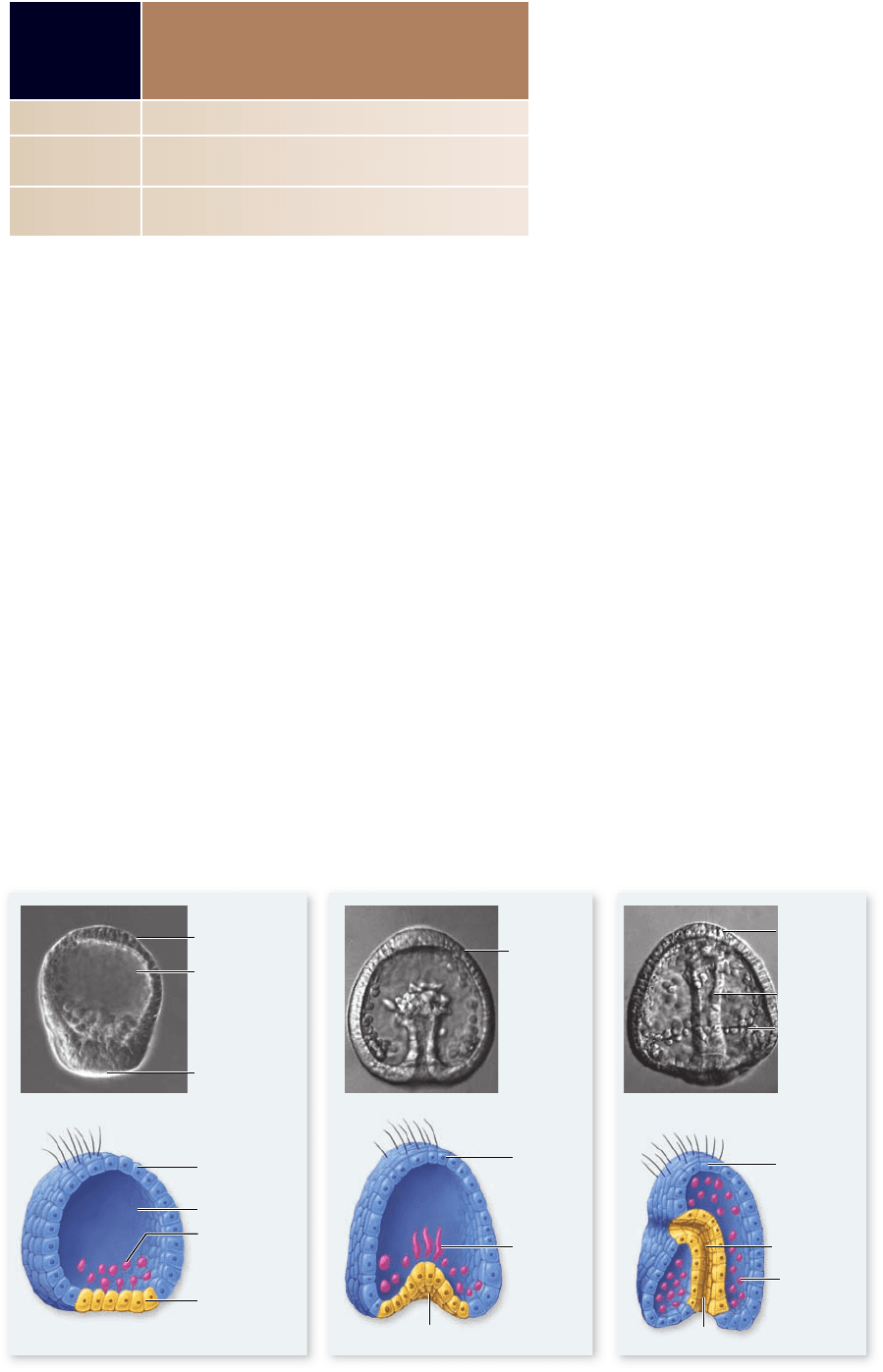
Gastrulation in sea urchins
Echinoderms such as sea urchins develop from relatively yolk-
poor eggs and form hollow, symmetrical blastulas. Gastrulation
begins when cells at the vegetal surface of the blastula change their
shape to form a flattened vegetal plate. In an example of ingres-
sion, a subset of cells in the vegetal plate breaks away from the
blastula wall and moves into the blastocoel cavity. These primary
mesenchyme cells are future mesoderm cells, and they use
filopodia to migrate through the blastocoel cavity (figure 54.11) .
Figure 54.11
Gastrulation in a sea
urchin. a. Gastrulation
begins with formation
of the vegetal plate and
ingression of primary
mesenchyme cells
(prospective mesoderm
cells) into the blastocoel
cavity. b. The endoderm
is then formed by
invagination of the
remaining vegetal plate
cells and extension of a
cellular tube to produce
the primitive gut, or
archenteron. c. Cells that
remain on the surface
form the ectoderm.
TABLE
54.3
Developmental Fates
of the Primary Germ Layers
in Vertebrates
Ectoderm Epidermis of skin, nervous system, sense organs
Mesoderm Skeleton, muscles, blood vessels, heart, blood, gonads, kidneys,
dermis of skin
Endoderm Lining of digestive and respiratory tracts, liver, pancreas,
thymus, thyroid
into a bilaterally symmetrical embryo with a central progenitor
gut and visible anterior–posterior and dorsal–ventral axes.
Gastrulation produces the three germ layers
Gastrulation creates the three primary germ layers : endoderm, ec-
toderm, and mesoderm. The cells in each germ layer have very
different developmental fates. The cells that move into the em-
bryo to form the tube of the primitive gut are endoderm ; they give
rise to the lining of the gut and its derivatives (pancreas, lungs,
liver, etc.). The cells that remain on the exterior are ectoderm , and
their derivatives include the epidermis on the outside of the body
and the nervous system. The cells that move into the space between
the endoderm and ectoderm are mesoderm ; they eventually form
the notochord, bones, blood vessels, connective tissues, muscles
and internal organs such as the kidneys and gonads (table 54.3).
Cells move during gastrulation using a variety of cell
shape changes. Some cells use broad, actin-filled extensions
called lamellipodia to crawl over neighboring cells. Other cells
send out narrow extensions called filopodia, which are used to
“feel out” the surfaces of other cells or the extracellular matrix.
Once a satisfactory attachment is made, the filopodia retract to
pull the cell forward. Contractions of actin filament bundles are
responsible for many of these cell shape changes. Cells that are
chapter
54
Animal Development
1113www.ravenbiology.com
rav32223_ch54_1105-1131.indd 1113rav32223_ch54_1105-1131.indd 1113 11/19/09 4:17:50 PM11/19/09 4:17:50 PM
Apago PDF Enhancer
Dorsal lip of
blastopore
Dorsal lip
Ventral lip
Archenteron
Yolk plug
Neural plate
Neural fold
Blastocoel
Blastocoel
Ectoderm
Ectoderm
Endoderm
Animal pole
Mesoderm
Mesoderm
Vegetal pole
a. b.
d.
e.
c.
Blastocoel
Ectoderm
Archenteron
Neural plate
Eventually, they become localized in the ventrolateral corners of
the blastocoel, where they form the larval skeleton.
The remaining cells of the vegetal plate then invagi-
nate into the blastocoel to form the endoderm layer, creating
a structure that looks something like an indented tennis ball.
Eventually, the inward-moving tube of cells contacts the oppo-
site side of the gastrula and stops moving. The hollow structure
resulting from the invagination is called the archenteron , and
it is the progenitor of the digestive tube. The opening of the
archenteron, the future anus, is known as the blastopore . A sec-
ondary opening develops at the point where the archenteron
contacts the opposite side of the gastrula, forming the mouth
(see figure 54.11). Animals in which the anus develops first and
the mouth second are termed deuterostomes, as was discussed in
chapter 32.
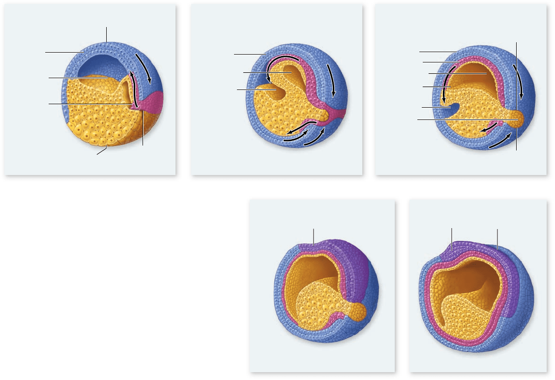
Gastrulation in frogs
The blastula of an amphibian has an asymmetrical yolk distri-
bution, and the yolk-laden cells of the vegetal pole are less nu-
merous but much larger than the yolk-free cells of the animal
pole. Consequently, gastrulation is more complex than it is in
sea urchins. In frogs, a layer of surface cells first invaginates to
form a small, crescent-shaped slit, which initiates formation of
the blastopore. Next, cells from the animal pole involute over
the dorsal lip of the blastopore (see figure 54.12a) , which forms
at the same location as the gray crescent of the fertilized egg
(see figure 54.5).
The involuting cell layer eventually presses against the
inner surface of the opposite side of the embryo, eliminating
the blastocoel and producing an archenteron with a blastopore.
In this case, however, the blastopore is filled with yolk-rich
cells, forming the yolk plug (figure 54.12b, c). The outer layer
of cells resulting from these movements is the ectoderm, and
the inner layer is the endoderm. Other cells that involute over
the dorsal lip and ventral lip (the two lips of the blastopore that
are separated by the yolk plug) migrate between the ectoderm
and endoderm to form the third germ layer—the mesoderm
(figure 54.12c–e).
Gastrulation in birds
At the end of cleavage in a bird or reptile, the developing em-
bryo is a small cap of cells called the blastoderm, which sits on
top of the large ball of yolk (figure 54.13a) . As a result, gastrula-
tion proceeds somewhat differently.
In birds, the blastoderm first separates into two layers,
and a blastocoel cavity forms between them (figure 54.13b).
Figure 54.12
Frog gastrulation. a. A layer of
cells from the animal pole moves toward the vegetal
pole, ultimately involuting through the dorsal lip of the
blastopore. b. Cells in the dorsal lip zone then involute into
the hollow interior, or blastocoel, eventually pressing against
the far wall. The three primary germ tissues (ectoderm,
mesoderm, and endoderm) become distinguished. Ectoderm
is shown in blue, mesoderm in red, and endoderm in yellow.
c. The movement of cells through the blastopore creates
a new internal cavity, the archenteron, which displaces
the blastocoel. d. Organogenesis begins when the neural
plate forms from dorsal ectoderm to begin the process of
neurulation. e. The neural plate next forms a neural groove
and then a neural tube. The cells of the neural ectoderm are
shown in purple.
1114
part
VII
Animal Form and Function
rav32223_ch54_1105-1131.indd 1114rav32223_ch54_1105-1131.indd 1114 11/19/09 4:17:53 PM11/19/09 4:17:53 PM
Apago PDF Enhancer
a.
b.
c.
Blastoderm
Yolk
Blastocoel
Yolk
Yolk
Ectoderm
Endoderm
Primitive streak
Mesoderm
Inner cell mass
Trophoblast
Amniotic cavity
Endoderm
Ectoderm
Endoderm
Ectoderm
Mesoderm
Primitive streak
Formation of
yolk sac
a. b. c. d.
The deep, internal layer of the bilayered blastoderm gives rise
to extraembryonic tissues only (described later on), whereas all
cells of the embryo proper are derived from the upper layer of
cells. Thus, the upper layer of the blastoderm gives rise to all
three germ layers.
Some of the surface cells begin moving to the midline,
where they break away from the surface sheet of cells and in-
gress into the blastocoel cavity. A furrow along the longitudinal
midline marks the site of this ingression (figure 54.13c). This
furrow, analogous to an elongated blastopore, is called the
primitive streak. Some cells migrate through the primitive
streak and across the blastocoel cavity to displace cells in the
lower layer. These deep-migrating cells form the endoderm.
Other cells that move through the primitive streak migrate
laterally into intermediate regions and form a new layer—the
mesoderm. Cells that remain on the surface and do not enter
the primitive streak form the ectoderm.
Gastrulation in mammals
Mammalian gastrulation proceeds much the same as it does
in birds. In both types of animals, the embryo develops from
a flattened collection of cells—the blastoderm in birds or the
inner cell mass in mammals. Although the blastoderm of a
bird is flattened because it is pressed against a mass of yolk,
the inner cell mass of a mammal is flat despite the absence of
a yolk mass.
In mammals, the placenta has made yolk dispensable; the
embryo obtains nutrients from its mother following implanta-
tion into the uterine wall. However, the embryo still gastrulates
as though it were sitting on top of a ball of yolk.
In mammals, a primitive streak forms, and cell movements
through the primitive streak give rise to the three primary germ
layers, much the same as in birds (figure 54.14) . Similarly, mam-
malian embryos envelop their “missing” yolk by forming a yolk
sac from extraembryonic cells that migrate away from the lower
layer of the blastoderm and line the blastocoel cavity.
Figure 54.13
Gastrulation in birds. a. The avian blastula
is made up of a disc of cells sitting atop the large yolk mass.
b. Gastrulation commences with the delamination of the
blastoderm into two layers. All three germ layers are derived from
the upper layer of the blastoderm. c. Cells that migrate through
the primitive streak into the interior of the embryo are future
endoderm or mesoderm cells. Cells that remain in the upper layer
form the ectoderm.
Figure 54.14
Mammalian gastrulation. a. Cross section of the mammalian blastocyst at the end of cleavage. b. The amniotic
cavity forms between the inner cell mass (ICM) and the pole of the embryo. Meanwhile, the ICM attens and delaminates into two layers
that will become ectoderm and endoderm. b. and c. Cells of the lower layer migrate out to line the blastocoel cavity to form the yolk sac. d. A
primitive streak forms the ectoderm layer, and cells destined to become mesoderm migrate into the interior, similar to gastrulation in birds.
chapter
54
Animal Development
1115www.ravenbiology.com
rav32223_ch54_1105-1131.indd 1115rav32223_ch54_1105-1131.indd 1115 11/19/09 4:17:55 PM11/19/09 4:17:55 PM
Apago PDF Enhancer
Chick Embryo Mammal Embryo
Umbilical
blood vessels
Chorion
Amnion
Yolk sac
Allantois
Villus of chorion
frondosum
Maternal blood
a. b.
Chorion
Amnion
Yolk sac
Extraembryonic membranes are
an adaptation to life on dry land
As an adaptation to terrestrial life, the embryos of reptiles, birds,
and mammals develop within a fluid-filled amniotic membrane, or
amnion (chapter 35). The amniotic membrane and several other
membranes form from embryonic cells, but they are located out-
side of the body of the embryo. For this reason, they are known as
extraembryonic membranes. The extraembryonic membranes
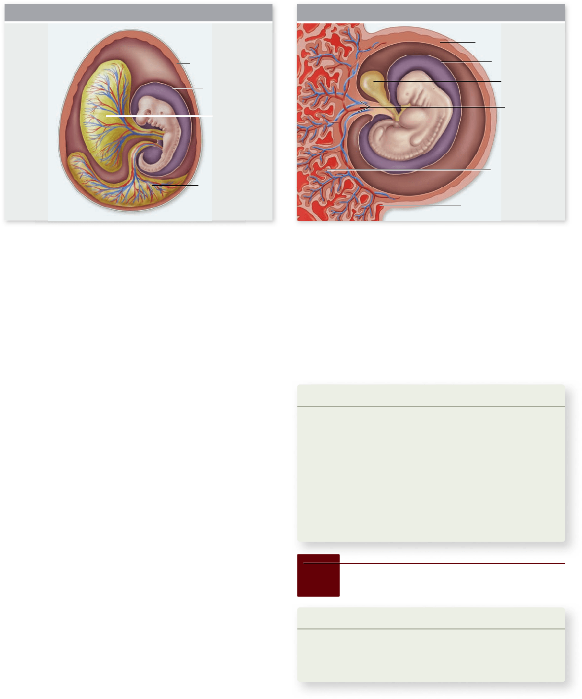
include the amnion, chorion, yolk sac, and allantois.
In birds, the amnion and chorion arise from two folds
that grow to completely surround the embryo (figure 54.15a) .
The amnion is the inner membrane that surrounds the embryo
and suspends it in amniotic fluid, thereby mimicking the aquatic
environments of fish and amphibian embryos. The chorion is
located next to the eggshell and is separated from the other
membranes by a cavity—the extraembryonic coelom.
The yolk sac plays a critical role in the nutrition of bird
and reptile embryos; it is also present in mammals, although it
does not nourish the embryo. The allantois is derived as an out-
pouching of the gut and serves to store the uric acid excreted
in the urine of birds. During development, the allantois of a
bird embryo expands to form a sac that eventually fuses with
the overlying chorion, just under the eggshell. The fusion of
the allantois and chorion form a functioning unit, the chorio-
allan toic membrane, in which embryonic blood vessels, carried
in the allantois, are brought close to the porous eggshell for gas
exchange. The chorio allan toic membrane is thus the respira-
tory membrane of a bird embryo.
In mammals, the trophoblast cells of the blastocyst implant
into the endometrial lining of the mother’s uterus and become
the chorionic membrane (figure 54.15b). The part of the chorion
in contact with endometrial tissue contributes to the placenta.
The other part of the placenta is composed of modified endome-
trial tissue of the mother’s uterus, as is described in more detail in
a later section. The allantois in mammals contributes blood ves-
sels to the structure that will become the umbilical cord, so that
fetal blood can be delivered to the placenta for gas exchange.
Learning Outcomes Review 54.3
Gastrulation involves cell rearrangement and migration to produce
ectoderm, mesoderm, and endoderm. In sea urchins, endoderm forms by
invagination of the blastula; mesodermal cells form from other surface cells.
In vertebrates with moderate to extensive amounts of yolk, surface cells
move through a blastopore or a primitive streak, respectively. Mammalian
gastrulation is similar to gastrulation in birds. Extraembryonic membranes
of amniote species form from embryonic cells outside the embryo’s body and
include the yolk sac, amnion, chorion, and allantois.
■ What kind of cellular behaviors are necessary
for gastrulation?
Figure 54.15
The extraembryonic membranes. The extraembryonic membranes in (a) a chick embryo and (b) a mammalian
embryo share some of the same characteristics. However, in the chick, the allantois continues to grow until it eventually unites with the
chorion just under the eggshell, where it is involved in gas exchange. In the mammalian embryo, the allantois contributes blood vessels to the
developing umbilical cord.
54.4
Organogenesis
Learning Outcomes
Describe examples of organogenesis.1.
Describe neurulation and somitogenesis.2.
Explain the migration and role of neural crest cells.3.
Gastrulation establishes the basic body plan and creates the
three primary germ layers of animal embryos. The stage is now
1116
part
VII
Animal Form and Function
rav32223_ch54_1105-1131.indd 1116rav32223_ch54_1105-1131.indd 1116 11/19/09 4:17:57 PM11/19/09 4:17:57 PM
Apago PDF Enhancer
Dpp
Salivary
gland
During Organogenesis
Prior to Organogenesis
Labium
a.
b.
set for organogenesis—the formation of the organs in their prop-
er locations—which occurs by interactions of cells within and
between the three germ layers. Thus, organogenesis follows
rapidly on the heels of gastrulation, and in many animals begins
before gastrulation is complete. Over the course of subsequent
development, tissues develop into organs and animal embryos
assume their unique body form (see table 54.1).
Changes in gene expression
lead to cell determination
All of the cells in an animal’s body, with the exception of a few
specialized ones that have lost their nuclei, have the same com-
plement of genetic information. Despite the fact that all of its
cells are genetically identical, an adult animal contains dozens
to hundreds of cell types, each expressing some unique aspect
of the total genetic information for that individual. The infor-
mation for other cell types is not lost, but most cells within a
developing organism progressively lose the capacity to express
ever-larger portions of their genomes. What factors determine
which genes are to be expressed in a particular cell?
To a large degree, a cell’s location in the developing em-
bryo determines its fate. By changing a cell’s location, an exper-
imenter can often alter its developmental destiny, as mentioned
in chapter 19. But this is only true up to a certain point in the
cell’s development. At some stage, every cell’s ultimate fate be-
comes fixed, a process referred to as cell determination .
A cell’s fate can be established by inheritance of cytoplasmic
determinants or by interactions with neighboring cells. The process
by which a cell or group of cells instructs neighboring cells to adopt
a particular fate is called induction . If a nonporous barrier, such as a
layer of cellophane, is imposed between the inducer and the target
tissue, no induction takes place. In contrast, a porous filter, through
which proteins can pass, does permit induction to occur.
In these experiments, researchers concluded that the in-
ducing cells secrete a paracrine signal molecule that binds to
the cells of the target tissue. Such signal molecules are capable
of producing changes in the patterns of gene transcription in
the target cells. You will learn more about the origin of embry-
onic induction a little later in this chapter.
Development of selected systems
in Drosophila illustrates organogenesis
In chapter 19, you saw how the creation of morphogen gradi-
ents in a fruit fly embryo leads to hierarchies of gene expres-
sion that direct cell fate decisions along both the anterior–
posterior and dorsal–ventral axes. These two axes form a co-
ordinate system to specify the position of tissues and organs
within the Drosophila embryo. In this section we look at devel-
opment of three different organs: salivary glands, the heart, and
the tracheae of the respiratory system.
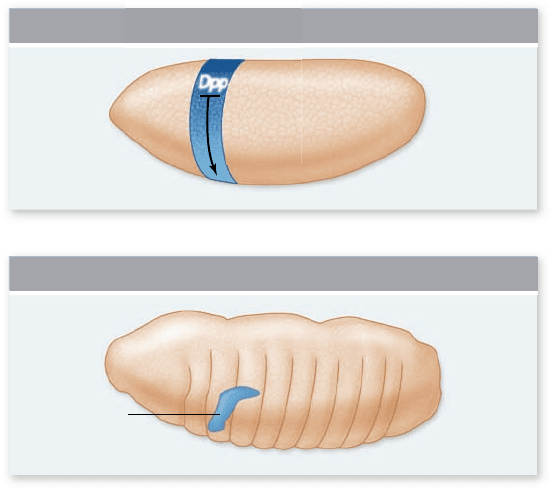
Salivary gland development
The fruit fly larva is a mobile eating machine, and thus it has
very active salivary glands. The primordia of the salivary glands
develop as simple tubular invaginations of ectodermal cells on
the ventral surface of the third head segment.
Salivary glands develop only from an anterior strip of cells
that express the sex combs reduced (scr) gene. No salivary glands
form in scr-deficient embryos, whereas experimental expansion
of scr expression along the anterior–posterior axis results in
the formation of additional salivary gland primordia along the
length of the embryo.
The scr gene is one of the homeotic genes in the Antenna-
pedia complex, which encode transcription factors that bind to
DNA via their homeodomains to regulate gene expression (see
chapter 19). One downstream target of the scr gene is the fork
head ( fkh) gene, which has Scr-binding sites in its enhancer. The
fkh gene is required for secretory cell development in salivary
gland rudiments, and it encodes a transcription factor that di-
rectly activates expression of salivary gland-specific genes. Thus,
action of the scr gene activates fkh expression at the proper ante-
rior location for salivary gland formation.
The inhibitory action of a dorsally expressed protein, De-
capentaplegic (Dpp), determines the ventral position of the sali-
vary glands. Activation of the Dpp-signaling pathway represses
salivary gland specification in neighboring cells. This restricts
development of salivary gland rudiments to their specific ventral
patch of ectoderm cells (figure 54.16) . In mutant embryos defi-
cient for Dpp or any of the downstream Dpp- signaling proteins,
Figure 54.16
Salivary gland formation in Drosophila.
Prospective salivary gland cells are determined by the intersection
of the anterior–posterior and dorsal–ventral axes. a. Prior to
organogenesis, the sex combs reduced (scr) gene is expressed
in an anterior band of cells (shaded blue). At the same time,
Decapentaplegic protein (Dpp) is released by cells on the dorsal side
of the embryo, forming a gradient in the dorsal–ventral direction.
Dpp speci es dorsal cell fates and inhibits formation of salivary
gland rudiments. b. During organogenesis, the salivary glands
develop in areas where Scr is expressed but Dpp is absent. Each
salivary gland rudiment forms as a ventral invagination of the surface
ectoderm on either side of the third head segment (the labium).
chapter
54
Animal Development
1117www.ravenbiology.com
rav32223_ch54_1105-1131.indd 1117rav32223_ch54_1105-1131.indd 1117 11/19/09 4:18:02 PM11/19/09 4:18:02 PM
Apago PDF Enhancer
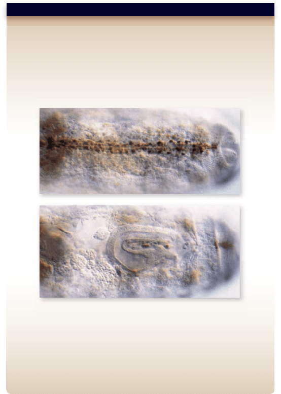
Hypothesis: The tinman gene is required for proper development of the
dorsal vessel in Drosophila.
Prediction: The tinman gene must be expressed in precursor cells for the
dorsal vessel. Loss of tinman function should result in loss of dorsal vessel.
Test: Analyze expression of tinman in whole mount embryos in wild
type (top) and mutant (bottom) embryos.
Result: In the wild type embryo, tinman is expressed in a line of cells
where the dorsal vessel forms. In mutant embryos with no expression, no
dorsal vessel forms.
Conclusion: The function of tinman is necessary for dorsal vessel formation.
Further Experiments: Tinman is a homeobox containing gene. What
does this suggest about its function? How could you follow up on this?
SCIENTIFIC THINKING
salivary gland rudiments are not restricted to this ventral patch,
and they form from the entire ectoderm of the third segment.
Heart development
The heart is a mesoderm-derived structure in all animals, and
it is the first organ to become functional during embryonic de-
velopment. The dorsal vessel is the heart-equivalent structure
in Drosophila melanogaster. The homeobox-containing gene tin-
man is expressed in the prospective heart mesoderm and in the
developing dorsal vessel, and its activity is required for dorsal
vessel development in Drosophila (figure 54.17) .
Dorsal vessel development in Drosophila is also dependent
on two other types of transcription factors (known as GATA
and T-box factors). In an illuminating case of evolutionary con-
servation, scientists have discovered gene families similar to
each of these three Drosophila genes in vertebrates. Moreover,
members of these gene families play important roles in verte-
brate heart specification.
This evolutionary conservation includes not just the
structure of these genes, but their function as well. Research-
ers have discovered that specification of cardiac mesoderm is
subject to inductive signals from adjoining germ layers in both
Drosophila and vertebrates. In vertebrates, the heart develops in
an internal location, and the inductive signals come from the
underlying anterior endoderm. In Drosophila, the dorsal vessel
forms in a more superficial location, and the signals come from
the overlying ectoderm.
Despite the different sources, the signals that regulate the ex-
pression of these three key types of transcription factors are them-
selves conserved between Drosophila and vertebrates. Given the
critical and conserved circulatory function of the heart, it is perhaps
not surprising that similar gene families mediate the specification
of heart mesoderm in both Drosophila and vertebrates.
Tracheae: Branching morphogenesis
As you learned in chapters 34 and 49, insects exchange gases
via a branching system of finer and finer tubes called tracheae .
The repeated branching of simple epithelial tubes that leads to
formation of the tracheal system is an example of branching
morphogenesis.
Mutations in the branchless gene in Drosophila result in
embryos with greatly reduced tracheal systems. The branch-
less gene encodes a member of the large family of fibroblast
growth factors (FGF), which bind to receptor tyrosine kinase
proteins (see chapter 9) to stimulate proliferation of target cells.
In another interesting case of evolutionary conservation, the
mammalian FGF homologue of the branchless gene is required
for branching morphogenesis that creates the alveolar passage-
ways in the mammalian lung.
In both animals, loose clusters of mesenchymal cells ad-
jacent to distal regions of the epithelial tube secrete FGF. The
FGF binds to a specific FGF receptor in the membrane of the
epithelial cells, stimulating them to proliferate and to grow out
into a new tube bud.
In vertebrates, organogenesis begins
with neurulation and somitogenesis
The process of organogenesis in vertebrates begins with the for-
mation of two morphological features found only in chordates:
the notochord and the hollow dorsal nerve cord (see chapter 35).
The development of the dorsal nerve cord is called neurulation .
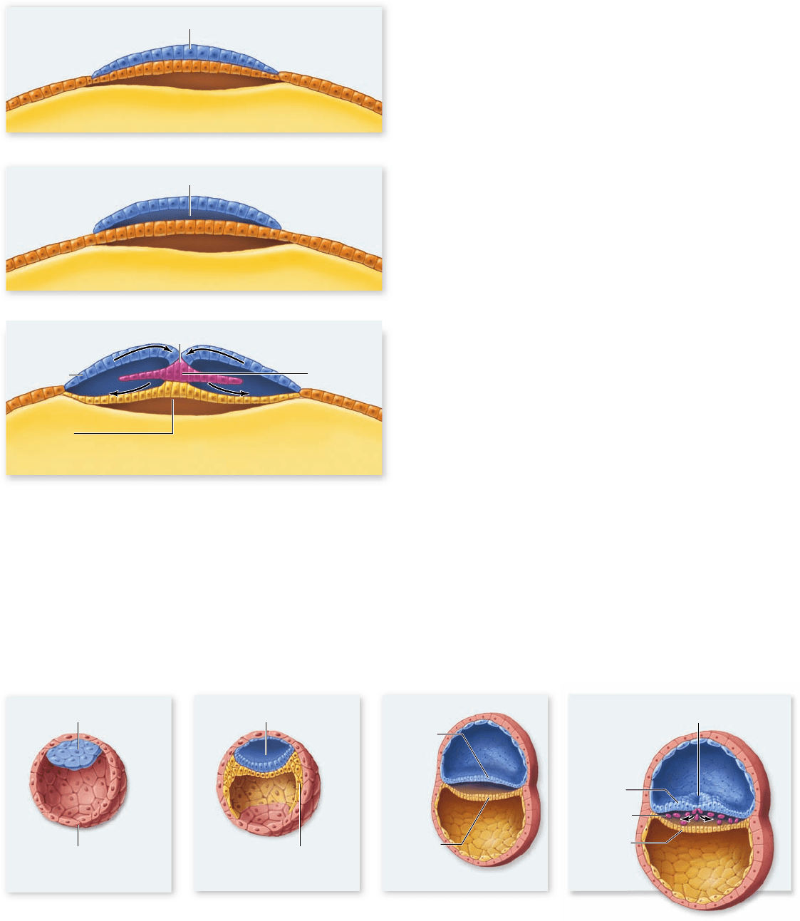
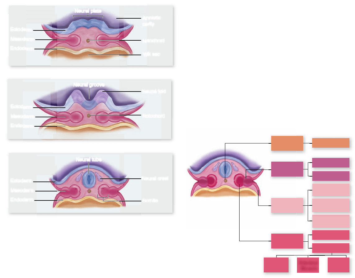
Development of the neural tube
The notochord forms from mesoderm and is first visible soon
after gastrulation is complete. It is a flexible rod located along
the dorsal midline in the embryos of all chordates, although
its function as a supporting structure is supplanted by the sub-
sequent development of the vertebral column in the verte-
brates. After the notochord has been laid down, the region of
dorsal ectodermal cells situated above the notochord begins
to thicken to form the neural plate.
The thickening is produced by the elongation of the dor-
sal ectoderm cells. Those cells then assume a wedge shape be-
cause of contracting bundles of actin filaments at their apical
end. This change in shape causes the neural tissue to roll up
into a neural groove running down the long axis of the em-
bryo. The edges of the neural groove then move toward each
Figure 54.17
A gene necessary for heart formation
in Drosophila.
1118
part
VII
Animal Form and Function
rav32223_ch54_1105-1131.indd 1118rav32223_ch54_1105-1131.indd 1118 11/19/09 4:18:02 PM11/19/09 4:18:02 PM
Apago PDF Enhancer
Neural fold
Notochord
Notochord
Yolk sac
Amniotic
cavity
Ectoderm
Mesoderm
Endoderm
Ectoderm
Mesoderm
Endoderm
Ectoderm
Mesoderm
Endoderm
a.
b.
c.
Neural
crest
Somite
Neural plate
Neural groove
Neural tube
Chorda-
mesoderm
Intermediate
mesoderm
Paraxial
mesoderm
Notochord
Kidney
Gonads
Circulatory
system
Extra-
embryonic
Linings of
body cavities
Head
Somite
Dermis Cartilage
Skeletal
muscle
Lateral plate
mesoderm
other and fuse, creating a long hollow cylinder, the neural tube
(figure 54.18) . The neural tube eventually pinches off from the
surface ectoderm to end up beneath the surface of the embryo’s
back. Regional changes, which are under control of the Hox
gene complexes (see chapter 19), then occur in the neural tube
as it differentiates into the spinal cord and brain.
Generation of somites
While the neural tube is forming from dorsal ectoderm, the rest
of the basic architecture of the body is being rapidly established
by changes in the mesoderm. The sheets of mesoderm on ei-
ther side of the developing notochord separate into a series of
rounded regions called somitomeres. The somitomeres then
separate into segmented blocks called somites (see figure 54.18 ).
The mesoderm in the head region does not separate into discrete
somites but remains connected as somitomeres, which form the
skeletal muscles of the face, jaws, and throat.
Somites form in an anterior–posterior wave with a regu-
lar periodicity that can be easily timed—for example, by using a
vital dye, which marks cells without killing them, to mark each
somite as it forms in a chick embryo. Cells at the presump-
tive boundary regions in the presomitic mesoderm instruct
cells anterior to them to condense and separate into somites at
specific times (for example, every 90 min in a chick embryo).
This “clock” appears to be regulated by contact-mediated cell
signaling between neighboring cells.
Somites themselves are transient embryonic structures,
and soon after their formation, cells disperse and start differen-
tiating along different pathways to ultimately form the skeleton,
skeletal musculature, and associated connective tissues. The to-
tal number of somites formed is species-specific; for example,
chickens form 50 somites, whereas some species of snakes form
as many as 400 somites.
Some body organs, including the kidneys, adrenal glands,
and gonads, develop within a strip of mesoderm that runs lateral
to each row of somites. The remainder of the mesoderm, which
is most ventrally located, moves out and around the endoderm
and eventually surrounds it completely. As a result of this move-
ment, the mesoderm becomes separated into two layers. The
outer layer is associated with the inner body wall, and the inner
layer is associated with the outer lining of the gut tube. Between
these two layers of mesoderm is the coelom (see chapter 32),
which becomes the body cavity of the adult. Figure 54.19 shows
the major mesoderm lineages of amniote embryos.
Migratory neural crest cells di erentiate
into many cell types
Neurulation occurs in all chordates, and the process in the sim-
ple lancelet, a nonvertebrate chordate, is much the same as it
is in a human. However, neurulation is accompanied by an ad-
ditional step in vertebrates. Just before the neural groove closes
Figure 54.18
Mammalian neural tube formation.
a. The neural plate forms from ectoderm above the notochord.
b. The cells of the neural plate fold together to form the neural
groove. c. The neural groove eventually closes to form a hollow
tube called the neural tube, which will become the brain and spinal
cord. As the tube closes, some of the cells from the dorsal margin
of the neural tube differentiate into the neural crest, migratory
cells that form a variety of structures and are characteristic
of vertebrates.
Figure 54.19
Mesoderm-derived structures of birds
and mammals.
chapter
54
Animal Development
1119www.ravenbiology.com
rav32223_ch54_1105-1131.indd 1119rav32223_ch54_1105-1131.indd 1119 11/19/09 4:18:04 PM11/19/09 4:18:04 PM
Apago PDF Enhancer
a.
c.
b.
Anterior
Posterior
Aorta Notochord
Neural
crest cells
Neural tube
Epidermis
Anterior somite
Posterior somite
Ventral Pathway
Cells travel
ventrally
through the
anterior half
of each somite
Dorsal root
ganglia
Ventral root
Schwann
cells
Sympathetic
ganglia
Adrenal
medulla
Melanocytes
Ventral Pathway Cell Fates Lateral Pathway Cell Fates
Lateral Pathway
Cells take a
dorsolateral
route between
the epidermis
and somites
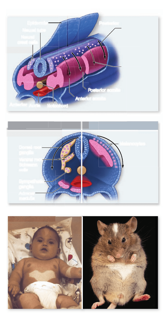
Figure 54.20
Migration pathways and cell fates of
trunk neural crest cells. a. The rst wave of trunk neural
crest cells migrates ventrally through the anterior half of each
somite, whereas the second wave of cells leaves dorsally and
migrates through the space between the epidermis and the somites.
b. Ventral pathway neural crest cells differentiate into a variety of
specialized cell types, but lateral pathway cells develop into the
melanocytes (pigment cells) of the skin. c. A mutation in a gene that
promotes survival of neural crest cells in all mammals leads
to white spotting on the bellies and foreheads of both human
babies and mice! Each individual is heterozygous for this
mutation and thus has only half as much of the survival factor
as unaffected individuals.
to form the neural tube, its edges pinch off, forming a small
cluster of cells—the neural crest — between the roof of the neu-
ral tube and the surface ectoderm (figure 54.18c).
In another example of extensive cell movements during
animal development, the neural crest cells then migrate away
from the neural tube to colonize many different regions of the
developing embryo. The appearance of the neural crest was a
key event in the evolution of the vertebrates because neural crest
cells, after reaching their final destinations, ultimately develop
into many structures characteristic of the vertebrate body.
The differentiation of neural crest cells depends on their
migration pathway and final location. Neural crest cells mi-
grate along one of three pathways in the embryo. Cranial neu-
ral crest cells are anterior cells that migrate into the head and
neck; trunk neural crest cells migrate along one of two different
pathways (to be described shortly). Each population of neural
crest cells develops into a variety of cell types.
Cranial neural crest cells’ migration
Cranial neural crest cells contribute significantly to development
of the skeletal and connective tissues of the face and skull, as well
as differentiating into nerve and glial cells of the nervous system,
and melanocyte pigment cells. Changes in the placement of cranial
neural crest cells during development have led to the evolution of
the great complexity and variety of vertebrate heads.
There are two waves of cranial neural crest cell migra-
tion. The first produces both dorsal and ventral structures, and
the second produces only dorsal structures and makes much
less cartilage and bone. Transplantation experiments indicate
that the developmental potential of the cells in these two waves
is identical. The differences in cell fate are due to the environ-
ment the migrating cells encounter and not due to prior deter-
mination of cell fate.
Trunk neural crest cells: Ventral pathway
Neural crest cells located in more posterior positions have very
different developmental fates depending on their migration
pathway. The first trunk neural crest cells that migrate away
from the neural tube pass through the anterior half of each ad-
joining somite to ventral locations (figure 54.20a).
Some of these cells form the sensory neurons of the dorsal
root ganglia, which send out projections to connect the periph-
ery of the animal with the spinal cord (see chapter 44). Others
become specialized as Schwann cells, which insulate nerve fibers
to facilitate the rapid conduction of impulses along peripheral
nerves. Still others form nerves of the autonomic ganglia, which
regulate the activity of internal organs, and endocrine cells of the
adrenal medulla (figure 54.20b). The chemical similarity of the
hormone epinephrine and the neurotransmitter norepinephrine,
which are released by sympathetic neurons of the autonomic
nervous systems, may result because both adrenal medullary cells
and sympathetic neurons derive from the neural crest.
Trunk neural crest cells: Lateral pathway
The second group of trunk neural crest cells migrate away from
the neural tube in the space just under the surface ectoderm,
to occupy this space around the entire body of the embryo.
There, they will differentiate into the pigment cells of the skin
1120
part
VII
Animal Form and Function
rav32223_ch54_1105-1131.indd 1120rav32223_ch54_1105-1131.indd 1120 11/19/09 4:18:05 PM11/19/09 4:18:05 PM
Apago PDF Enhancer
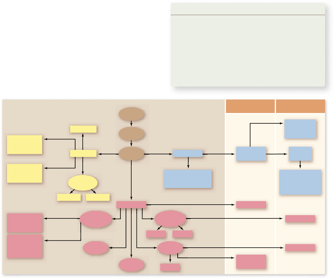
Blood Vessels
Dermis
Epidermis, skin, hair,
epithelium, inner
ear, lens of eye
Circulatory
system
Ectoderm
Gill arches,
sensory ganglia,
Schwann cells,
adrenal medulla
Brain,
spinal cord,
spinal nerves
Heart
Skeleton
Neural
crest
Notochord
Striated
muscles
Dorsal
nerve cord
Pancreas Liver
Gonads
Integuments
Kidney
Gastrula
Blastula
Zygote
Major
glands
Endoderm
Pharynx
Mesoderm
Lining of
respiratory
tract
Lining of
digestive
tract
Outer covering
of internal
organs
Lining of
thoracic and
abdominal
cavities
Chordates Vertebrates
Somites
(figure 54.20a, b). Mutations in genes that affect the survival
and migration of neural crest cells lead to white spotting in the
skin on ventral surfaces, as well as internal problems in other
neural crest-derived tissues (figure 54.20c).
Because the fate of a neural crest cell is dictated by its
migration pathway, many studies have been done to identify
the molecules that control the migration pathways of neural
crest cells. Cell adhesion molecules on cell surfaces and in the
extracellular matrix are expected to play prominent roles. For
example, prospective neural crest cells down-regulate the ex-
pression of N-cadherin on their surfaces, which enables them
to break away from the neural tube. Then, soon after leaving
the neural tube, integrin receptors appear on the surfaces of
neural crest cells, allowing them to interact with proteins in the
extracellular matrix pathways along which they will migrate.
Neural crest derivatives are important
in vertebrate evolution
Primitive chordates such as lancelets are filter feeders, using the
rapid beating of cilia to draw water into their mouths, which then
exits through slits in their pharynx. These pharyngeal slits evolved
into the vertebrate gill chamber, a structure that provides a greatly
improved means of gas exchange. Thus, evolution of the gill cham-
ber was certainly a key event in the transition from filter feeding to
active predation, which requires a much higher metabolic rate.
In the development of the gill chamber, some of the cra-
nial neural crest cells form cartilaginous bars between the em-
bryonic pharyngeal slits. Other cranial neural crest cells induce
portions of the mesoderm to form muscles along the cartilage,
and still others to form neurons that carry impulses between
the central nervous system and these muscles.
Many of the unique vertebrate adaptations that contribute to
their varied ecological roles involve structures that arise from neural
crest cells. The vertebrates became fast-swimming predators with
much higher metabolic rates. This accelerated metabolism permit-
ted a greater level of activity than was possible among the more
primitive chordates. Other evolutionary changes associated with the
derivatives of the neural crest provided better detection of prey, a
greatly improved ability to orient spatially during prey capture, and
the means to respond quickly to sensory information. The evolution
of the neural crest and of the structures derived from it were thus
crucial steps in the evolution of the vertebrates (figure 54.21).
Learning Outcomes Review 54.4
Genetic control of organogenesis relies on conserved families of cell-
signaling molecules and transcription factors. The control of heart
development in Drosophila and mammals uses some of the same proteins.
The process of neurulation forms the basic nervous system in vertebrates.
Somitogenesis is the division of mesoderm into somites. Neural crest cells
arise from the neural tube and migrate to many sites to form a variety of
cell types. The evolution of the neural crest led to the appearance of many
vertebrate-specifi c adaptations.
■ Are neural crest cells determined prior to migration?
Figure 54.21
Germ-layer derivation of the major tissue types in animals. The three germ layers that form during gastrulation
give rise to all the organs and tissues in the body, but the neural crest cells that form from ectodermal tissue give rise to structures that are
prevalent in vertebrates, such as gill arches and bones of the face and skull.
chapter
54
Animal Development
1121www.ravenbiology.com
rav32223_ch54_1105-1131.indd 1121rav32223_ch54_1105-1131.indd 1121 11/19/09 4:18:10 PM11/19/09 4:18:10 PM