Raven P.H., Johnson G.B., Mason K.A. Biology (Ninth Edition)
Подождите немного. Документ загружается.

Apago PDF Enhancer
Recipient embryoDonor embryo Primary
neural fold
Secondary
neural fold
Primary notochord, somites,
and neural development
Secondary
notochord,
somites,
and neural
development
Dorsal lip
54.5
Vertebrate Axis Formation
Learning Outcomes
Describe the Spemann-Mangold experiment.1.
Explain the function of the organizer.2.
Distinguish between primary and secondary 3.
inductive events.
In animal development, the relative position of cells in particu-
lar germ layers determines, to a large extent, the organs that
develop from them. In Drosophila, you have seen that formation
of morphogen gradients in the syncytial blastoderm establishes
the anterior–posterior and dorsal–ventral axes of the embryo.
The Hox gene complexes in vertebrates function similarly to
the homeotic genes of Drosophila to specify the position of or-
gans along the anterior–posterior axis. But how is cell fate se-
lection along the dorsal–ventral axis accomplished in vertebrate
embryos? Put another way, how do cells of the dorsal ectoderm
“know” they are above the mesoderm-derived notochord, and
thus fated to develop into the neural tube? The solution to this
puzzle is one of the outstanding accomplishments of experi-
mental embryology.
The Spemann organizer determines
dorsal–ventral axis
The renowned German biologist Hans Spemann and his
student Hilde Mangold solved this puzzle early in the 20th
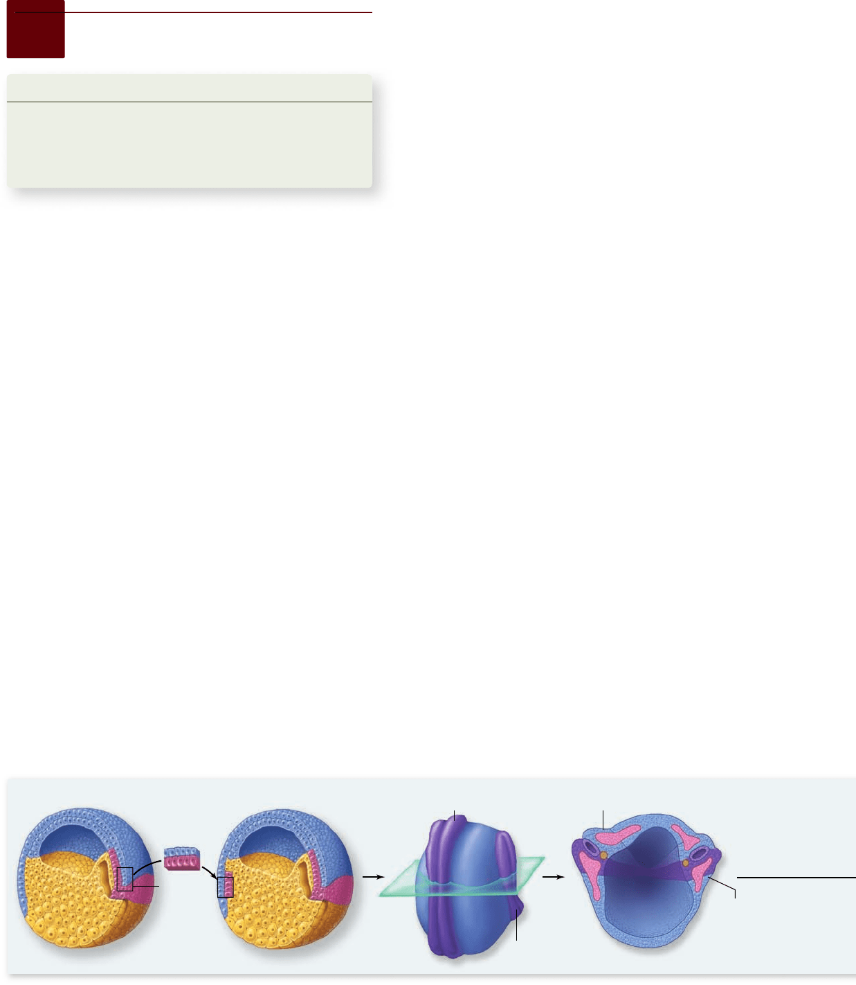
century. Normally, cells derived from the dorsal lip of the
blastopore of a gastrulating amphibian embryo give rise to the
notochord. Spemann and Mangold removed cells of the dor-
sal lip from one embryo and transplanted them to a different
location on another embryo (figure 54.22) . The new location
corresponded to that of the animal’s future belly. They found
that some of the embryos developed two notochords: a nor-
mal dorsal one, and a second one along the belly. Moreover, a
complete set of dorsal axial structures (e.g., notochord, neural
tube, and somites) formed at the ventral transplantation site
in most of these embryos.
By using genetically different donor and host blastulas,
Spemann and Mangold were able to show that the second no-
tochord produced by transplanting dorsal lip cells contained
host cells as well as transplanted ones. The transplanted dorsal
lip cells had thus acted as organizers, stimulating cells that would
normally form skin and belly structures to develop into dorsal
axial structures. The belly cells must clearly contain the genetic
information for dorsal axial developmental program, but they
do not express it in the normal course of their development.
Signals from the transplanted dorsal lip cells, however, must
have caused them to do so.
How the organizer works
An organizer is a cluster of cells that release diffusible signal
molecules, which then convey positional information to other
cells. As seen earlier, organizers can have a profound influence
on the development of surrounding tissues. Working as signal
beacons, they inform surrounding cells of their distance from
the organizer. The closer a particular cell is to an organizer,
the higher the concentration of the signal molecule (morphogen)
it experiences. Organizers and the diffusible morphogens that
they release are thought to be part of a widespread mechanism
for determining relative position and cell fates during verte-
brate development.
The action of morphogens
The action of morphogens can be studied by using isolated por-
tions of the blastula. The blastula can be bisected into an animal
half (the animal cap) and vegetal half (the vegetal cap). If animal
caps are removed from a frog blastula and cultured alone, they
will form only ectoderm-derived epidermal cells. Similarly, cul-
tured vegetal caps will form only endodermal cells. However, if
animal caps are cultured combined with vegetal caps, the ani-
mal caps will form mesodermal structures.
The molecules involved in this induction have not been
unambiguously identified. Members of the transforming growth
factor beta (TGF-β) family have been implicated. These include
activin, and Xenopus nodal-related proteins (Xnrs). Evidence for
the inducing action of these molecules ranges from indirect: the
Figure 54.22
Spemann and Mangold’s dorsal lip transplant experiment. Tissue from the dorsal lip of a donor embryo
induced the formation of a second axis in the future belly region of a second, recipient embryo.
1122
part
VII
Animal Form and Function
rav32223_ch54_1105-1131.indd 1122rav32223_ch54_1105-1131.indd 1122 11/19/09 4:18:11 PM11/19/09 4:18:11 PM
Apago PDF Enhancer
Animal pole
Pigmented
cortical cytoplasm
Gray crescent
Organizer
Dorsal
mesoderm-
inducing signal
Mesoderm-
inducing signals
(TGF-β family proteins)
Nieuwkoop center
Point of
sperm entry
Clear cortical
cytoplasm
Inner
cytoplasm
Diffuse black
pigment
Microtubule
array
Microtubules
Dorsal determinants
Shifted dorsal
determinants
Vegetal pole
a.
b.
c.
Primary embryo
Secondary embryo
timing and pattern of expression correlates with inducing tissue,
to depleting developing embryos of these proteins with specific
reagents that block gene expression.
The origin of the organizer
How do cells of the frog blastopore’s dorsal lip become the Spe-
mann organizer and how do they acquire their ability to specify
cell fate along the dorsal–ventral axis? In frogs, as in fruit flies,
this process starts during oogenesis in the mother. At that time,
maternally encoded dorsal determinants are put into the de-
veloping oocyte, one of which accumulates at the vegetal pole
of the unfertilized egg. At fertilization, cytoplasmic rearrange-
ments cause this determinant to shift to the future dorsal side of
the egg.
First, a signal from the point of sperm entry initiates the
assembly of a microtubule array, which enables the egg’s plas-
ma membrane and the underlying cortical cytoplasm to rotate
over the surface of the deeper cytoplasm. This physical rotation
shifts this maternally encoded dorsal determinant to the opposite
side of the egg from the point of sperm entry (figure 54.23a, b) .
In some frogs, a gray crescent forms opposite the sperm entry
point, as mentioned earlier, and this crescent marks the future
site of the dorsal lip.
Cells that form in this area during cleavage (called the
Nieuwkoop center for the scientist who did the previously
mentioned animal cap studies) receive the dorsal determinants
that moved during cortical rotation. The dorsal determinants
cause a change in gene expression in these cells, producing a
signaling molecule that induces the cells above them to develop
into the dorsal lip of the blastopore (figure 54.23c).
Maternally encoded dorsal determinants
activate Wnt signaling
Experiments carried out over the last 15 years suggest that the
maternally encoded dorsal determinants in Xenopus are mRNAs
for proteins that function in the intracellular Wnt signaling
pathway. Wnt genes encode a large family of cell-signaling pro-
teins that affect the development of a number of structures in
both vertebrates and invertebrates. Turning on the Wnt path-
way in the dorsal vegetal cells of the Nieuwkoop center leads
ultimately to activation of a transcription factor, which moves
into the nucleus to activate the expression of genes necessary
for organizer specification.
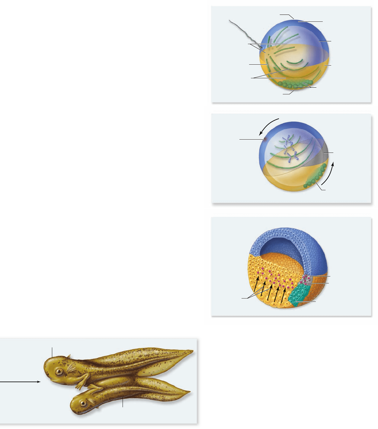
Figure 54.23
Creation of the Spemann organizer.
a. Dorsal determinants are localized at the vegetal pole of the
unfertilized frog egg. At fertilization, a microtubule array forms
at the site of sperm entry. These microtubules organize parallel
microtubules to line the vegetal half of the egg between the cortex
and cytoplasm. b. The cortical cytoplasm and dorsal determinants
ride on this parallel array of microtubules, shifting to a site
opposite sperm entry. c. Cells that inherit these shifted dorsal
determinants form the Nieuwkoop center, which releases diffusible
signaling molecules that specify the cells in the overlying dorsal
marginal zone to become the organizer. The organizer forms at
the area of the gray crescent, visible following the cytoplasmic
rearrangements at fertilization.
chapter
54
Animal Development
112 3www.ravenbiology.com
rav32223_ch54_1105-1131.indd 1123rav32223_ch54_1105-1131.indd 1123 11/19/09 4:18:12 PM11/19/09 4:18:12 PM
Apago PDF Enhancer
(
(
–
–
)
Animal pole
Vegetal pole
Organizer molecules:
Chordin, Noggin, and others
Mesoderm
Epidermal ectoderm
Neural ectoderm
Endoderm
Dorsal
Ventral
Signaling molecules from the Spemann
organizer inhibit ventral development
It has taken decades to establish the identity and function of
the molecules that are synthesized by cells of the Spemann or-
ganizer to subsequently specify dorsal mesoderm cell fates in
frogs. A surprising finding of recent experiments indicates that
dorsal lip cells do not directly activate dorsal development. In-
stead, dorsal mesoderm development is a result of the inhibition
of ventral development.
A protein called bone morphogenetic protein 4
(BMP4) is expressed in all marginal zone cells (the prospective
mesoderm) of a frog embryo. Cells with receptors for BMP4
have the potential to develop into mesodermal derivatives. The
specific mesodermal fate depends on how many receptors bind
BMP4: More BMP4 binding induces a more ventral meso-
dermal fate.
The organizer functions by secreting a host of inhibitory
molecules that can bind to BMP4 and prevent its binding to
receptor. Such molecules are referred to as BMP4 antagonists.
Up to 13 different proteins have been identified in the Spe-
mann organizer, most of which appear to function as BMP4
antagonists. These include the proteins Noggin, Chordin,
Dickkopf, and Cerebrus. Noggin and BMP4 are also involved
in toe and finger joint formation, so humans homozygous for a
Noggin mutation have fused joints.
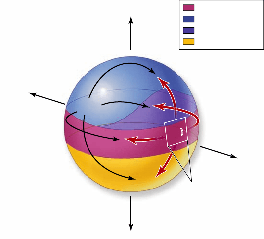
Thus, the gradient of inhibitory molecules that emanates
from the Spemann organizer leads to a declining level of BMP4
function in the ventral-to-dorsal direction. Cells farthest from
the organizer bind the highest levels of BMP4 and differenti-
ate into ventral mesoderm structures such as blood and con-
nective tissues. Cells that are midway from the organizer bind
intermediate amounts of BMP4, differentiate into intermediate
mesoderm, and form organs such as the kidneys and gonads.
BMP4 binding is completely inhibited by the high levels of
antagonists in the organizer itself. Thus, these cells adopt the
most dorsal of mesoderm fates and develop into somites. The
influence of the organizer also extends to ectoderm as inhibi-
tion of BMP4 in ectoderm leads to formation of neural tissue
instead of epidermis (figure 54.24) .
Evidence indicates that organizers
are present in all vertebrates
In chicks, a group of cells at the anterior limit of the primi-
tive streak called Hensen’s node functions similarly to the dor-
sal lip of the blastopore: Hensen’s node induces a second axis
when transplanted to another area of a chick embryo. Recent
studies have shown that cells of Hensen’s node act like the
Spemann organizer, secreting molecules that inhibit ventral
development. These molecules are the same as those found
in frog embryos. Therefore, these experiments once again il-
lustrate the evolutionary conservation of particular genes in
animal development.
In addition, notochord signaling acts to pattern the neu-
ral tube. The notochord produces the signaling molecule sonic
hedgehog (Shh), which is related to a signaling molecule in
Drosophila called hedgehog. Signaling by Shh specifies ventral
cell fate with dose-related effects similar to those described for
the TGF-β family proteins discussed earlier. In this way, induc-
tion by the notochord causes somites to form vertebrae, ribs,
muscle, and skin, depending on the levels of Shh cells are ex-
posed to.
Induction can be primary or secondary
The process of induction that Spemann initially discovered
appears to be a fundamental mode of development in verte-
brates. Inductions between the three primary germ layers—
ectoderm, mesoderm, and endoderm—are referred to as
primary inductions. The differentiation of the central ner-
vous system during neurulation by the interaction of dorsal
ectoderm and dorsal mesoderm to form the neural tube is an
example of primary induction.
Inductions between tissues that have already been spec-
ified to develop along a particular developmental pathway
are called secondary inductions. An example of secondary
induction is the development of the lens of the vertebrate
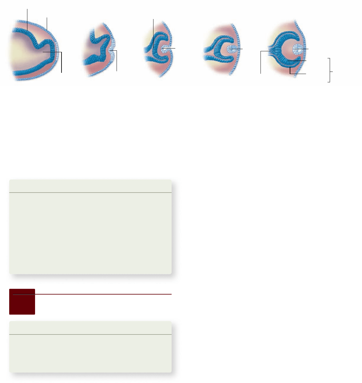
eye. The eye develops as an extension of the forebrain, a
stalk that grows outward until it comes into contact with the
surface ectoderm (figure 54.25) . At a point directly above
the growing stalk, a layer of the surface ectoderm pinches
off, forming a transparent lens. The formation of lens from
the surface ectoderm requires induction by the underlying
neural ectoderm.
This was shown by transplantation experiments per-
formed by Spemann. When the optic stalks of the two eyes
have just started to project from the brain prior to lens
Figure 54.24
Function of the Spemann organizer.
The organizer is a hotbed of secreted molecules that bind to and
antagonize the action of BMP4, a morphogen that at high levels
speci es ventral mesoderm cell fates.
1124
part
VII
Animal Form and Function
rav32223_ch54_1105-1131.indd 1124rav32223_ch54_1105-1131.indd 1124 11/19/09 4:18:14 PM11/19/09 4:18:14 PM
Apago PDF Enhancer
Neural
cavity
Ectoderm
Wall of forebrain
Optic stalk
Lens
invagination
Optic cup
Lens
vesicle
Lens
Optic nerve
Sensory
layer
Pigment
layer
Retina
Lens
formation, one of the budding stalks can be removed and
transplanted underneath surface ectoderm in a region that
would normally develop into the epidermis of the skin (such
as that of the belly). When this is done, a lens forms from
belly ectoderm cells in the region above where the budding
stalk was transplanted. This lens forms due to inductive sig-
nals from the underlying optic stalk.
Learning Outcomes Review 54.5
The Spemann-Mangold experiment showed that transplanted cells of the
dorsal lip of the blastopore act as organizers stimulating development
of a notochord. Hensen’s node plays an equivalent role in vertebrates. By
inhibiting BMP4, the organizer induces ectoderm to form neural tissue and
mesoderm to form dorsal mesoderm. Primary inductions between germ
layers lead to development of the vertebrate nervous system, whereas
secondary inductions result in formation of structures such as the lens of
the eye.
■ How can the organizer function by inhibiting the action
of other molecules?
During the rst trimester, the zygote undergoes
rapid development and di erentiation
About 30 hr after fertilization, the zygote undergoes its first
cleavage; the second cleavage occurs about 30 hr after that. By
the time the embryo reaches the uterus, 6 to 7 days after fer-
tilization, it has differentiated into a blastocyst. As mentioned
earlier, the blastocyst consists of an inner cell mass, which will
become the body of the embryo, and a surrounding layer of
trophoblast cells (see figure 54.10).
The trophoblast cells of the blastocyst digest their way
into the endometrial lining of the uterus in the process known
as implantation. The blastocyst begins to grow rapidly and ini-
tiates the formation of the amnion and the chorion.
Development in the first month
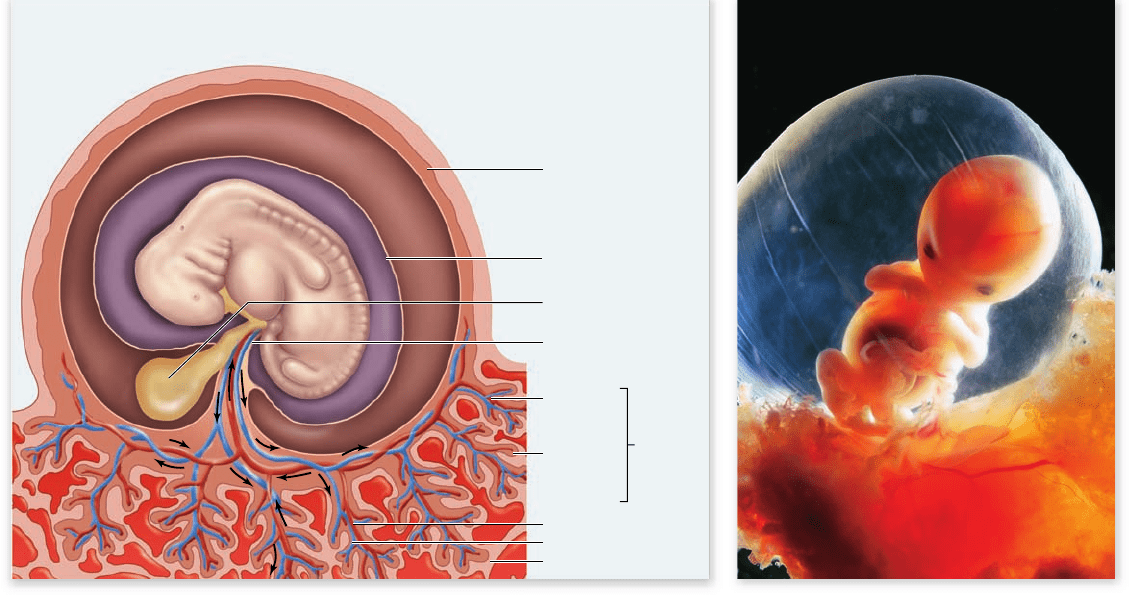
During the second week after fertilization, the developing cho-
rion and the endometrial tissues of the mother engage to form
the placenta (figure 54.26) . Within the placenta, the mother’s
blood and the blood of the embryo come into close proximity
but do not mix. Gases are exchanged, however, and the pla-
centa provides nourishment for the embryo, detoxifies certain
molecules that may pass into the embryonic circulation, and
secretes hormones. Certain substances, such as alcohol, drugs,
and antibiotics, are not stopped by the placenta and pass from
the mother’s bloodstream into the embryo.
One of the hormones released by the placenta is hu-
man chorionic gonadotropin (hCG), which was discussed in
chapter 53. This hormone is secreted by the trophoblast cells
even before they become the chorion, and it is the hormone
assayed in pregnancy tests. Human chorionic gonadotropin
maintains the mother’s corpus luteum. The corpus luteum, in
turn, continues to secrete estradiol and progesterone, thereby
preventing menstruation and further ovulations.
Gastrulation also takes place in the second week after fer-
tilization, and the three germ layers are formed. Neurulation
occurs in the third week. The first somites appear, which give
rise to the muscles, vertebrae, and connective tissues. By the
end of the third week, over a dozen somites are evident, and
the blood vessels and gut have begun to develop. At this point,
the embryo is about 2 mm long.
Figure 54.25
Development of the vertebrate eye by induction. An extension of the optic stalk grows until it contacts the
surface ectoderm, where it induces a section of the ectoderm to pinch off and form the lens. Other structures of the eye develop from the
optic stalk, with lens cells reciprocally inducing the formation of photoreceptors in the optic cup.
54.6
Human Development
Learning Outcomes
Describe the major developmental events in 1.
first trimester.
Explain the role of the placenta.2.
Describe the hormonal control of the birth process.3.
Human development from fertilization to birth takes an aver-
age of 266 days, or about 9 months. This time is commonly
divided into three periods called trimesters. We describe here
the development of the embryo as it takes place during these
trimesters. Later, we summarize the process of birth, nursing of
the infant, and postnatal development.
chapter
54
Animal Development
112 5www.ravenbiology.com
rav32223_ch54_1105-1131.indd 1125rav32223_ch54_1105-1131.indd 1125 11/19/09 4:18:15 PM11/19/09 4:18:15 PM
Apago PDF Enhancer
Chorion
Chorionic
frondosum
(fetal)
Amnion
Yolk sac
Decidua
basalis
(maternal)
Placenta
Umbilical
cord
Uterine wall
Umbilical artery
Umbilical vein
a.
b.
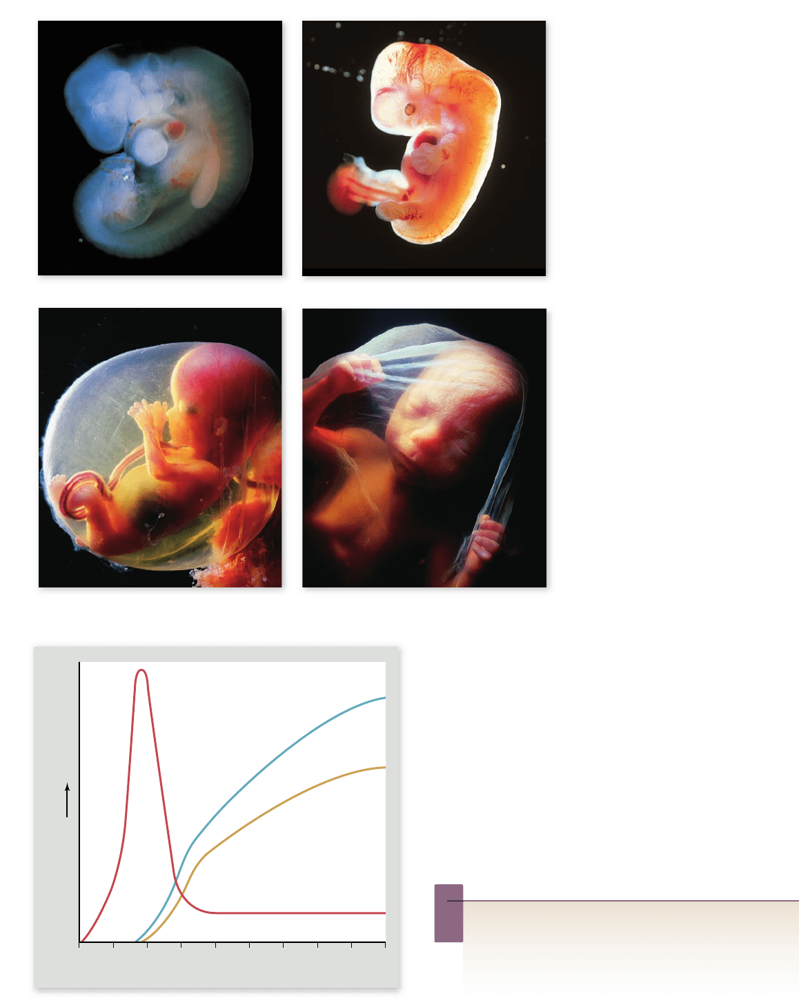
Organogenesis begins during the fourth week (figure 54.27a).
The eyes form. The tubular heart develops its four chambers and
starts to pulsate rhythmically, as it will for the rest of the individu-
al’s life. At 70 beats per minute, the heart is destined to beat more
than 2.5 billion times during a lifetime of 70 years. Over 30 pairs of
somites are visible by the end of the fourth week, and the arm and
leg buds have begun to form. The embryo has increased in length
to about 5 mm. Although the developmental scenario is now far
advanced, many women are still unaware they are pregnant at this
stage. Most spontaneous abortions (miscarriages), which frequently
occur in the case of a defective embryo, occur during this period.
The second month
Organogenesis continues during the second month (figure 54.27b).
The miniature limbs of the embryo assume their adult shapes. The
arms, legs, knees, elbows, fingers, and toes can all be seen—as well
as a short bony tail. The bones of the embryonic tail, an evolution-
ary reminder of our past, later fuse to form the coccyx.
Within the abdominal cavity, the major organs, including
the liver, pancreas, and gallbladder, become evident. By the end
of the second month, the embryo has grown to about 25 mm in
length, weighs about 1 g, and begins to look distinctly human.
The ninth week marks the transition from embryo to fetus. At
this time, all of the major organs of the body have been estab-
lished in their proper locations.
The third month
The nervous system develops during the third month, and the
arms and legs start to move (figure 54.27 c) . The embryo be-
gins to show facial expressions and carries out primitive reflexes
such as the startle reflex and sucking.
At around 10 weeks, the secretion of hCG by the placenta
declines, and the corpus luteum regresses as a result. However,
menstruation does not occur because the placenta itself secretes
estradiol and progesterone (figure 54.28) .
The high levels of estradiol and progesterone in the blood
during pregnancy continue to inhibit the release of FSH and
LH, thereby preventing ovulation. They also help maintain the
uterus and eventually prepare it for labor and delivery, and they
stimulate the development of the mammary glands in prepara-
tion for lactation after delivery.
During the second trimester, the
basic body plan develops further
Bones actively enlarge during the fourth month (figure 54.27d),
and by the end of the month, the mother can feel the baby kick-
ing. By the end of the fifth month, the rapid heartbeat of the
fetus can be heard with a stethoscope, although it can also be
detected as early as 10 weeks with a fetal monitor.
Growth begins in earnest in the sixth month; by the end
of that month, the fetus weighs 600 g (1.3 lb) and is over 300
mm (1 ft) long. Most of its prebirth growth is still to come,
however. The fetus cannot yet survive outside the uterus with-
out special medical intervention.
During the third trimester, organs mature
to the point at which the baby can survive
outside the womb
The third trimester is predominantly a period of growth and
maturation of organs. The weight of the fetus doubles several
Figure 54.26
Structure of the placenta. a. The placenta contains a fetal component, the chorionic frondosum, and a maternal
component, the decidua basalis. Deoxygenated fetal blood from the umbilical arteries (shown in blue) enters the placenta, where it picks up
oxygen and nutrients from the mother’s blood. Oxygenated fetal blood returns in the umbilical vein (shown in red) to the fetus. b. Note that
the 7-week embryo is surrounded by a uid- lled amniotic sac.
1126
part
VII
Animal Form and Function
rav32223_ch54_1105-1131.indd 1126rav32223_ch54_1105-1131.indd 1126 11/19/09 4:18:16 PM11/19/09 4:18:16 PM
Apago PDF Enhancer
a.
b.
c.
d.
Months of Pregnancy
Increasing Hormone Concentration
0 1 2 3 4 5 6 7 8 9
hCG Estrogen
Progesterone
Figure 54.27
The developing
human. (a) 4 weeks, (b) end of 5th
week, (c) 3 months, and (d) 4 months.
Figure 54.28
Hormonal secretion by the placenta.
The placenta secretes human chorionic gonadotropin (hCG), which
peaks in the second month and then declines. After 5 weeks, it
secretes increasing amounts of estrogen and progesterone.
Inquiry question
?
The high levels of estradiol and progesterone secreted by
the placenta prevent ovulation and thus formation of any
additional embryos during pregnancy. What would be the
expected effect of these high hormone levels in the absence
of pregnancy?
times, but this increase in bulk is not the only kind of growth
that occurs. Most of the major nerve tracts in the brain, as well
as many new neurons (nerve cells), are formed during this pe-
riod. Neurological growth is far from complete when birth
takes place, however. If the fetus remained in the uterus until
its neurological development was complete, it would grow too
large for safe delivery through the pelvis. Instead, the infant
chapter
54
Animal Development
112 7www.ravenbiology.com
rav32223_ch54_1105-1131.indd 1127rav32223_ch54_1105-1131.indd 1127 11/19/09 4:18:19 PM11/19/09 4:18:19 PM
Apago PDF Enhancer
Intestine
Placenta
Umbilical
cord
Wall of
uterus
Vagina
Cervix
is born as soon as the probability of its survival is high, and
its brain continues to develop and produce new neurons for
months after birth.
Critical changes in hormones bring on birth
In some mammals, changing hormone levels in the developing
fetus initiate the process of birth. The fetuses of these mam-
mals have an extra layer of cells in their adrenal cortex, which
secrete corticosteroids that induce the uterus of the mother to
manufacture prostaglandins. Prostaglandins trigger powerful
contractions of the uterine smooth muscles.
In humans, fetal secretion of cortisol increases during
late pregnancy, which appears to stimulate estradiol secretion
by the placenta. The mother’s uterus releases prostaglandins,
possibly as a result of the high levels of estradiol secreted by the
placenta. Estradiol also stimulates the uterus to produce more
oxytocin receptors, and as a result, the uterus becomes increas-
ingly sensitive to oxytocin.
Prostaglandins begin the uterine contractions, but then
sensory feedback from the uterus stimulates the release of oxy-
tocin from the mother’s posterior-pituitary gland. Working to-
gether, oxytocin and prostaglandins further stimulate uterine
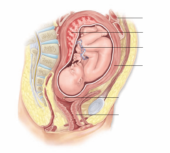
contractions, forcing the fetus downward (figure 54.29) . This
positive feedback mechanism accelerates during labor. Initially,
only a few contractions occur each hour, but the rate eventually
increases to one contraction every 2 to 3 min. Finally, strong
contractions, aided by the mother’s voluntary pushing, expel
the fetus, which is now a newborn baby, or neonate.
After birth, continuing uterine contractions expel the placen-
ta and associated membranes, collectively called the afterbirth. The
umbilical cord is still attached to the baby, and to free the newborn,
a doctor or midwife clamps and cuts the cord. Blood clotting and
contraction of muscles in the cord prevent excessive bleeding.
Nursing of young is a distinguishing
feature of mammals
Milk production, or lactation, occurs in the alveoli of mammary
glands when they are stimulated by the anterior-pituitary hor-
mone prolactin. Milk from the alveoli is secreted into a series
of alveolar ducts, which are surrounded by smooth muscle and
lead to the nipple.
During pregnancy, high levels of progesterone stimulate
the development of the mammary alveoli, and high levels of es-
tradiol stimulate the development of the alveolar ducts. How-
ever, estradiol blocks the actions of prolactin on the mammary
glands, and it inhibits prolactin secretion by promoting the re-
lease of prolactin-inhibiting hormone from the hypothalamus.
During pregnancy, therefore, the mammary glands are prepared
for, but prevented from, lactating. The growth of mammary
glands is also stimulated by the placental hormones human cho-
rionic somatomammotropin, a prolactin-like hormone, and hu-
man somatotropin, a growth hormone-like hormone.
When the placenta is discharged after birth, the concen-
trations of estradiol and progesterone in the mother’s blood
decline rapidly. This decline allows the anterior-pituitary gland
to secrete prolactin, which stimulates the mammary alveoli
to produce milk. Sensory impulses associated with the baby’s
suckling trigger the posterior-pituitary gland to release oxyto-
cin. Oxytocin stimulates contraction of the smooth muscle sur-
rounding the alveolar ducts, thus causing milk to be ejected by
the breast. This pathway is known as the milk let-down reflex,
and it is found in other mammals as well. The secretion of oxy-
tocin during lactation also causes some uterine contractions, as
it did during labor. These contractions help restore the tone of
uterine muscles in mothers who are breast-feeding.
The first milk produced after birth is a yellowish fluid
called colostrum, which is both nutritious and rich in maternal
antibodies. Milk synthesis begins about 3 days following the
birth and is referred to as the milk “coming in.” Many mothers
nurse for a year or longer. When nursing stops, the accumula-
tion of milk in the breasts signals the brain to stop secreting
prolactin, and milk production ceases.
Postnatal development in
humans continues for years
Growth of the infant continues rapidly after birth. Babies typi-
cally double their birth weight within 2 months. Because differ-
ent organs grow at different rates and cease growing at different
times, the body proportions of infants are different from those of
adults. The head, for example, is disproportionately large in new-
borns, but after birth it grows more slowly than the rest of the
body. Such a pattern of growth, in which different components
grow at different rates, is referred to as allometric growth.
In most mammals, brain growth is mainly a fetal phenom-
enon. In chimpanzees, for instance, the brain and the cerebral
Figure 54.29
Position of the fetus just before birth.
A developing fetus causes major changes in a woman’s anatomy.
The stomach and intestines are pushed far up, and considerable
discomfort often results from pressure on the lower back. In a
normal vaginal delivery, the fetus exits through the cervix, which
must dilate (expand) considerably to permit passage.
1128
part
VII
Animal Form and Function
rav32223_ch54_1105-1131.indd 1128rav32223_ch54_1105-1131.indd 1128 11/19/09 4:18:26 PM11/19/09 4:18:26 PM

Apago PDF Enhancer
portion of the skull grow very little after birth, whereas the bones
of the jaw continue to grow. As a result, the head of an adult chim-
panzee looks very different from that of a fetal or infant chimpan-
zee. In human infants, by contrast, the brain and cerebral skull
grow at the same rate as the jaw. Therefore, the jaw–skull propor-
tions do not change after birth, and the head of a human adult
looks very similar to that of a human fetus or infant.
The fact that the human brain continues to grow signifi-
cantly for the first few years of postnatal life means that ad-
equate nutrition and a safe environment are particularly crucial
during this period for the full development of a person’s intel-
lectual potential.
Learning Outcomes Review 54.6
The critical stages of human development occur in the fi rst trimester of
gestation; the subsequent 6 months involve growth and maturation. Growth
of the brain is not complete at birth and must be completed postnatally.
Hormones in the mother’s blood maintain the nutritive uterine environment
for the developing fetus; changes in hormone secretion and levels stimulate
birth (prostaglandins and oxytocin) and lactation (oxytocin and prolactin).
■ Why are teratogens (agents that cause birth defects)
most potent in the first trimester?
54.1 Fertilization
A sperm must penetrate to the plasma membrane of the egg for
membrane fusion to occur.
The sperm’s acrosome releases digestive enzymes to penetrate
the egg’s external layers (see gure 54.1). Fusion with the egg’s
membrane allows the sperm nucleus to pass into the egg’s cytoplasm.
Membrane fusion activates the egg.
Fusion of membranes triggers egg activation by the release of
calcium (see gure 54.2). Blocks to polyspermy include changes in
membrane potential and alterations to the external coat of the egg.
Upon egg activation, meiosis is completed (see gures 54.4 and 54.5).
The fusion of nuclei restores the diploid state.
Fertilization is complete when the haploid sperm nucleus fuses with
the haploid egg nucleus.
54.2 Cleavage and the Blastula Stage
The blastula is a hollow mass of cells.
Cleavage is a rapid series of cell divisions that produces blastomeres,
which form a hollow ball of cells called a blastula.
Cleavage patterns are highly diverse and distinctive.
Cleavage patterns are primarily in uenced by the amount of yolk (see
table 54.2). With little or no yolk, cleavage is holoblastic (involving
the whole egg); where more yolk is present, cleavage is meroblastic
(involving the blastodisc only). Cleavage in mammals is holoblastic.
Blastomeres may or may not be committed to developmental paths.
In many animals, unequal segregation of cytoplasmic determinants
commits each blastomere to a different path. Mammals exhibit
regulative development in which the fate of early blastomeres is
not predetermined.
54.3 Gastrulation
Gastrulation produces the three germ layers.
During gastrulation the three germ layers differentiate: endoderm,
ectoderm, and mesoderm (see table 54.3). Cells move during
gastrulation using a variety of cell shape changes.
Gastrulation patterns also vary according to the amount of yolk.
The amount of yolk also in uences cell movement. In frogs, a layer
of cells involutes through the dorsal lip of the blastopore. In birds,
surface cells migrate through the primitive streak. Mammalian
gastrulation is similar to that of birds.
Extraembryonic membranes are an adaptation to life on dry land.
The yolk sac, amnion, chorion, and allantois prevent dessication and
nourish and protect the developing embryo (see gure 54.15).
54.4 Organogenesis
Changes in gene expression lead to cell determination.
A cell’s location in the developing embryo often determines its fate.
Differentiation can be established by inheritance of cytoplasmic
determinants and by interactions with other cells (induction).
Development of selected systems in Drosophila illustrates
organogenesis.
The development of salivary glands, the dorsal vessel, and tracheae
all demonstrate the action of gene expression on development.
In vertebrates, organogenesis begins with neurulation and
somitogenesis (see gures 55.18–55.20).
Neurulation is the formation of the neural tube from ectoderm near
the notochord; somitogenesis is the establishment of mesoderm into
units called somites.
Migratory neural crest cells di erentiate into many cell types.
Neural crest cells migrate widely to become connective tissue, nerve
and glial cells, melanocytes, sensory neurons, and other cells.
Neural crest derivatives are important in vertebrate evolution.
Many of the unique adaptations of vertebrates have arisen from
neural crest cells (see gure 54.21).
54.5 Vertebrate Axis Formation
The Spemann organizer determines dorsal–ventral axis.
Organizers are a cluster of cells that produce gradients of diffusible
signal molecules, conveying positional information to other cells.
Chapter Review
chapter
54
Animal Development
112 9www.ravenbiology.com
rav32223_ch54_1105-1131.indd 1129rav32223_ch54_1105-1131.indd 1129 11/19/09 4:18:28 PM11/19/09 4:18:28 PM
Apago PDF Enhancer
Maternally encoded dorsal determinants activate Wnt signaling.
Turning on the Wnt pathway activates organizer speci cation.
Signaling molecules from the Spemann organizer inhibit
ventral development.
Morphogens can either activate or inhibit development along a
certain path. The Spemann organizer induces formation of the
dorsum by inhibiting ventral development (see gure 54.24 ).
Evidence indicates that organizers are present in all vertebrates.
Cells at the anterior edge of the primitive streak, termed Hensen’s
node, function similarly to the Spemann organizer.
Induction can be primary or secondary.
Primary induction occurs between the three germ layers; secondary
induction occurs between already determined tissues.
54.6 Human Development
During the rst trimester, the zygote undergoes rapid development
and di erentiation.
Implantation of the blastocyst occurs at the end of the rst week
of pregnancy. During the second week, the embryonic chorion and
the mother’s endometrial tissues form the placenta, and gastrulation
occurs. Organogenesis begins during the fourth week. The eighth
week marks the transition from embryo to fetus.
During the second trimester, the basic body plan develops further.
During the third trimester, organs mature to the point at which the
baby can survive outside the womb.
Critical changes in hormones bring on birth.
Birth is initiated by secretions of steroids from the fetal adrenal
cortex that induce prostaglandins, which cause contractions.
Nursing of young is a distinguishing feature of mammals.
Nursing involves a neuroendocrine re ex, causing the release of
oxytocin and the milk let-down response.
Postnatal development in humans continues for years.
Postnatal development continues with different organs growing at
different rates—called allometric growth .
UNDERSTAND
1. Which of the following events occur immediately after fertilization?
a. Egg activation
b. Polyspermy defense
c. Cytoplasm changes
d. All of these occur after fertilization
2. Which of the following plays the greatest role in determining
how cytoplasmic division occurs during cleavage?
a. Number of chromosomes
b. Amount of yolk
c. Orientation of the vegetal pole
d. Sex of the zygote
3. Gastrulation is a critical event during development. Why?
a. Gastrulation converts a hollow ball of cells into a bilaterally
symmetrical structure.
b. Gastrulation causes the formation of a primitive
digestive tract.
c. Gastrulation causes the blastula to develop a
dorsal–ventral axis.
d. All of these are signi cant events that occur
during gastrulation.
4. Gastrulation in a mammal would be most similar to
gastrulation in
a. a gecko.
b. a tuna.
c. an eagle.
d. no other species; mammalian gastrulation is unique.
5. Somites
a. begin forming at the tail end of the embryo and then move
forward in a wavelike fashion.
b. are derived from endoderm.
c. develop into only one type of tissue per somite.
d. may vary in number from one species to the next.
6. Of the following processes, which occurs last?
a. Cleavage c. Gastrulation
b. Neurulation d. Fertilization
APPLY
1. Your cousin just had twins. She tells you that twinning occurs
when two sperm fertilize the same egg. You reply that
a. yes, she is right, that is the most common source of
twinning.
b. no, only one sperm survives passage through the uterine
cervix, so two sperm are never present at fertilization.
c. no, cortical granules are used to prevent additional sperm
penetration.
d. no, twinning occurs when unfertilized eggs divide
spontaneously and thus is parthenogenic in nature.
2. In the Spemann experiment, when the dorsal lip is transplanted,
the recipient embryo then has a second source of molecules that
a. speci es ventral fate.
b. inhibits the molecules that specify ventral fate.
c. speci es dorsal fate.
d. inhibits the molecules that specify dorsal fate.
Review Questions
1130
part
VII
Animal Form and Function
rav32223_ch54_1105-1131.indd 1130rav32223_ch54_1105-1131.indd 1130 11/19/09 4:18:29 PM11/19/09 4:18:29 PM

Apago PDF Enhancer
3. Suppose that a burst of electromagnetic radiation were to strike
the blastomeres of only the animal pole of a frog embryo.
Which of the following would be most likely to occur?
a. A change or mutation relevant to the epidermis or skin
b. A switching of the internal organs so that reverse orientation
(left/right) occurs along the midline of the body
c. The migration of the nervous system to form outside
of the body
d. Failure of the reproductive system to develop
4. Which of the following would qualify as a secondary induction?
a. The formation of the lens of the eye due to induction by
the neural ectoderm
b. Differentiation during neurulation by the dorsal ectoderm
and mesoderm
c. Both of these
d. Neither of these
5. Your Aunt Ida thinks that babies can stimulate the onset of their
own labor. You tell her that
a. among mammals the onset of labor has been most closely
linked to a change in the phases of the Moon.
b. it is the mother’s circadian clock that determines the onset
of labor.
c. body weight determines the onset of labor.
d. changes in fetal hormone levels can affect the onset
of labor.
6. Drug or alcohol exposure during which of the following
stages is most likely to have a profound effect on the neural
development of the fetus?
a. Preimplantation c. Second trimester
b. First trimester d. Third trimester
7. Axis formation in amniotic embryos could be affected by
a. mutations in cells in the dorsal lip of the blastopore.
b. mutations in cells in the primitive streak.
c. both of these.
d. neither of these.
SYNTHESIZE
1. Suppose you discover a new species whose development
mechanisms have not been documented before. How could you
determine at what stage the cell fate is determined?
2. You look up from your studying to see your dog, Fi , acting
silly again. Using this as a teachable moment, compare and
contrast the homeoboxes in your dog and the fruit y she
just ate.
3. Why doesn’t a woman menstruate while she is pregnant?
4. Spemann and Mangold were able to demonstrate that some
cells act as “organizers” during development. What types of
cells did they use? How did they determine that these cells
were organizers?
ONLINE RESOURCE
www.ravenbiology.com
Understand, Apply, and Synthesize—enhance your study with
animations that bring concepts to life and practice tests to assess
your understanding. Your instructor may also recommend the
interactive eBook, individualized learning tools, and more.
chapter
54
Animal Development
1131www.ravenbiology.com
rav32223_ch54_1105-1131.indd 1131rav32223_ch54_1105-1131.indd 1131 11/19/09 4:18:29 PM11/19/09 4:18:29 PM