Raven P.H., Johnson G.B., Mason K.A. Biology (Ninth Edition)
Подождите немного. Документ загружается.

Apago PDF Enhancer
Photo-
system
I
Electron
Transport
System
Photo-
system
II
O
2
Heat
ATP
NADPH
ATP
NADH
ATP
ATP
Sunlight
Pyruvate
CO
2
Glucose
ADP+P
i
ADP+P
i
ADP+P
i
NAD
;
NADP
;
Calvin
Cycle
Krebs
Cycle
H
2
O
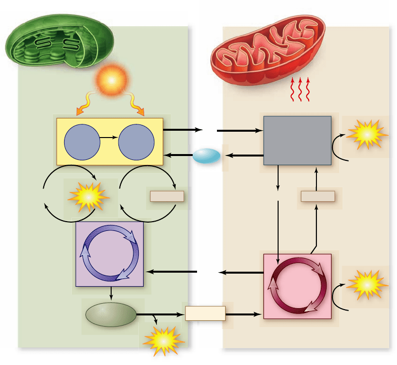
Figure 8.19
Chloroplasts and
mitochondria: completing an
energy cycle. Water and O
2
cycle
between chloroplasts and
mitochondria within a plant cell, as
do glucose and CO
2
. Cells with
chloroplasts require an outside
source of CO
2
and H
2
O and generate
glucose and O
2
. Cells without
chloroplasts, such as animal cells,
require an outside source of glucose
and O
2
and generate CO
2
and H
2
O.
around the cycle incorporate enough carbon to produce a new
molecule of G3P, and six turns incorporate enough carbon to
synthesize one glucose molecule.
We now know that light is required indirectly for different
segments of the CO
2
reduction reactions. Five of the Calvin
cycle enzymes—including rubisco—are light-activated; that is,
they become functional or operate more efficiently in the pres-
ence of light. Light also promotes transport of required
3-carbon intermediates across chloroplast membranes. And fi-
nally, light promotes the influx of Mg
2+
into the chloroplast
stroma, which further activates the enzyme rubisco.
Output of the Calvin cycle
Glyceraldehyde 3-phosphate is a 3-carbon sugar, a key intermedi-
ate in glycolysis. Much of it is transported out of the chloroplast to
the cytoplasm of the cell, where the reversal of several reactions in
glycolysis allows it to be converted to fructose 6-phosphate and
glucose 1-phosphate. These products can then be used to form
sucrose, a major transport sugar in plants. (Sucrose, table sugar, is
a disaccharide made of fructose and glucose.)
In times of intensive photosynthesis, G3P levels rise in the
stroma of the chloroplast. As a consequence, some G3P in the chlo-
roplast is converted to glucose 1-phosphate. This takes place in a set
of reactions analogous to those occurring in the cytoplasm, by re-
versing several reactions similar to those of glycolysis. The glucose
1-phosphate is then combined into an insoluble polymer, forming
long chains of starch stored as bulky starch grains in the cytoplasm.
These starch grains represent stored glucose for later use.
PGA (containing 12 × 3 = 36 carbon atoms in all, 6 from CO
2
and 30 from RuBP). The 36 carbon atoms then undergo a cycle
of reactions that regenerates the six molecules of RuBP used in
the initial step (containing 6 × 5 = 30 carbon atoms). This leaves
two molecules of glyceraldehyde 3-phosphate (G3P) (each with
three carbon atoms) as the net gain. (You may recall G3P as also
being the product of the first half of glycolysis, described in
chapter 7. ) These two molecules of G3P can then be used to
make one molecule of glucose.
The net equation of the Calvin cycle is:
6 CO
2
+ 18 ATP + 12 NADPH + water
→
2 glyceraldehyde 3-phosphate + 16 P
i
+ 18 ADP + 12 NADP
+
With six full turns of the cycle, six molecules of carbon dioxide
enter, two molecules of G3P are produced, and six molecules of
RuBP are regenerated. Thus six turns of the cycle produce two
G3P that can be used to make a single glucose molecule. The
six turns of the cycle also incorporated six CO
2
molecules, pro-
viding enough carbon to synthesize glucose, although the six
carbon atoms do not all end up in this molecule of glucose.
Phases of the cycle
The Calvin cycle can be thought of as divided into three
phases: (1) carbon fixation, (2) reduction, and (3) regeneration
of RuBP. The carbon fixation reaction generates two mole-
cules of the 3- carbon acid PGA; PGA is then reduced to G3P
by reactions that are essentially a reverse of part of glycolysis;
finally, the PGA is used to regenerate RuBP. Three turns
162
part
II
Biology of the Cell
rav32223_ch08_147-167.indd 162rav32223_ch08_147-167.indd 162 11/6/09 2:07:36 PM11/6/09 2:07:36 PM
Apago PDF Enhancer
Heat
H
2
O
H
2
O
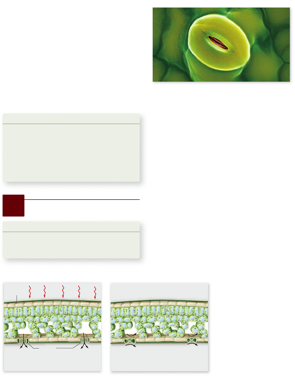
Leaf
epidermis
Stomata
Under hot, arid conditions, leaves lose water by
evaporation through openings in the leaves
called stomata.
The stomata close to conserve water but as a
result, O
2
builds up inside the leaves, and CO
2
cannot enter the leaves.
O
2
O
2
CO
2
CO
2
Figure 8.20
Stoma. A closed stoma in the leaf of a tobacco
plant. Each stoma is formed from two guard cells whose shape
changes with turgor pressure to open and close. Under dry
conditions plants close their stomata to conserve water.
Figure 8.21
Conditions
favoring photorespiration. In
hot, arid environments, stomata
close to conserve water, which also
prevents CO
2
from entering and O
2
from exiting the leaf. The high-O
2
/
low-CO
2
conditions favor
photorespiration.
tures that already exist. Photosynthesis is no exception. Rubisco,
the enzyme that catalyzes the key carbon-fixing reaction of
photo synthesis, provides a decidedly suboptimal solution. This
enzyme has a second enzymatic activity that interferes with car-
bon fixation, namely that of oxidizing RuBP. In this process, called
photorespiration, O
2
is incorporated into RuBP, which under-
goes additional reactions that actually release CO
2
. Hence, photo-
respiration releases CO
2
, essentially undoing carbon fixation.
Photorespiration reduces
the yield of photosynthesis
The carboxylation and oxidation of RuBP are catalyzed at the
same active site on rubisco, and CO
2
and O
2
compete with each
other at this site. Under normal conditions at 25°C, the rate of
the carboxylation reaction is four times that of the oxidation
reaction, meaning that 20% of photosynthetically fixed carbon
is lost to photorespiration.
This loss rises substantially as temperature increases, be-
cause under hot, arid conditions, specialized openings in the
leaf called stomata (singular, stoma) (figure 8.20) close to con-
serve water. This closing also cuts off the supply of CO
2
enter-
ing the leaf and does not allow O
2
to exit (figure 8.21). As a
result, the low-CO
2
and high-O
2
conditions within the leaf
favor photorespiration.
The energy cycle
The energy-capturing metabolisms of the chloroplasts studied
in this chapter and the mitochondria studied in chapter 7 are
intimately related (figure 8.19). Photosynthesis uses the prod-
ucts of respiration as starting substrates, and respiration uses
the products of photosynthesis as starting substrates. The pro-
duction of glucose from G3P even uses part of the ancient gly-
colytic pathway, run in reverse. Also, the principal proteins
involved in electron transport and ATP production in plants
are evolutionarily related to those in mitochondria.
Photosynthesis is but one aspect of plant biology, although
it is an important one. In chapters 36 through 42 , we examine
plants in more detail. We have discussed photosynthesis as a part
of cell biology because photosynthesis arose long before plants
did, and because most organisms depend directly or indirectly on
photosynthesis for the energy that powers their lives.
Learning Outcomes Review 8.6
Carbon fi xation takes place in the stroma of the chloroplast, where inorganic
CO
2
is incorporated into an organic molecule. The key intermediate is the
5-carbon sugar RuBP that combines with CO
2
in a reaction catalyzed by the
enzyme rubisco. The cycle can be broken down into three stages: carbon
fi xation, reduction, and regeneration of RuBP. ATP and NADPH from the light
reactions provide energy and electrons for the reduction reactions, which
produce G3P. Glucose is synthesized when two molecules of G3P are combined.
■ How does the Calvin cycle compare with glycolysis?
8.7
Photorespiration
Learning Outcomes
Explain the action of rubisco in oxidizing RuBP.1.
Compare the function of carbon fixation in the C2.
3
, C
4
, and
CAM pathways.
Evolution does not necessarily result in optimum solutions.
Rather, it favors workable solutions that can be derived from fea-
chapter
8
Photosynthesis
163www.ravenbiology.com
rav32223_ch08_147-167.indd 163rav32223_ch08_147-167.indd 163 11/6/09 2:07:37 PM11/6/09 2:07:37 PM
Apago PDF Enhancer
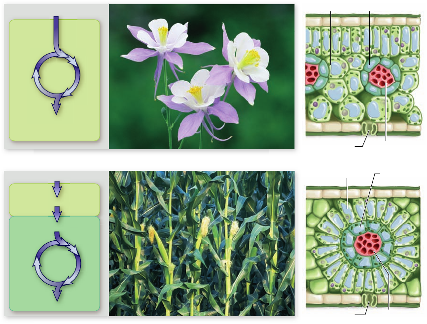
Calvin
Cycle
Mesophyll
cell
CO
2
RuBP
3PG
(C
3
)
a. C
3
pathway
Bundle-sheath cell Mesophyll cell
Stoma Vein
G3P
b. C
4
pathway
Bundle-
sheath cell
Stoma Vein
Mesophyll cell
Mesophyll
cell
Bundle-
sheath
cell
Calvin
Cycle
G3P
CO
2
CO
2
C
4
Figure 8.22
Comparison of C
3
and
C
4
pathways of carbon
xation. a. The C
3
pathway uses the Calvin
cycle to x carbon. All
reactions occur in
mesophyll cells using CO
2
that diffuses in through
stomata. b. The C
4
pathway incorporates CO
2
into a 4-carbon molecule
of malate in mesophyll
cells. This is transported
to the bundle sheath cells
where it is converted back
into CO
2
and pyruvate,
creating a high level of
CO
2
. This allows ef cient
carbon xation by the
Calvin cycle.
Plants that fix carbon using only C
3
photosynthesis
(the Calvin cycle) are called C
3
plants (figure 8.22a). Other
plants add CO
2
to phosphoenolpyruvate (PEP) to form a
4- carbon molecule. This reaction is catalyzed by the enzyme
PEP carboxylase. This enzyme has two advantages over rubis-
co: it has a much greater affinity for CO
2
than rubisco, and
it does not have oxidase activity.
The 4-carbon compound produced by PEP carboxy-
lase undergoes further modification, only to be eventually
decarboxylated. The CO
2
released by this decarboxylation is
then used by rubisco in the Calvin cycle. This allows CO
2
to
be pumped directly to the site of rubisco, which increases
the local concentration of CO
2
relative to O
2
, minimizing
photorespiration. The 4-carbon compound produced by
PEP carboxylase allows CO
2
to be stored in an organic form,
to then be released in a different cell, or at a different time
to keep the level of CO
2
high relative to O
2
.
The reduction in the yield of carbohydrate as a result
of photorespiration is not trivial. C
3
plants lose between
25% and 50% of their photosynthetically fixed carbon in
this way. The rate depends largely on temperature. In tropi-
cal climates, especially those in which the temperature is of-
ten above 28°C, the problem is severe, and it has a major
effect on tropical agriculture.
The two main groups of plants that initially capture
CO
2
using PEP carboxylase differ in how they maintain high
levels of CO
2
relative to O
2
. In C
4
plants (figure 8.22b), the
capture of CO
2
occurs in one cell and the decarboxylation
occurs in an adjacent cell. This represents a spatial solution
to the problem of photorespiration. The second group,
CAM plants, perform both reactions in the same cell, but
capture CO
2
using PEP carboxylase at night, then decarboxy-
late during the day. CAM stands for crassulacean acid
metabolism, after the plant family Crassulaceae (the
stonecrops, or hens-and-chicks), in which it was first discov-
ered. This mechanism represents a temporal solution to the
photorespiration problem.
C
4
plants have evolved
to minimize photorespiration
The C
4
plants include corn, sugarcane, sorghum, and a
number of other grasses. These plants initially fix carbon
using PEP carboxylase in mesophyll cells. This reaction
produces the organic acid oxaloacetate, which is converted
to malate and transported to bundle-sheath cells that
surround the leaf veins. Within the bundle-sheath cells,
malate is decarboxylated to produce pyruvate and CO
2
(figure 8.23). Because the bundle-sheath cells are imper-
meable to CO
2
, the local level of CO
2
is high and carbon
fixation by rubisco and the Calvin cycle is efficient. The
164
part
II
Biology of the Cell
rav32223_ch08_147-167.indd 164rav32223_ch08_147-167.indd 164 11/6/09 2:07:40 PM11/6/09 2:07:40 PM
Apago PDF Enhancer
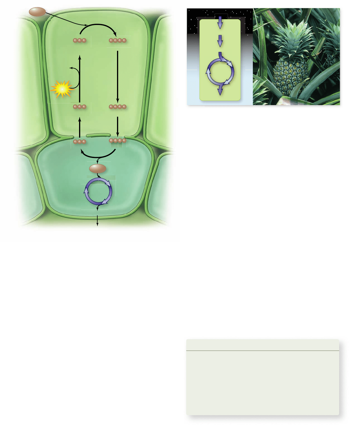
Phosphoenolpyruvate
(PEP)
Oxaloacetate
Mesophyll
cell
CO
2
Pyruvate Malate
Bundle-sheath
cell
Glucose
Malate Pyruvate
Calvin
Cycle
CO
2
ATP
AMP+
PP
i
+P
i
night
day
Calvin
Cyc
le
CO
2
CO
2
C
4
G3P
The Crassulacean acid pathway splits
photosynthesis into night and day
A second strategy to decrease photorespiration in hot regions
has been adopted by the CAM plants. These include many suc-
culent (water-storing) plants, such as cacti, pineapples, and
some members of about two dozen other plant groups.
In these plants, the stomata open during the night and
close during the day (figure 8.24). This pattern of stomatal open-
ing and closing is the reverse of that in most plants. CAM plants
initially fix CO
2
using PEP carboxylase to produce oxaloacetate.
The oxaloacetate is often converted into other organic acids, de-
pending on the particular CAM plant. These organic compounds
accumulate during the night and are stored in the vacuole. Then
during the day, when the stomata are closed, the organic acids are
decarboxylated to yield high levels of CO
2
. These high levels of
CO
2
drive the Calvin cycle and minimize photorespiration.
Like C
4
plants, CAM plants use both C
3
and C
4
pathways.
They differ in that they use both of these pathways in the same
cell: the C
4
pathway at night and the C
3
pathway during the
day. In C
4
plants the two pathways occur in different cells.
Learning Outcomes Review 8.7
Rubisco can also oxidize RuBP under conditions of high O
2
and low CO
2
. In
plants that use only C
3
metabolism (Calvin cycle), up to 20% of fi xed carbon
is lost to this photorespiration. Plants adapted to hot, dry environments
are capable of storing CO
2
as a 4-carbon molecule and avoiding some of this
loss; they are called C
4
plants. In CAM plants, CO
2
is fi xed at night into a C
4
organic compound; in the daytime, this compound is used as a source of CO
2
C
3
metabolism when stomata are closed to prevent water loss.
■ How do C
4
plants and CAM plants differ?
pyruvate produced by decarboxylation is transported back
to the mesophyll cells, where it is converted back to PEP,
thereby completing the cycle.
The C
4
pathway, although it overcomes the problems of
photorespiration, does have a cost. The conversion of pyru-
vate back to PEP requires breaking two high-energy bonds in
ATP. Thus each CO
2
transported into the bundle-sheath cells
cost the equivalent of two ATP. To produce a single glucose,
this requires 12 additional ATP compared with the Calvin
cycle alone. Despite this additional cost, C
4
photosynthesis is
advantageous in hot dry climates where photorespiration
would remove more than half of the carbon fixed by the usual
C
3
pathway alone.
Figure 8.23
Carbon xation in C
4
plants. This process is
called the C
4
pathway because the rst molecule formed,
oxaloacetate, contains four carbons. The oxaloacetate is converted
to malate, which moves into bundle-sheath cells where it is
decarboxylated back to CO
2
and pyruvate. This produces a high
level of CO
2
in the bundle-sheath cells that can be xed by the usual
C
3
Calvin cycle with little photorespiration. The pyruvate diffuses
back into the mesophyll cells, where it is converted back to PEP to
be used in another C
4
xation reaction.
Figure 8.24
Carbon xation in CAM plants. CAM plants
also use both C
4
and C
3
pathways to x carbon and minimize
photorespiration. In CAM plants, the two pathways occur in the
same cell but are separated in time: The C
4
pathway is utilized to x
carbon at night, then CO
2
is released from these accumulated stores
during the day to drive the C
3
pathway. This achieves the same
effect of minimizing photorespiration while also minimizing loss of
water by opening stomata at night when temperatures are lower.
chapter
8
Photosynthesis
165www.ravenbiology.com
rav32223_ch08_147-167.indd 165rav32223_ch08_147-167.indd 165 11/6/09 2:07:42 PM11/6/09 2:07:42 PM

Apago PDF Enhancer
energy to the reaction center. The reaction center is composed of
two chlorophyll a molecules in a protein matrix that pass an excited
electron to an electron acceptor.
8.5 The Light-Dependent Reactions
The light reactions can be broken down into four processes: primary
photoevent, charge separation, electron transport, and chemiosmosis.
Some bacteria use a single photosystem ( gure 8.12 ) .
An excited electron moves along a transport chain and eventually
returns to the photosystem. This cyclic process is used to generate a
proton gradient. In some bacteria, this can also produce NADPH.
Chloroplasts have two connected photosystems.
Photosystem I transfers electrons to NADP
+
, reducing it to NADPH.
Photosystem II replaces electrons lost by photosystem I. Electrons
lost from photosystem II are replaced by electrons from oxidation of
water, which also produces O
2
.
The two photosystems work together in noncyclic
photophosphorylation.
Photosystem II and photosystem I are linked by an electron transport
chain; the b
6
-f complex in this chain pumps protons into the
thylakoid space.
ATP is generated by chemiosmosis .
ATP synthase is a channel enzyme; as protons ow through the
channel down their gradient, ADP is phosphorylated producing
ATP, similar to the mechanism in mitochondria. Plants can make
additional ATP by cyclic photophosphorylation.
Thylakoid structure reveals components’ locations.
Imaging studies suggest that photosystem II is primarily found in the
grana, while photosystem I and ATP synthase are found in the
stroma lamella.
8.6 Carbon Fixation: The Calvin Cycle (see gure 8.18 )
Calvin cycle reactions convert inorganic carbon into organic molecules.
The Calvin cycle,
also known as C
3
photosynthesis, uses CO
2
, ATP,
and NADPH to build simple sugars.
Carbon is transferred through cycle intermediates, eventually
producing glucose.
The Calvin cycle occurs in three stages: carbon xation via the enzyme
rubisco’s action on RuBP and CO
2
; reduction of the resulting 3-carbon
PGA to G3P, generating ATP and NADPH; and regeneration of RuBP.
Six turns of the cycle x enough carbon to produce two excess G3Ps
used to make one molecule of glucose.
8.7 Photorespiration
Photorespiration reduces the yield of photosynthesis.
Rubisco can catalyze the oxidation of RuBP, reversing carbon
xation. Dry, hot conditions tend to increase this reaction.
C
4
plants have evolved to minimize photorespiration.
C
4
plants x carbon by adding CO
2
to a 3-carbon molecule, forming
oxaloacetate. Carbon is xed in one cell by the C
4
pathway, then CO
2
is released in another cell for the Calvin cycle (see gure 8.23 ).
The Crassulacean acid pathway splits photosynthesis
into night and day.
CAM plants use the C
4
pathway during the day when stomata are
closed, and the Calvin cycle at night in the same cell.
8.1 Overview of Photosynthesis
Photosynthesis is the conversion of light energy into chemical energy
(see gure 8.2 ).
Photosynthesis combines CO
2
and H
2
O, producing glucose and O
2
.
Photosynthesis has three stages: absorbing light energy, using this energy
to synthesize ATP and NADPH, and using the ATP and NADPH to
convert CO
2
to organic molecules. The rst two stages consist of light-
dependent reactions, and the third stage of light-independent reactions.
In plants, photosynthesis takes place in chloroplasts.
Chloroplasts contain internal thylakoid membranes and a uid
matrix called stroma. The photosystems involved in energy capture
are found in the thylakoid membranes, and enzymes for assembling
organic molecules are in the stroma.
8.2 The Discovery of Photosynthetic Processes
Plants do not increase mass from soil and water alone.
Early investigations revealed that plants produce O
2
from carbon
dioxide and water in the presence of light.
Photosynthesis includes both light-dependent
and light-independent reactions.
The light-dependent reactions require light; the light-
independent reactions occur in both daylight and darkness. The
rate of photosynthesis depends on the amount of light, the CO
2
concentration, and temperature.
O
2
comes from water, not from CO
2
.
The use of isotopes revealed the individual origins and fates of
different molecules in photosynthetic reactions.
ATP and NADPH from light-dependent reactions reduce
CO
2
to make sugars.
Carbon xation requires ATP and NADPH, which are products of
the light-dependent reactions. As long as these are available, CO
2
is
reduced by enzymes in the stroma to form simple sugars.
8.3 Pigments
Light is a form of energy.
Light exists both as a wave and as a particle (photon). Light can
remove electrons from some metals by the photoelectric effect, and
in photosynthesis, chloroplasts act as photoelectric devices.
Each pigment has a characteristic absorption spectrum.
Chlorophyll a is the only pigment that can convert light energy into
chemical energy. Chlorophyll b is an accessory pigment that increases
the harvest of photons for photosynthesis.
Carotenoids and other accessory pigments further increase a plant’s
ability to harvest photons.
8.4 Photosystem Organization (see gure 8.10 )
Production of one O
2
molecule requires many chlorophyll molecules.
Measurement of O
2
output led to the idea of photosystems— clusters
of pigment molecules that channel energy to a reaction center.
A generalized photosystem contains an antenna complex
and a reaction center.
A photosystem is a network of chlorophyll a, accessory pigments,
and proteins embedded in the thylakoid membrane. Pigment
molecules of the antenna complex harvest photons and feed light
Chapter Review
166
part
II
Biology of the Cell
rav32223_ch08_147-167.indd 166rav32223_ch08_147-167.indd 166 11/6/09 2:07:44 PM11/6/09 2:07:44 PM

Apago PDF Enhancer
c. In the intermembrane space
d. In the antenna complex
3. How does the reaction center of photosystem I regain
an electron during noncyclic photosynthesis?
a. The electron is recycled directly back to the reaction
center pigment.
b. The electron is donated from H
2
O.
c. The electron is donated from photosystem II.
d. The electron is donated from NADPH.
4. If the Calvin cycle runs through six turns
a. all of the xed carbon will end up in the same
glucose molecule.
b. 12 carbons will be xed by the process.
c. enough carbon will be xed to make one glucose, but they
will not all be in the same molecule.
d. one glucose will be converted into six CO
2
.
5. Which of the following are similarities between the structure
and function of mitochondria and chloroplasts?
a. They both create internal proton gradients by
electron transport.
b. They both generate CO
2
by oxidation reactions.
c. They both have an outer membrane and an inner
membrane system.
d. Both a and c are correct.
6. Carbon xation by the C
4
pathway produces
a. the same product as is produced by the Calvin cycle.
b. an organic acid, but a 4-carbon one not a 3-carbon.
c. a 3-carbon organic acid that is converted to the
4-carbon malate.
d. RuBP.
7. If the thylakoid membrane became leaky to ions, what would
you predict to be the result on the light reactions?
a. It would stop ATP production.
b. It would stop NADPH production.
c. It would stop the oxidation of H
2
O.
d. All of the above are correct.
8. The overall process of photosynthesis
a. results in the reduction of CO
2
and the oxidation of H
2
O.
b. results in the reduction of H
2
O and the oxidation of CO
2
.
c. consumes O
2
and produces CO
2
.
d. produces O
2
from CO
2
.
SYNTHESIZE
1. Compare and contrast the xation of carbon in C
3
, C
4
, and
CAM plants.
2. Diagram the relationship between the reactants and products of
photosynthesis and respiration.
3. Do plant cells need mitochondria? Explain your answer.
UNDERSTAND
1. The light-dependent reactions of photosynthesis are responsible
for the production of
a. glucose. c. ATP and NADPH.
b. CO
2
.
d. H
2
O.
2. Which region of a chloroplast is associated with the capture
of light energy?
a. Thylakoid membrane c. Stroma
b. Outer membrane d. Both a and c
3. The colors of light that are most effective for photosynthesis are
a. red, blue, and violet.
b. green, yellow, and orange.
c. infrared and ultraviolet.
d. All colors of light are equally effective.
4. During noncyclic photosynthesis, photosystem I functions to
___________, and photosystem II functions to ______________.
a. synthesize ATP; produce O
2
b. reduce NADP
+
; oxidize H
2
O
c. reduce CO
2
; oxidize NADPH
d. restore an electron to its reaction center; gain an electron
from water
5. How is a reaction center pigment in a photosystem different
from a pigment in the antenna complex?
a. The reaction center pigment is a chlorophyll molecule.
b. The antenna complex pigment can only re ect light.
c. The reaction center pigment loses an electron when
it absorbs light energy.
d. The antenna complex pigments are not attached to proteins.
6. The ATP and NADPH from the light reactions are used
a. in glycolysis in roots.
b. directly in most biochemical reactions in the cell.
c. during the reactions of the Calvin cycle to produce glucose.
d. to synthesize chlorophyll.
7. The carbon xation reaction converts
a. inorganic carbon into an organic acid.
b. CO
2
into glucose.
c. inactive rubisco into active rubisco.
d. an organic acid into CO
2
.
8. C
4
plants initially x carbon by
a. the same pathway as C
3
plants, but they modify this
product.
b. incorporating CO
2
into oxaloacetate, which is converted
to malate.
c. incorporating CO
2
into citrate via the Krebs cycle.
d. incorporating CO
2
into glucose via reverse glycolysis.
APPLY
1. The overall ow of electrons in the light reactions is from
a. antenna pigments to the reaction center.
b. H
2
O to CO
2
.
c. photosystem I to photosystem II.
d. H
2
O to NADPH.
2. Where in a chloroplast would you nd the highest
concentration of protons?
a. In the stroma
b. In the lumen of the thylakoid
Review Questions
ONLINE RESOURCE
www.ravenbiology.com
Understand, Apply, and Synthesize—enhance your study with
animations that bring concepts to life and practice tests to assess
your understanding. Your instructor may also recommend the
interactive eBook, individualized learning tools, and more.
chapter
8
Photosynthesis
167www.ravenbiology.com
rav32223_ch08_147-167.indd 167rav32223_ch08_147-167.indd 167 11/6/09 2:07:45 PM11/6/09 2:07:45 PM
Apago PDF Enhancer
S
Chapter
9
Cell Communication
Chapter Outline
9.1 Overview of Cell Communication
9.2 Receptor Types
9.3 Intracellular Receptors
9.4 Signal Transduction Through Receptor Kinases
9.5 Signal Transduction Through G Protein-
Coupled Receptors
Introduction
Springtime is a time of rebirth
and renewal. Trees that have appeared dead produce new leaves and buds, and owers sprout
from the ground. For su erers of seasonal allergy, this is not quite such a pleasant time. The pollen in the micrograph and other
allergens produced stimulate the immune system to produce the molecule histamine and other molecules that form cellular
signals. These signals cause in ammation, mucus secretion, vasodilation, and other responses that together cause the runny nose,
itching watery eyes, and other symptoms that make up the allergic reaction. We treat allergy symptoms by using drugs called
antihistamines that interfere with this cellular signaling. The popular drug loratadine (better known as Claritin), for example, acts
by blocking the receptor for histamine, thus preventing its action.
We will begin this chapter with a general overview of signaling, and the kinds of receptors cells use to respond to
signals. Then we will look in more detail at how these different types of receptors can elicit a response from cells, and finally,
how cells make connections with one another.
9.1
Overview of Cell
Communication
Learning Outcomes
Explain the different ways that cells communicate.1.
Describe how cells use signal transduction pathways. 2.
Communication between cells is common in nature. Cell sig-
naling occurs in all multicellular organisms, providing an indis-
pensable mechanism for cells to influence one another. Effective
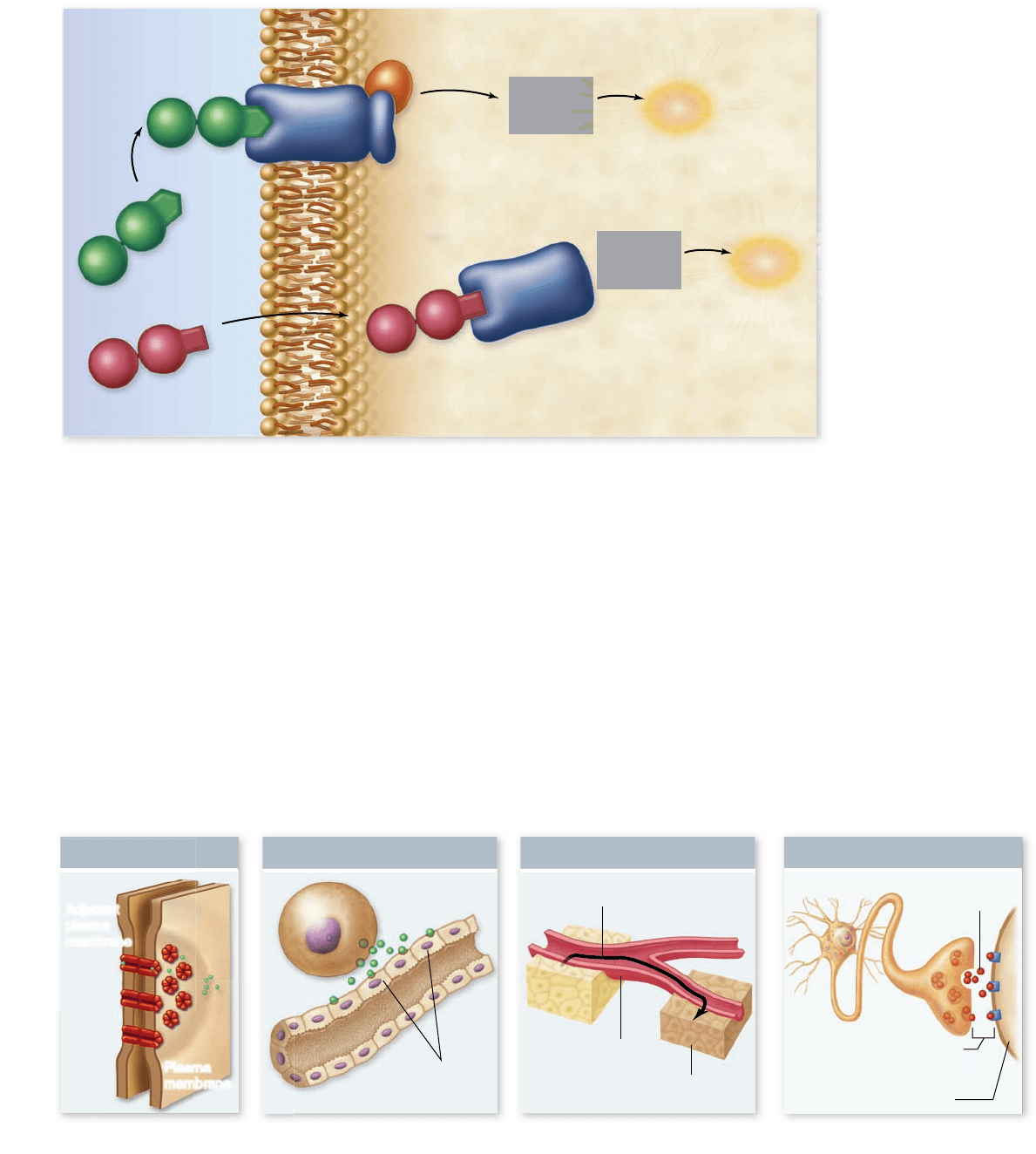
signaling requires a signaling molecule, called a ligand, and a
molecule to which the signal binds, called a receptor protein.
The interaction of these two components initiates the process of
signal transduction, which converts the information in the signal
into a cellular response (figure 9.1).
The cells of multicellular organisms use a variety of mol-
ecules as signals, including but not limited to, peptides, large
CHAPTER
rav32223_ch09_168-185.indd 168rav32223_ch09_168-185.indd 168 11/6/09 2:32:45 PM11/6/09 2:32:45 PM
Apago PDF Enhancer
Cellular
response
Cellular
response
Cytoplasm External environment
Membrane receptor
Intracellular receptor
Plasma membrane
Hydrophilic
ligand
Hydrophobic
ligand
Signal
transduction
pathway
Signal
transduction
pathway
a.
b.
c. d.
Synaptic Signaling Endocrine Signaling Paracrine Signaling
Nerve cell
Neurotransmitter
Synaptic gap
Target cell
Hormone secretion into
blood by endocrine gland
Blood vessel
Distant target cells
Adjacent
target cells
Secretory cell
Direct Contact
Plasma
membrane
Adjacent
plasma
membrane
Figure 9.1
Overview
of cell signaling. Cell
signaling involves a signal
molecule called a ligand,
a receptor, and a signal
transduction pathway
that produces a cellular
response. The location
of the receptor can either
be intracellular, for
hydrophobic ligands that
can cross the membrane, or
in the plasma membrane,
for hydrophilic ligands that
cannot cross
the membrane.
proteins, individual amino acids, nucleotides, and steroids and
other lipids. Even dissolved gases such as NO (nitric oxide) are
used as signals.
Any cell of a multicellular organism is exposed to a con-
stant stream of signals. At any time, hundreds of different
chemical signals may be present in the environment surround-
ing the cell. Each cell responds only to certain signals, however,
and ignores the rest, like a person following the conversation of
one or two individuals in a noisy, crowded room.
How does a cell “choose” which signals to respond
to? The number and kind of receptor molecules determine
this. When a ligand approaches a receptor protein that has
a complementary shape, the two can bind, forming a com-
plex. This binding induces a change in the receptor pro-
tein’s shape, ultimately producing a response in the cell via
a signal transduction pathway. In this way, a given cell re-
sponds to the signaling molecules that fit the particular set
of receptor proteins it possesses and ignores those for which
it lacks receptors.
Signaling is de ned by the distance
from source to receptor
Cells can communicate through any of four basic mechanisms,
depending primarily on the distance between the signaling and
responding cells (figure 9.2). These mechanisms are (1) direct
contact, (2) paracrine signaling, (3) endocrine signaling, and
(4) synaptic signaling.
In addition to using these four basic mechanisms, some
cells actually send signals to themselves, secreting signals that
bind to specific receptors on their own plasma membranes.
This process, called autocrine signaling, is thought to play an
Figure 9.2
Four kinds of cell signaling. Cells communicate in several ways. a. Two cells in direct contact with each other may
send signals across gap junctions. b. In paracrine signaling, secretions from one cell have an effect only on cells in the immediate area. c. In
endocrine signaling, hormones are released into the organism’s circulatory system, which carries them to the target cells. d. Chemical synapse
signaling involves transmission of signal molecules, called neurotransmitters, from a neuron over a small synaptic gap to the target cell.
www.ravenbiology.com
chapter
9
Cell Communication
169
rav32223_ch09_168-185.indd 169rav32223_ch09_168-185.indd 169 11/6/09 2:32:49 PM11/6/09 2:32:49 PM
Apago PDF Enhancer
transduction. These events form discrete pathways that lead
to a cellular response to the signal received by receptors.
Knowledge of these signal transduction pathways has exploded
in recent years and indicates a high degree of complexity that
explains how in some cases different cell types can have the
same response to different signals, and in other cases different
cell types can have a different response to the same signal.
For example, a variety of cell types respond to the hor-
mone glucagon by mobilizing glucose as part of the body’s
mechanism to control blood glucose (chapter 46) . This involves
breaking down stored glycogen into glucose and turning on the
genes that encode the enzymes necessary to synthesize glucose.
In contrast, the hormone epinephrine has diverse effects on dif-
ferent cell types. We have all been startled or frightened by a
sudden event. Your heart beats faster, you feel more alert, and
you can even feel the hairs on your skin stand up. All of this is
due in part to your body releasing the hormone epinephrine
(also called adrenaline) into the bloodstream. This leads to the
heightened state of alertness and increased heart rate and en-
ergy that prepare us to respond to extreme situations.
These differing effects of epinephrine depend on the dif-
ferent cell types with receptors for this hormone. In the liver,
cells are stimulated to mobilize glucose while in the heart mus-
cle cells contract more forcefully to increase blood flow. In ad-
dition, blood vessels respond by expanding in some areas and
contracting in others to redirect blood flow to the liver, heart,
and skeletal muscles. These different reactions depend on the
fact that each cell type has a receptor for epinephrine, but dif-
ferent sets of proteins that respond to this signal.
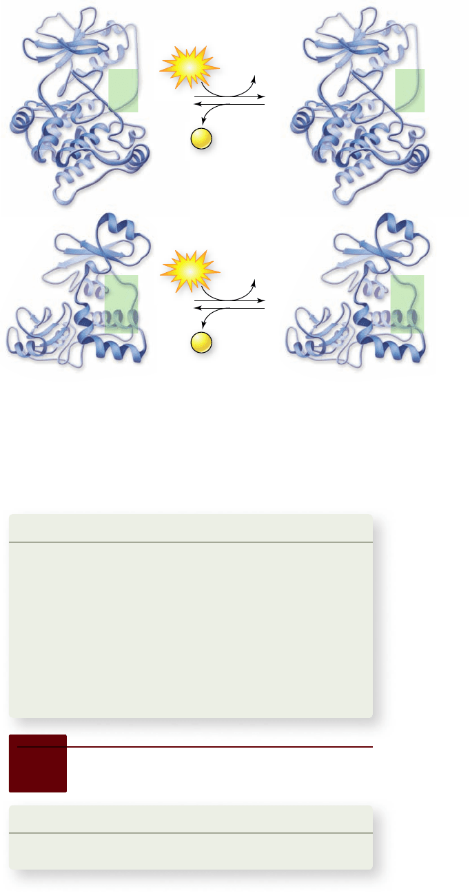
Phosphorylation is key in control
of protein function
The function of a signal transduction pathway is to change the
behavior or nature of a cell. This action may require changing the
composition of proteins that make up a cell or altering the activ-
ity of cellular proteins. Many proteins are inactive or nonfunc-
tional as they are initially synthesized and require modification
after synthesis for activation. In other cases, a protein may re-
quire modification for deactivation. A major source of control for
protein function is the addition or removal of phosphate groups,
called phosphorylation or dephosphorylation, respectively.
As you learned in preceding chapters, the end result of the
metabolic pathways of cellular respiration and photosynthesis
was the phosphorylation of ADP to ATP. The ATP synthesized
by these processes can donate phosphate groups to proteins.
The phosphorylation of proteins alters their function, which
allows them to transmit information from an extracellular sig-
nal through a signal transduction pathway.
Protein kinases
The class of enzyme that adds phosphate groups from ATP to
proteins is called a protein kinase. These phosphate groups can
be added to the three amino acids that have an OH as part
of their R group, namely serine, threonine, and tyrosine. We
categorize protein kinases based on which of these three sub-
strates they alter (figure 9.3). Most cytoplasmic protein kinases
fall into the serine/threonine kinase class.
important role in reinforcing developmental changes, and it is
an important component of signaling in the immune system
(chapter 52) .
Direct contact
As you saw in chapter 5, the surface of a eukaryotic cell is richly
populated with proteins, carbohydrates, and lipids attached to
and extending outward from the plasma membrane. When cells
are very close to one another, some of the molecules on the
plasma membrane of one cell can be recognized by receptors
on the plasma membrane of an adjacent cell. Many of the im-
portant interactions between cells in early development occur
by means of direct contact between cell surfaces. Cells also sig-
nal through gap junctions (figure 9.2a). We’ll examine contact-
dependent interactions more closely later in this chapter.
Paracrine signaling
Signal molecules released by cells can diffuse through the extra-
cellular fluid to other cells. If those molecules are taken up
by neighboring cells, destroyed by extracellular enzymes, or
quickly removed from the extracellular fluid in some other way,
their influence is restricted to cells in the immediate vicinity of
the releasing cell. Signals with such short-lived, local effects are
called paracrine signals (figure 9.2b).
Like direct contact, paracrine signaling plays an important
role in early development, coordinating the activities of clusters
of neighboring cells. The immune response in vertebrates also
involves paracrine signaling between immune cells (chapter 52) .
Endocrine signaling
A released signal molecule that remains in the extracellular fluid
may enter the organism’s circulatory system and travel widely
throughout the body. These longer-lived signal molecules,
which may affect cells very distant from the releasing cell, are
called hormones, and this type of intercellular communication
is known as endocrine signaling (figure 9.2c). Chapter 46 dis-
cusses endocrine signaling in detail. Both animals and plants
use this signaling mechanism extensively.
Synaptic signaling
In animals, the cells of the nervous system provide rapid commu-
nication with distant cells. Their signal molecules, neurotrans-
mitters, do not travel to the distant cells through the circulatory
system as hormones do. Rather, the long, fiberlike extensions of
nerve cells release neurotransmitters from their tips very close to
the target cells (figure 9.2d ). The association of a neuron and its
target cell is called a chemical synapse, and this type of inter-
cellular communication is called synaptic signaling. Whereas
paracrine signals move through the fluid between cells, neuro-
transmitters cross the synaptic gap and persist only briefly. We
will examine synaptic signaling more fully in chapter 44.
Signal transduction pathways
lead to cellular responses
The types of signaling outlined earlier are descriptive and say
nothing about how cells respond to signals. The events that
occur within the cell on receipt of a signal are called signal
170
part
II
Biology of the Cell
rav32223_ch09_168-185.indd 170rav32223_ch09_168-185.indd 170 11/6/09 2:32:51 PM11/6/09 2:32:51 PM
Apago PDF Enhancer
Kinase
Phosphatase
Kinase
Phosphatase
O
J
J
P
J
O
:
O
J
O
:
J
O
J
J
P
J
O
:
O
J
O
:
J
J
OH
J
Ser
or
Thr
OH
J
Tyr
J
Tyr
Ser
or
Thr
ADP
P
i
ADP
P
i
ATP
ATP
plementary shape with another molecule. This interaction causes
subtle changes in the structure of the receptor, thereby activating it.
This is the beginning of any signal transduction pathway.
Receptors are de ned by location
The nature of these receptor molecules depends on their lo-
cation and on the kind of ligands they bind. Intracellular re-
ceptors bind hydro-phobic ligands, which can easily cross the
membrane, inside the cell. In contrast, cell surface or mem-
brane receptors bind hydrophilic ligands, which cannot easily
cross the membrane, outside the cell (figure 9.1). Membrane
receptors consist of transmembrane proteins that are in con-
tact with both the cytoplasm and the extracellular environment.
Table 9.1 summarizes the types of receptors and communica-
tion mechanisms discussed in this chapter.
Membrane receptors include three subclasses
When a receptor is a transmembrane protein, the ligand binds
to the receptor outside of the cell and never actually crosses
the plasma membrane. In this case, the receptor itself, and not
the signaling molecule is responsible for information crossing
the membrane. Membrane receptors can be categorized based
on their structure and function.
Channel-linked receptors
Chemically gated ion channels are receptor proteins that al-
low the passage of ions (figure 9.4a). The receptor proteins that
bind many neurotransmitters have the same basic structure.
Each is a membrane protein with multiple transmembrane do-
mains, meaning that the chain of amino acids threads back and
forth across the plasma membrane several times. In the center
of the protein is a pore that connects the extracellular fluid with
the cytoplasm. The pore is big enough for ions to pass through,
so the protein functions as an ion channel.
Phosphatases
Part of the reason for the versatility of phosphorylation as a form
of protein modification is that it is reversible. Another class of en-
zymes called phosphatases removes phosphate groups, reversing
the action of kinases (see figure 9.3). Thus, a protein activated by
a kinase can be deactivated by a phosphatase, or the reverse.
Learning Outcomes Review 9.1
Cell communication involves chemical signals, or ligands, that bind
to cellular receptors. Binding of ligand to receptor initiates signal
transduction pathways that lead to a cellular response. Diff erent cells
may have the same response to one signal and the same signal can
also elicit diff erent responses in diff erent cells. The phosphorylation–
dephosphorylation of proteins is a common mechanism of controlling
protein function found in signaling pathways.
■ How are receptor ligand interactions similar to enzyme
substrate interactions?
Figure 9.3
Phosphorylation
of proteins. Many proteins are
controlled by their phosphorylation
state: that is, they are activated by
phosphorylation and deactivated by
dephosphorylation or the reverse. The
enzymes that add phosphate groups
are called kinases. These form two
classes depending on the amino acid
the phosphate is added to, either serine/
threonine kinases or tyrosine kinases.
The action of kinases is reversed by
protein phosphatase enzymes.
9.2
Receptor Types
Learning Outcome
Contrast the different types of receptors.1.
The first step in understanding cell signaling is to consider the re-
ceptors themselves. Cells must have a specific receptor to be able
to respond to a particular signaling molecule. The interaction of a
receptor and its ligand is an example of molecular recognition, a
process in which one molecule fits specifically based on its com-
www.ravenbiology.com
chapter
9
Cell Communication
171
rav32223_ch09_168-185.indd 171rav32223_ch09_168-185.indd 171 11/6/09 2:32:51 PM11/6/09 2:32:51 PM