Raven P.H., Johnson G.B., Mason K.A. Biology (Ninth Edition)
Подождите немного. Документ загружается.

Apago PDF Enhancer
G
1
G
2
S
Interphase
Mitosis
M Phase
Cytokinesis
M Phase
G
2
S
G
1
Metaphase
Prophase
Anaphase
Telophase
Prometaphase
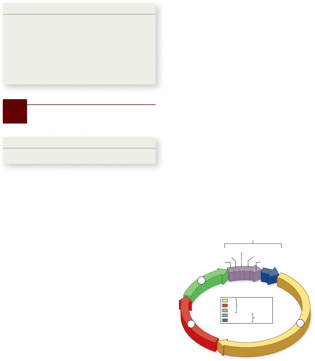
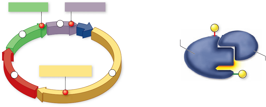
Figure 10.8
The cell cycle. The cell cycle is depicted as a
circle. The rst gap phase, G
1
, involves growth and preparation for
DNA synthesis. During S phase, a copy of the genome is
synthesized. The second gap phase, G
2
, prepares the cell for
mitosis. During mitosis, replicated chromosomes are partitioned.
Cytokinesis divides the cell into two cells with identical genomes.
cells, the microtubule spindle helps position a contracting
ring of actin that constricts like a drawstring to pinch the
cell in two. In cells with a cell wall, such as plant cells, a
plate forms between the dividing cells.
Mitosis and cytokinesis together are usually referred to
collectively as M phase, to distinguish the dividing phase from
interphase.
The duration of the cell cycle varies
depending on cell type
The time it takes to complete a cell cycle varies greatly. Cells in
animal embryos can complete their cell cycle in under 20 min;
the shortest known animal nuclear division cycles occur in fruit
fly embryos (8 min). These cells simply divide their nuclei as
quickly as they can replicate their DNA, without cell growth.
Half of their cycle is taken up by S, half by M, and essentially
none by G
1
or G
2
.
Because mature cells require time to grow, most of their
cycles are much longer than those of embryonic tissue. Typi-
cally, a dividing mammalian cell completes its cell cycle in about
24 hr, but some cells, such as certain cells in the human liver,
have cell cycles lasting more than a year. During the cycle,
growth occurs throughout the G
1
and G
2
phases, as well as dur-
ing the S phase. The M phase takes only about an hour, a small
fraction of the entire cycle.
Most of the variation in the length of the cell cycle be-
tween organisms or cell types occurs in the G
1
phase. Cells of-
ten pause in G
1
before DNA replication and enter a resting
state called the G
0
phase; cells may remain in this phase for
Learning Outcome
Describe the eukaryotic cell cycle.1.
Compared with prokaryotes, the increased size and more com-
plex organization of eukaryotic genomes required radical
changes in the partitioning of replicated genomes into daugh-
ter cells. The cell cycle requires the duplication of the genome,
its accurate segregation, and the division of cellular contents.
The cell cycle is divided into ve phases
The cell cycle is divided into phases based on the key events of
genome duplication and segregation. The cell cycle is usually
diagrammed using the metaphor of a clock face (figure 10.8).
■ G
1
(gap phase 1) is the primary growth phase of the cell.
The term gap phase refers to its filling the gap between
cytokinesis and DNA synthesis. For most cells, this
is the longest phase.
■ S (synthesis) is the phase in which the cell synthesizes
a replica of the genome.
■ G
2
(gap phase 2) is the second growth phase, and
preparation for separation of the newly replicated
genome. This phase fills the gap between DNA
synthesis and the beginning of mitosis. During this
phase, mitochondria and other organelles replicate, and
microtubules begin to assemble at a spindle.
G
1
, S, and G
2
together constitute interphase, the
portion of the cell cycle between cell divisions.
■ Mitosis is the phase of the cell cycle in which the spindle
apparatus assembles, binds to the chromosomes, and
moves the sister chromatids apart. Mitosis is the essential
step in the separation of the two daughter genomes. It is
traditionally subdivided into five stages: prophase,
prometaphase, metaphase, anaphase, and telophase.
■ Cytokinesis is the phase of the cell cycle when the
cytoplasm divides, creating two daughter cells. In animal
10.3
Overview of the Eukaryotic
Cell Cycle
Learning Outcomes Review 10.2
Eukaryotic chromosomes are complex structures that can be compacted
for cell division. During interphase, DNA is coiled around proteins into a
structure called a nucleosome. The string of nucleosomes is further coiled
into a solenoid (30-nm fi ber). Diploid cells contain a maternal and paternal
copy, or homologue, for each chromosome. After chromosome replication,
each homologue consists of two sister chromatids. The chromatids are held
together by proteins called cohesins.
■ Is chromosome number related to organismal
complexity?
192
part
II
Biology of the Cell
rav32223_ch10_186-206.indd 192rav32223_ch10_186-206.indd 192 11/6/09 2:14:38 PM11/6/09 2:14:38 PM
Apago PDF Enhancer
Chromatid
Kinetochore
microtubules
Centromere
region of
chromosome
Metaphase
chromosome
Cohesin
proteins
Kinetochore
2.0 µm
duced. The cell’s DNA replicates only during the S phase of
the cell cycle.
After the chromosomes have replicated in S phase, they
remain fully extended and uncoiled, although cohesin proteins
are associated with them at this stage. In G
2
phase, they begin
Learning Outcomes
Describe the events that take place during interphase.1.
Explain the structure of the centromere after S phase. 2.
The events that occur during interphase—the G
1
, S, and G
2
phases—are very important for the successful completion of
mitosis. During G
1
, cells undergo the major portion of their
growth. During the S phase, each chromosome replicates to
produce two sister chromatids, which remain attached to each
other at the centromere. In the G
2
phase, the chromosomes coil
even more tightly.
The centromere is a point of constriction on the chro-
mosome containing certain repeated DNA sequences that bind
specific proteins. These proteins make up a disklike structure
called the kinetochore. This disk functions as an attachment
site for microtubules necessary to separate the chromosomes
during cell division (figure 10.9). As seen in figure 10.6, each
chromosome’s centromere is located at a characteristic site
along the length of the chromosome.
After the S phase, the sister chromatids appear to share a
common centromere, but at the molecular level the DNA of
the centromere has actually already replicated, so there are two
complete DNA molecules. Functionally, however, the two
chromatids have a single centromere due to their being at-
tached by cohesin proteins at the centromere site (figure 10.10).
In metazoan animals, most of the cohesins that hold sister chro-
matids together after replication appear to be replaced by con-
densin during the process of chromosome compaction. This
leaves the chromosomes still attached tightly at the centro-
mere, but loosely attached elsewhere.
The cell grows throughout interphase. The G
1
and G
2
segments of interphase are periods of active growth, during
which proteins are synthesized and cell organelles are pro-
10.4
Interphase: Preparation
for Mitosis
days to years before resuming cell division. At any given time,
most of the cells in an animal’s body are in G
0
phase. Some,
such as muscle and nerve cells, remain there permanently; oth-
ers, such as liver cells, can resume G
1
phase in response to fac-
tors released during injury.
Learning Outcome Review 10.3
Cell division in eukaryotes is a complex process that involves fi ve phases:
a fi rst gap phase (G
1
); a DNA synthesis phase (S); a second gap phase (G
2
);
mitosis (M), during which chromatids are separated; and cytokinesis in
which a cell becomes two separate cells.
■ When during the cycle is a cell irreversibly committed
to dividing?
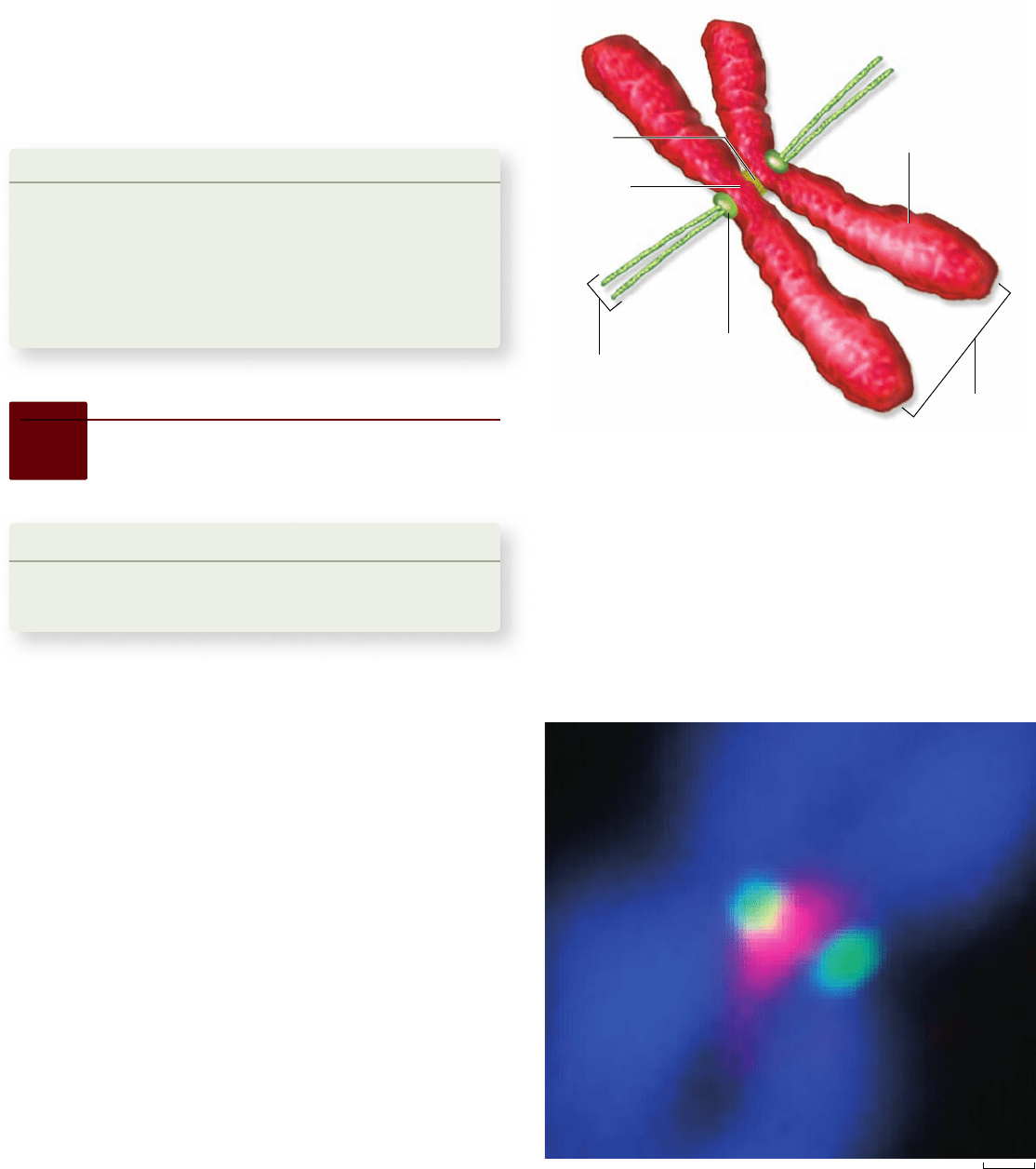
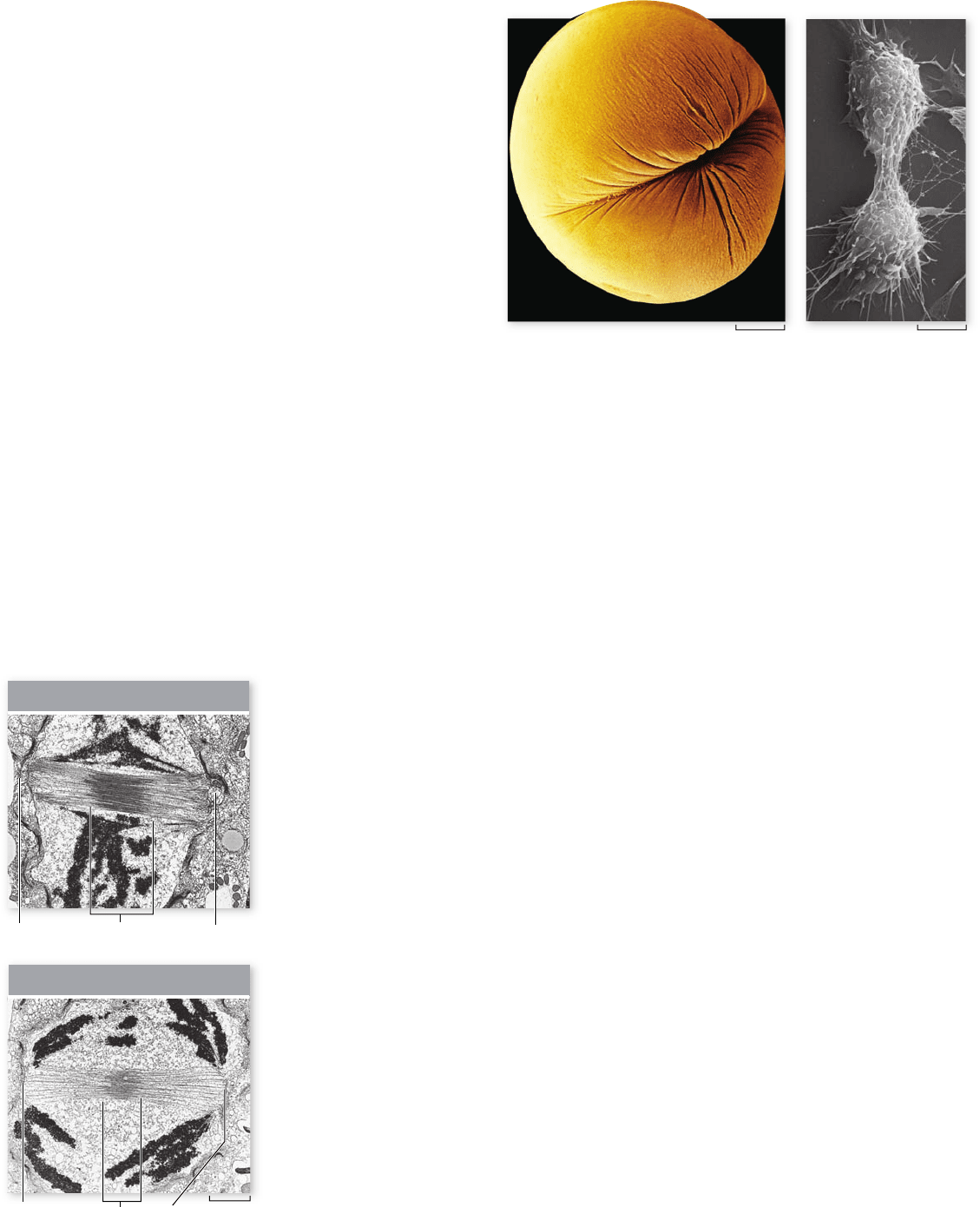
Figure 10.9
Kinetochores. Separation of sister chromatids
during mitosis depends on microtubules attaching to proteins found
in the kinetochore. These kinetochore proteins are assembled on
the centromere of chromosomes. The centromeres of the two sister
chromatids are held together by cohesin proteins.
Figure 10.10
Proteins found at the centromere. In this
image DNA, a cohesin protein and a kinetochore protein have all
been labeled with a different colored uorescent dye. Cohesin (red),
which holds centromeres together, lies between the sister chromatids
(blue). Each sister chromatid has its own separate kinetochore (green).
chapter
10
How Cells Divide
193www.ravenbiology.com
rav32223_ch10_186-206.indd 193rav32223_ch10_186-206.indd 193 11/6/09 2:14:39 PM11/6/09 2:14:39 PM
Apago PDF Enhancer
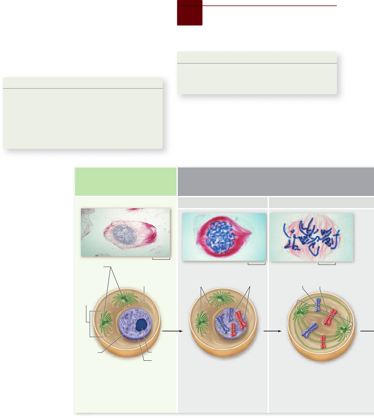
INTERPHASE G
2
MITOSIS
Prophase Prometaphase
Nucleus
Nucleolus
Aster
Centrioles
(replicated;
animal
cells only)
Chromatin
(replicated)
Nuclear
membrane
• DNA has been replicated
• Centrioles replicate (animal cells)
• Cell prepares for division
Centromere and
kinetochore
Mitotic spindle
beginning to form
Condensed
chromosomes
Mitotic
spindle
• Chromosomes condense and
become visible
• Chromosomes appear as two sister
chromatids held together at the
centromere
• Cytoskeleton is disassembled: spindle
begins to form
• Golgi and ER are dispersed
• Nuclear envelope breaks down
• Chromosomes attach to
microtubules at the kinetochores
• Each chromosome is oriented
such that the kinetochores
of sister chromatids are
attached to microtubules
from opposite poles.
• Chromosomes move to
equator of the cell
80 µm 80 µm
80 µm
Learning Outcomes
Describe the phases of mitosis.1.
Understand the importance of chromatid cohesion.2.
Compare cytokinesis in plants and animals.3.
The process of mitosis is one of the most dramatic and beautiful
biological processes that can be readily observed. In our attempts
to understand this process, we have divided it into discrete phases
but it should always be remembered that this is a dynamic, con-
tinuous process, not a set of discrete steps. This process is shown
both schematically and in micrographs in figure 10.11.
10.5
M Phase: Chromosome
Segregation and the Division
of Cytoplasmic Contents
the process of condensation, coiling ever more tightly. Special
motor proteins are involved in the rapid final condensation of the
chromosomes that occurs early in mitosis. Also during G
2
phase, the cells begin to assemble the machinery they will later
use to move the chromosomes to opposite poles of the cell. In
animal cells, a pair of microtubule-organizing centers called
centrioles replicate, producing one for each pole. All eukaryotic
cells undertake an extensive synthesis of tubulin, the protein
that forms microtubules.
Learning Outcomes Review 10.4
Interphase includes the G
1
, S, and G
2
phases of the cell cycle. During
interphase, the cell grows; replicates chromosomes, organelles, and
centrioles; and synthesizes components needed for mitosis, including
tubulin. Cohesin proteins hold chromatids together at the centromere of
each chromosome.
■ How would a mutation that deleted cohesin proteins
affect cell division?
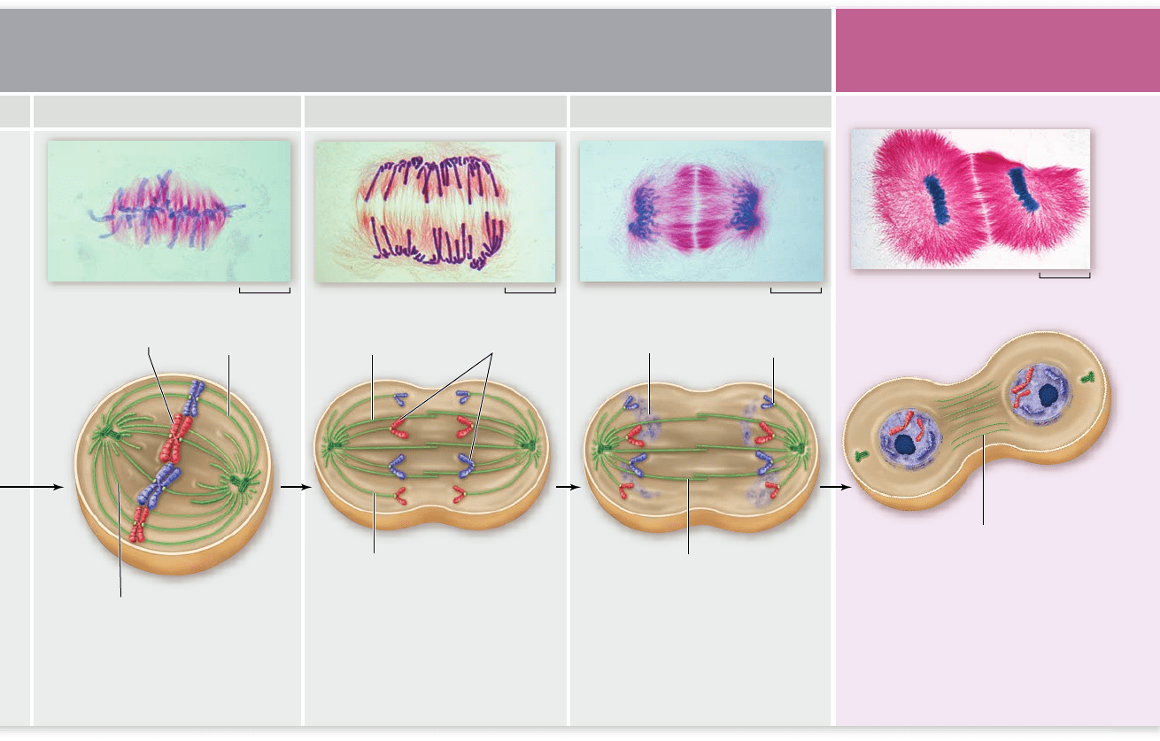
Figure 10.11
Mitosis
and cytokinesis.
Mitosis is conventionally
divided into ve stages—
prophase, prometaphase,
metaphase, anaphase, and
telophase—which together
act to separate duplicated
chromosomes. This is
followed by cytokinesis,
which divides the cell into
two separate cells. Photos
depict mitosis and
cytokinesis in a plant, the
African blood lily
(Haemanthus katharinae),
with chromosomes stained
blue and microtubules
stained red. Drawings
depict mitosis and
cytokinesis in animal cells.
194
part
II
Biology of the Cell
rav32223_ch10_186-206.indd 194rav32223_ch10_186-206.indd 194 11/6/09 2:14:41 PM11/6/09 2:14:41 PM
Apago PDF Enhancer
CYTOKINESIS
Metaphase Anaphase Telophase
Polar microtubule
Kinetochore
microtubule
Chromosomes
aligned on
metaphase plate
Polar
microtubule
Chromosomes
Kinetochore
microtubule
Polar microtubule
Nucleus reforming
Cleavage furrow
• All chromosomes are aligned
at equator of the cell, called
the metaphase plate
• Chromosomes are attached
to opposite poles and are
under tension
• Proteins holding centromeres of sister
chromatids are degraded, freeing
individual chromosomes
• Chromosomes are pulled to opposite
poles (anaphase A)
• Spindle poles move apart
(anaphase B)
• Chromosomes are clustered at
opposite poles and decondense
• Nuclear envelopes re-form around
chromosomes
• Golgi complex and ER re-form
• In animal cells, cleavage furrow
forms to divide the cells
• In plant cells, cell plate forms
to divide the cells
80 µm 80 µm 80 µm
80 µm
Kinetochore
microtubule
called the spindle apparatus, between them. In plant cells, a
similar bridge of microtubular fibers forms between opposite
poles of the cell, although centrioles are absent in plant cells.
In animal cell mitosis, the centrioles extend a radial array
of microtubules toward the nearby plasma membrane when
they reach the poles of the cell. This arrangement of micro-
tubules is called an aster. Although the aster’s function is not
fully understood, it probably braces the centrioles against the
membrane and stiffens the point of microtubular attachment
during the retraction of the spindle. Plant cells, which have
rigid cell walls, do not form asters.
Breakdown of the nuclear envelope
During the formation of the spindle apparatus, the nuclear en-
velope breaks down, and the endoplasmic reticulum reabsorbs
its components. At this point, the microtubular spindle fibers
extend completely across the cell, from one pole to the other.
Their orientation determines the plane in which the cell will
subsequently divide, through the center of the cell at right
angles to the spindle apparatus.
During prophase, the mitotic apparatus forms
When the chromosome condensation initiated in G
2
phase
reaches the point at which individual condensed chromosomes
first become visible with the light microscope, the first stage of
mitosis, prophase, has begun. The condensation process con-
tinues throughout prophase; consequently, chromosomes that
start prophase as minute threads appear quite bulky before its
conclusion. Ribosomal RNA synthesis ceases when the portion
of the chromosome bearing the rRNA genes is condensed.
The spindle and centrioles
The assembly of the spindle apparatus that will later separate
the sister chromatids occurs during prophase. The normal mi-
crotubule structure in the cell disassembled in the G
2
phase is
replaced by the spindle. In animal cells, the two centriole pairs
formed during G
2
phase begin to move apart early in prophase,
forming between them an axis of microtubules referred to as
spindle fibers. By the time the centrioles reach the opposite
poles of the cell, they have established a bridge of microtubules,
chapter
10
How Cells Divide
195www.ravenbiology.com
rav32223_ch10_186-206.indd 195rav32223_ch10_186-206.indd 195 11/6/09 2:14:45 PM11/6/09 2:14:45 PM
Apago PDF Enhancer
57 µm
Centrioles
Metaphase
plate
Sister chromatids
Aster
Polar
microtubule
Kinetochore
microtubule
During prometaphase, chromosomes
attach to the spindle
The transition from prophase to prometaphase occurs follow-
ing the disassembly of the nuclear envelope. During prometa-
phase the condensed chromosomes become attached to the
spindle by their kinetochores. Each chromosome possesses two
kinetochores, one attached to the centromere region of each
sister chromatid (see figure 10.9).
Microtubule attachment
As prometaphase continues, a second group of microtubules
grow from the poles of the cell toward the centromeres. These
microtubules are captured by the kinetochores on each pair of
sister chromatids. This results in the kinetochores of each sister
chromatid being connected to opposite poles of the spindle.
This bipolar attachment is critical to the process of mito-
sis; any mistakes in microtubule positioning can be disastrous.
For example, the attachment of the kinetochores of both sister
chromatids to the same pole leads to a failure of sister chroma-
tid separation, and they will be pulled to the same pole ending
up in the same daughter cell, with the other daughter cell miss-
ing that chromosome.
Movement of chromosomes to the cell center
With each chromosome attached to the spindle by micro-
tubules from opposite poles to the kinetochores of sister chro-
matids, the chromosomes begin to move to the center of the
cell. This movement is jerky, as if a chromosome is being pulled
toward both poles at the same time. This process is called con-
gression, and it eventually leads to all of the chromosomes being
arranged at the equator of the cell with the sister chromatids of
each chromosome oriented to opposite poles by their kineto-
chore microtubules.
The force that moves chromosomes has been of great in-
terest since the process of mitosis was first observed. Two basic
mechanisms have been proposed to explain this: (1) assembly
and disassembly of microtubules provides the force to move
chromosomes, and (2) motor proteins located at the kineto-
chore and poles of the cell pull on microtubules to provide
force. Data have been obtained that support both mechanisms.
In support of the microtubule-shortening proposal, iso-
lated chromosomes can be pulled by microtubule disassembly.
The spindle is a very dynamic structure, with microtubules be-
ing added to at the kinetochore and shortened at the poles, even
during metaphase. In support of the motor protein proposal,
multiple motor proteins have been identified as kinetochore
proteins, and inhibition of the motor protein dynein slows chro-
mosome separation at anaphase. Like many phenomena that we
analyze in living systems, the answer is not a simple either–or
choice; both mechanisms are probably at work.
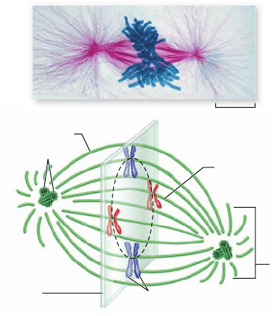
In metaphase, chromosomes
align at the equator
The alignment of the chromosomes in the center of the cell
signals the third stage of mitosis, metaphase. When viewed
with a light microscope, the chromosomes appear to array
Figure 10.12
Metaphase. In metaphase, the chromosomes
are arrayed at the midpoint of the cell. The imaginary plane
through the equator of the cell is called the metaphase plate. As the
spindle itself is a three dimensional structure, the chromosomes are
arrayed in a rough circle on the metaphase plate.
themselves in a circle along the inner circumference of the cell,
just as the equator girdles the Earth (figure 10.12). An imagi-
nary plane perpendicular to the axis of the spindle that passes
through this circle is called the metaphase plate. The metaphase
plate is not an actual structure, but rather an indication of the
future axis of cell division.
Positioned by the microtubules attached to the kineto-
chores of their centromeres, all of the chromosomes line up
on the metaphase plate. At this point their centromeres are
neatly arrayed in a circle, equidistant from the two poles of
the cell, with microtubules extending back toward the oppo-
site poles of the cell. The cell is prepared to properly separate
sister chromatids, such that each daughter cell will receive a
complete set of chromosomes. Thus metaphase is really a
transitional phase in which all the preparations are checked
before the action continues.
At anaphase, the chromatids separate
Of all the stages of mitosis, shown in figure 10.11, anaphase is
the shortest and the most amazing to watch. It begins when the
centromeres split, freeing the two sister chromatids from each
other. Up to this point in mitosis, sister chromatids have been
held together by cohesin proteins concentrated at the cen tro-
mere, as mentioned earlier. The key event in anaphase, then, is
the simultaneous removal of these proteins from all of the chro-
mosomes. The control and details of this process are discussed
later on in the context of control of the entire cell cycle.
Freed from each other, the sister chromatids are pulled
rapidly toward the poles to which their kinetochores are attached.
In the process, two forms of movement take place simultaneously,
each driven by microtubules. These movements are often called
anaphase A and anaphase B to distinguish them.
196
part
II
Biology of the Cell
rav32223_ch10_186-206.indd 196rav32223_ch10_186-206.indd 196 11/6/09 2:14:47 PM11/6/09 2:14:47 PM
Apago PDF Enhancer
Metaphase
Late Anaphase
Pole
Overlapping
microtubules
Pole
Pole
Overlapping
microtubules
Pole
2 µm
a.
b.
333.3 µm 16.6 µm
First, during anaphase A, the kinetochores are pulled toward
the poles as the microtubules that connect them to the poles
shorten. This shortening process is not a contraction; the
microtubules do not get any thicker. Instead, tubulin subunits
are removed from the kinetochore ends of the microtubules.
As more subunits are removed, the chromatid-bearing micro-
tubules are progressively disassembled, and the chromatids are
pulled ever closer to the poles of the cell.
Second, during anaphase B, the poles move apart as micro-
tubular spindle fibers physically anchored to opposite poles slide
past each other, away from the center of the cell (figure 10.13).
Because another group of microtubules attach the chromosomes
to the poles, the chromosomes move apart, too. If a flexible
membrane surrounds the cell, it becomes visibly elongated.
When the sister chromatids separate in anaphase, the ac-
curate partitioning of the replicated genome—the essential ele-
ment of mitosis—is complete.
During telophase, the nucleus re-forms
In telophase, the spindle apparatus disassembles as the micro-
tubules are broken down into tubulin monomers that can be
used to construct the cytoskeletons of the daughter cells. A
nuclear envelope forms around each set of sister chromatids,
which can now be called chromosomes because they are no
longer attached at the centromere. The chromosomes soon be-
gin to uncoil into the more extended form that permits gene
expression. One of the early group of genes expressed after mi-
tosis is complete are the rRNA genes, resulting in the reappear-
ance of the nucleolus.
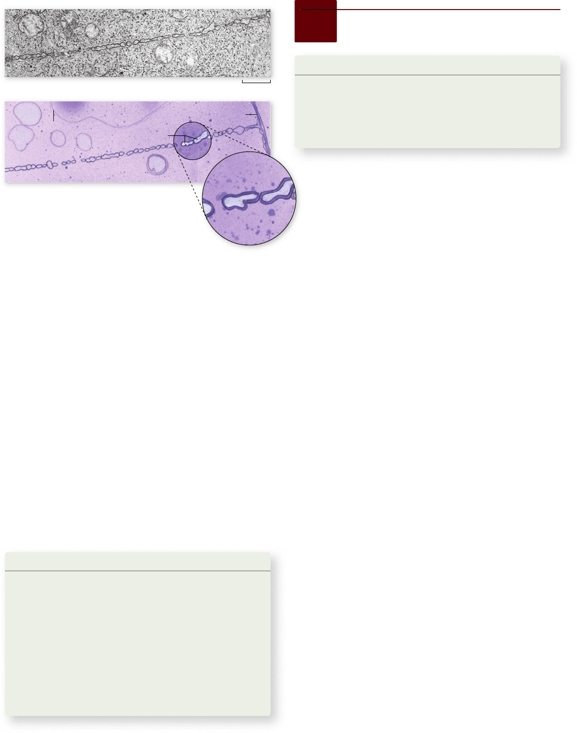
Figure 10.13
Microtubules slide
past each other as the
chromosomes separate.
In these electron
micrographs of dividing
diatoms, the overlap of the
microtubules lessens
markedly during spindle
elongation as the cell passes
from metaphase to
anaphase. During
anaphase B the poles move
farther apart as the
chromosomes move toward
the poles.
Telophase can be viewed as a reversal of the process of
prophase, bringing the cell back to the state of interphase. Mi-
tosis is complete at the end of telophase. The eukaryotic cell
has partitioned its replicated genome into two new nuclei po-
sitioned at opposite ends of the cell. Other cytoplasmic organ-
elles, including mitochondria and chloroplasts (if present),
were reassorted to areas that will separate and become the
daughter cells.
Cell division is still not complete at the end of mitosis,
however, because the division of the cell body proper has not
yet begun. The phase of the cell cycle when the cell actually
divides is called cytokinesis. It generally involves the cleavage
of the cell into roughly equal halves.
In animal cells, a belt of actin pinches
o the daughter cells
In animal cells and the cells of all other eukaryotes that lack
cell walls, cytokinesis is achieved by means of a constricting
belt of actin filaments. As these filaments slide past one an-
other, the diameter of the belt decreases, pinching the cell and
creating a cleavage furrow around the cell’s circumference
(figure 10.14a).
As constriction proceeds, the furrow deepens until it
eventually slices all the way into the center of the cell. At this
point, the cell is divided in two (figure 10.14b).
In plant cells, a cell plate divides
the daughter cells
Plant cell walls are far too rigid to be squeezed in two by actin fila-
ments. Instead, these cells assemble membrane components in
their interior, at right angles to the spindle apparatus. This ex-
panding membrane partition, called a cell plate, continues to
grow outward until it reaches the interior surface of the plasma
membrane and fuses with it, effectively dividing the cell in two
Figure 10.14
Cytokinesis in animal cells. a. A cleavage
furrow forms around a dividing frog egg. b. The completion of
cytokinesis in an animal cell. The two daughter cells are still joined
by a thin band of cytoplasm occupied largely by microtubules.
chapter
10
How Cells Divide
197www.ravenbiology.com
rav32223_ch10_186-206.indd 197rav32223_ch10_186-206.indd 197 11/6/09 2:14:48 PM11/6/09 2:14:48 PM
Apago PDF Enhancer
Cell wall
Nucleus
Vesicles containing
membrane components
fusing to form cell plate
0.7 µm
Figure 10.15
Cytokinesis in plant cells.
In this photomicrograph and companion drawing,
a cell plate is forming between daughter nuclei. The
cell plate forms from the fusion of Golgi-derived
vesicles. Once the plate is complete, there will be two cells.
Learning Outcomes
Distinguish the role of checkpoints in the control1.
of the cell cycle.
Understand the role of the anaphase-promoting 2.
complex/cyclosome in mitosis.
Describe cancer in terms of cell cycle control.3.
Our knowledge of how the cell cycle is controlled, although
still incomplete, has grown enormously in the past 30 years.
Our current view integrates two basic concepts. First, the
cell cycle has two irreversible points: the replication of ge-
netic material and the separation of the sister chromatids.
Second, the cell cycle can be put on hold at specific points
called checkpoints. At any of these checkpoints, the process is
checked for accuracy and can be halted if there are errors. This
leads to extremely high fidelity overall for the entire process.
The checkpoint organization also allows the cell cycle to re-
spond to both the internal state of the cell, including nutritional
state and integrity of genetic material, and to signals from the
environment, which are integrated at major checkpoints.
Research uncovered cell cycle
control factors
The history of investigation into control of the cell cycle is in-
structive in two ways. First, it allows us to place modern obser-
vations into context; second, we can see how biologists using
very different approaches often end up at the same place. The
following brief history introduces three observations and then
shows how they can be integrated into a single mechanism.
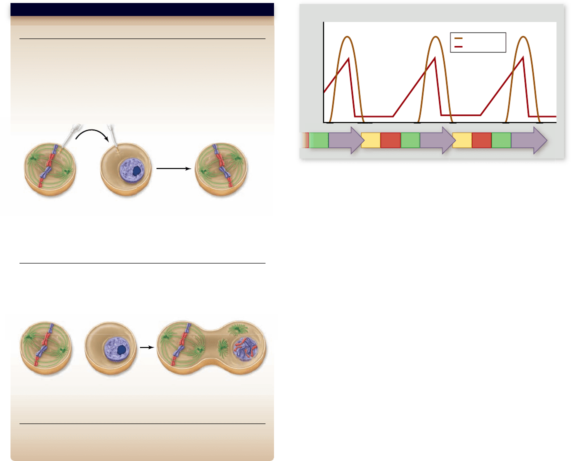
Discovery of MPF
Research on the activation of frog oocytes led to the discovery
of a substance that was first called maturation-promoting factor
(MPF ). Frog oocytes, which go on to become egg cells, be-
come arrested near the end of their development at the G
2
stage before meiosis I, which is the division leading to the
production of gametes (chapter 11). They remain in this ar-
rested state and await hormonal signaling to complete this
division process.
Cytoplasm taken from a variety of actively dividing cells
could prematurely induce cell division when injected into oo-
cytes (figure 10.16). These experiments indicated the presence
of a positive regulator of cell cycle progression in the cytoplasm
of dividing cells: MPF. These experiments also fit well with cell
fusion experiments done with mitotic and interphase cells that
also indicated a cytoplasmic positive regulator that could in-
duce mitosis (figure 10.16).
Further studies highlighted two key aspects of MPF.
First, MPF activity varied during the cell cycle: low in early G
2
,
rising throughout this phase, and then peaking in mitosis
( fig ure 10.17). Second, the enzymatic activity of MPF involved
10.6
Control of the Cell Cycle
(figure 10.15). Cellulose is then laid down on the new membranes,
creating two new cell walls. The space between the daughter cells
becomes impregnated with pectins and is called a middle lamella .
In fungi and some protists, daughter nuclei
are separated during cytokinesis
In most fungi and some groups of protists, the nuclear mem-
brane does not dissolve, and as a result, all the events of mitosis
occur entirely within the nucleus. Only after mitosis is complete
in these organisms does the nucleus divide into two daughter
nuclei; then, during cytokinesis, one nucleus goes to each
daughter cell. This separate nuclear division phase of the cell
cycle does not occur in plants, animals, or most protists.
After cytokinesis in any eukaryotic cell, the two daughter
cells contain all the components of a complete cell. Whereas
mitosis ensures that both daughter cells contain a full comple-
ment of chromosomes, no similar mechanism ensures that or-
ganelles such as mitochondria and chloroplasts are distributed
equally between the daughter cells. But as long as at least one of
each organelle is present in each cell, the organelles can repli-
cate to reach the number appropriate for that cell.
Learning Outcomes Review 10.5
Mitosis is divided into phases: prophase, prometaphase, metaphase,
anaphase, and telophase. The early phases involve restructuring the cell to
create the microtubule spindle that pulls chromosomes to the equator of
the cell in metaphase. Chromatids for each chromosome remain attached at
the centromere by cohesin proteins. Chromatids are then pulled to opposite
poles during anaphase when cohesin proteins are destroyed. The nucleus is
re-formed in telophase, and cytokinesis then divides the cell cytoplasm and
organelles. In animal cells, actin pinches the cell in two; in plant cells, a cell
plate forms in the middle of the dividing cell.
■ What would happen to a chromosome that loses cohesin
protein between sister chromatids before metaphase?
198
part
II
Biology of the Cell
rav32223_ch10_186-206.indd 198rav32223_ch10_186-206.indd 198 11/6/09 2:14:49 PM11/6/09 2:14:49 PM
Apago PDF Enhancer
Prediction: Frog oocytes are arrested in G
2
of meiosis I. They can be
induced to mature (undergo meiosis) by progesterone treatment. If
maturing oocytes contain a positive regulator of cell division, injection of
cytoplasm should induce an immature oocyte to undergo meiosis.
Test: Oocytes are induced with progesterone, then cytoplasm from these
maturing cells is injected into immature oocytes.
Hypothesis: There are positive regulators of cell division.
Prediction: If mitosis is driven by positive regulators, then cytoplasm
from a mitotic cell should cause a G
1
cell to enter mitosis.
Test: M phase cells are fused with G
1
phase cells, then the nucleus from
the G
1
phase cell is monitored microscopically.
Result: Injected oocytes progress from G
2
into meiosis I.
Conclusion: The progesterone treatment causes production of a
positive regulator of maturation: Maturation Promoting Factor (MPF).
Further Experiments: How can both of these experiments be
rationalized? What would be the next step in characterizing these factors?
Conclusion: Cytoplasm from M phase cells contains a positive regulator
that causes a cell to enter mitosis.
SCI E NT I FIC THINKING
Progesterone-
treated oocyte
Remove
cytoplasm
Inject
cytoplasm
Arrested oocyte Oocyte in meiosis I
M phase cell G
1
phase cell Fused cells
G
1
S
G
2
M
G
2
M
G
1
S
G
2
M
Concentration
Low
High
MPF activity
Cyclin
the phosphorylation of proteins. This second point is not sur-
prising given the importance of phosphorylation as a reversible
switch on the activity of proteins (see chapter 9). The first
observation indicated that MPF itself was not always active, but
rather was being regulated with the cell cycle, and the second
showed the possible enzymatic activity of MPF.
Discovery of cyclins
Other researchers examined proteins produced during the early
divisions in sea urchin embryos. They identified proteins that
were produced in synchrony with the cell cycle, and named
them cyclins (see figure 10.17). These observations were
extended in another marine invertebrate, the surf clam. Two
forms of cyclin were found that cycled at slightly different
times, reaching peaks at the G
1
/S and G
2
/M boundaries.
Figure 10.16
Discovery of positive regulator of cell
division.
Figure 10.17
Correlation of MPF activity, amount
of cyclin protein, and stages of the cell cycle. Cyclin
concentration and MPF activity are shown plotted vs. stage of the
cell cycle. MPF activity changes in a repeating pattern through the
cell cycle. This also correlates with the level of mitotic cyclin in the
cell, which shows a similar pattern. The reason for this correlation
is that cyclin is actually one component of MPF, the other being a
cyclin-dependent kinase (Cdk). Together these act as a positive
regulator of cell division.
Despite much effort, no identified enzymatic activity was asso-
ciated with these proteins. Their hallmark was the timing of
their production and not any intrinsic activity.
Genetic analysis of the cell cycle
Geneticists using two different yeasts, budding yeast and fission
yeast, as model systems set out to determine the genes neces-
sary for control of the cell cycle. By isolating mutants that were
halted during division, they identified genes that were neces-
sary for cell cycle progression. These studies indicated that in
yeast, there were two critical control points: the commitment
to DNA synthesis, called START, as it meant committing to
divide, and the commitment to mitosis. One particular gene,
named cdc2, from fission yeast, was shown to be critical for
passing both of these boundaries.
MPF is cyclin plus cdc2
All of these findings came together in an elegant fashion with
the following observations. First, the protein encoded by the
cdc2 gene was shown to be a protein kinase. Second, the purifi-
cation and identification of MPF showed that it was composed
of both a cyclin component and a kinase component. Last, the
kinase itself was the cdc2 protein!
The cdc2 protein was the first identified cyclin-
dependent kinase (Cdk), that is, a protein kinase enzyme that
is only active when complexed with cyclin. This finding led to
the renaming of MPF as mitosis-promoting factor, as its role
was clearly more general than simply promoting the matura-
tion of frog oocytes.
These Cdk enzymes are the key positive drivers of the
cell division cycle. They are often called the engine that drives
cell division. The control of the cell cycle in higher eukaryotes
chapter
10
How Cells Divide
199www.ravenbiology.com
rav32223_ch10_186-206.indd 199rav32223_ch10_186-206.indd 199 11/6/09 2:14:51 PM11/6/09 2:14:51 PM
Apago PDF Enhancer
G
2
M
S
G
1
G
1
/S checkpoint
(Start or restriction point)
Spindle checkpoint
G
2
/M checkpoint
Cyclin
Cyclin-dependent kinase
(Cdk)
P
P
is much more complex than the simple single-engine cycle of
yeast, but the yeast model remains a useful framework for un-
derstanding more complex regulation. The discovery of Cdks
and their role in the cell cycle is an excellent example of the
progressive nature of science.
The cell cycle can be halted
at three checkpoints
Although we have divided the cell cycle into phases and subdi-
vided mitosis into stages, the cell recognizes three points at
which the cycle can be delayed or halted. The cell uses these
three checkpoints to both assess its internal state and integrate
external signals (figure 10.18): G
1
/S, G
2
/M, and late metaphase
(the spindle checkpoint). Passage through these checkpoints is
controlled by the Cdk enzymes described earlier and also in the
following section.
G
1
/S checkpoint
The G
1
/S checkpoint is the primary point at which the cell
“decides” whether or not to divide. This checkpoint is there-
fore the primary point at which external signals can influence
events of the cycle. It is the phase during which growth factors
(discussed later on) affect the cycle and also the phase that links
cell division to cell growth and nutrition.
In yeast systems, where the majority of the genetic analy-
sis of the cell cycle has been performed, this checkpoint is called
START. In animals, it is called the restriction point (R point).
In all systems, once a cell has made this irreversible commit-
ment to replicate its genome, it has committed to divide. Dam-
age to DNA can halt the cycle at this point, as can starvation
conditions or lack of growth factors.
Figure 10.18
Control of the cell cycle. Cells use a
centralized control system to check whether proper conditions
have been achieved before passing three key checkpoints in the
cell cycle.
G
2
/M checkpoint
The G
2
/M checkpoint has received a large amount of atten-
tion because of its complexity and its importance as the stimu-
lus for the events of mitosis. Historically, Cdks active at this
checkpoint were first identified as MPFs, a term that has now
evolved into M phase-promoting factor (MPF).
Passage through this checkpoint represents the commit-
ment to mitosis. This checkpoint assesses the success of DNA
replication and can stall the cycle if DNA has not been accu-
rately replicated. DNA-damaging agents result in arrest at this
checkpoint as well as at the G
1
/S checkpoint.
Spindle checkpoint
The spindle checkpoint ensures that all of the chromosomes
are attached to the spindle in preparation for anaphase. The
second irreversible step in the cycle is the separation of chro-
mosomes during anaphase, and therefore it is critical that they
are properly arrayed at the metaphase plate.
Cyclin-dependent kinases drive the cell cycle
The primary molecular mechanism of cell cycle control is phos-
phorylation, which you may recall is the addition of a phosphate
group to the amino acids serine, threonine, and tyrosine in pro-
teins (chapter 9). The enzymes that accomplish this phosphory-
lation are the Cdks (figure 10.19).
The action of Cdks
The first important cell cycle kinase was identified in fission
yeast and named Cdc2 (now also called Cdk1). In yeast, this
Cdk can partner with different cyclins at different points in the
cell cycle (figure 10.20).
Even in the simplified cycle of the yeasts, we are left with
the important question of what controls the activity of the Cdks
during the cycle. For many years, a common view was that cy-
clins drove the cell cycle—that is, the periodic synthesis and
destruction of cyclins acted as a clock. More recently, it has
become clear that the Cdc2 kinase is also itself controlled by
Figure 10.19
Cdk enzyme forms a complex with cyclin.
Cdk is a protein kinase that activates numerous cell proteins by
phosphorylating them. Cyclin is a regulatory protein required to
activate Cdk. This complex is also called mitosis-promoting factor
(MPF). The activity of Cdk is also controlled by the pattern of
phosphorylation: phosphorylation at one site (represented by the red
site) inactivates the Cdk, and phosphorylation at another site
(represented by the green site) activates the Cdk.
200
part
II
Biology of the Cell
rav32223_ch10_186-206.indd 200rav32223_ch10_186-206.indd 200 11/6/09 2:14:52 PM11/6/09 2:14:52 PM
Apago PDF Enhancer
G
1
/S Checkpoint
Cdc2/G
1
Cyclin
• Growth factors
• Nutritional state
of cell
• Size of cell
G
2
M
S
G
1
Spindle Checkpoint
APC
G
2
/M Checkpoint
Cdc2/Mitotic Cyclin
• Replication
completed
• DNA integrity
• Chromosomes
attached at
metaphase plate
I
n
a
c
t
i
v
e
C
d
c
2
A
c
t
i
v
e
C
d
c
2
A
c
t
i
v
e
C
d
c
2
I
n
a
c
t
i
v
e
C
d
c
2
I
n
a
c
t
i
v
e
A
P
C
A
c
t
i
v
e
A
P
C
phosphorylation: Phosphorylation at one site activates Cdc2, and
phosphorylation at another site inactivates it (see figure 10.19).
Full activation of the Cdc2 kinase requires complexing with a cy-
clin and the appropriate pattern of phosphorylation.
As the G
1
/S checkpoint is approached, the triggering sig-
nal in yeast appears to be the accumulation of G
1
cyclins. These
form a complex with Cdc2 to create the active G
1
/S Cdk, which
phosphorylates a number of targets that bring about the in-
creased enzyme activity for DNA replication.
The action of MPF
MPF and its role at the G
2
/M checkpoint has been extensively
analyzed in a number of different experimental systems. The
control of MPF is sensitive to agents that disrupt or delay
replication and to agents that damage DNA. It was once
thought that MPF was controlled solely by the level of the
M phase-specific cyclins, but it has now become clear that this
is not the case.
Although M phase cyclin is necessary for MPF function,
activity is controlled by inhibitory phosphorylation of the ki-
nase component, Cdc2. The critical signal in this process is the
removal of the inhibitory phosphates by a protein, phosphatase.
This action forms a molecular switch based on positive feed-
back because the active MPF further activates its own activat-
ing phosphatase.
Figure 10.20
Checkpoints of the yeast cell cycle. The
simplest cell cycle that has been studied in detail is the ssion
yeast. This is controlled by three main checkpoints and a single
Cdk enzyme, called Cdc2. The Cdc2 enzyme partners with
different cyclins to control the G
1
/S and G
2
/M checkpoints. The
spindle checkpoint is controlled by the anaphase-promoting
complex (APC).
The checkpoint assesses the balance of the kinase that
adds inhibitory phosphates with the phosphatase that removes
them. Damage to DNA acts through a complex pathway that
includes damage sensing and a response to tip the balance to-
ward the inhibitory phosphorylation of MPF. Later on, we de-
scribe how some cancers overcome this inhibition.
The anaphase-promoting complex
The molecular details of the sensing system at the spindle
checkpoint are not clear. The presence of all chromosomes at
the metaphase plate and the tension on the microtubules be-
tween opposite poles are both important. The signal is trans-
mitted through the anaphase-promoting complex, also called
the cyclosome (APC/C).
The function of the APC/C is to trigger anaphase itself.
As described earlier, the sister chromatids at metaphase are still
held together by the protein complex cohesin. The APC does
not act directly on cohesin, but rather acts by marking a protein
called securin for destruction. The securin protein acts as an
inhibitor of another protease called separase that appears to be
specific for the cohesin complex. Once inhibition is lifted, sepa-
rase destroys cohesin.
This process has been analyzed in detail in budding yeast,
where it has been shown that the separase enzyme specifically
degrades a component of cohesin called Scc1. This leads to the
release of the sister chromatids and results in their sudden
movement toward opposite poles during anaphase.
In vertebrates, most cohesin is removed from the sister
chromatids during chromosome condensation, possibly with
cohesin being replaced by condensin. At metaphase, the major-
ity of the cohesin that remains on vertebrate chromatids is con-
centrated at the centromere (figure 10.10). The destruction of
this cohesin explains the anaphase movement of chromosomes
and the apparent “division” of the centromeres.
The APC/C has a number of roles in mitosis: it activates
the protease that removes the cohesins holding sister chromatids
together, and it is necessary for the destruction of mitotic cyclins
to drive the cell out of mitosis. The APC/C complex marks pro-
teins for destruction by the proteosome, the organelle responsi-
ble for the controlled degradation of proteins (chapter 16). The
signal to degrade a protein is the addition of a molecule called
ubiquitin, and the APC/C acts as a ubiquitin ligase. As we learn
more about the APC/C and its functions, it is clear that the con-
trol of its activity is a key regulator of the cell cycle.
In multicellular eukaryotes, many Cdks
and external signals act on the cell cycle
The major difference between more complex animals and
single-celled eukaryotes such as fungi and protists is twofold:
First, multiple Cdks control the cycle as opposed to the single
Cdk in yeasts; and second, animal cells respond to a greater
variety of external signals than do yeasts, which primarily re-
spond to signals necessary for mating.
In higher eukaryotes there are more Cdk enzymes and
more cyclins that can partner with these multiple Cdks, but
their basic role is the same as in the yeast cycle. A more complex
chapter
10
How Cells Divide
201www.ravenbiology.com
rav32223_ch10_186-206.indd 201rav32223_ch10_186-206.indd 201 11/6/09 2:14:53 PM11/6/09 2:14:53 PM