Raven P.H., Johnson G.B., Mason K.A. Biology (Ninth Edition)
Подождите немного. Документ загружается.

Apago PDF Enhancer
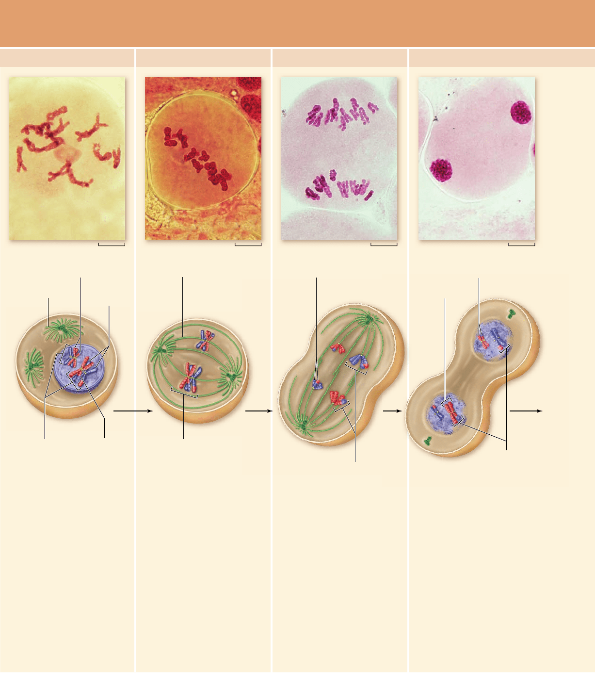
40 µm 40 µm 40 µm 40 µm
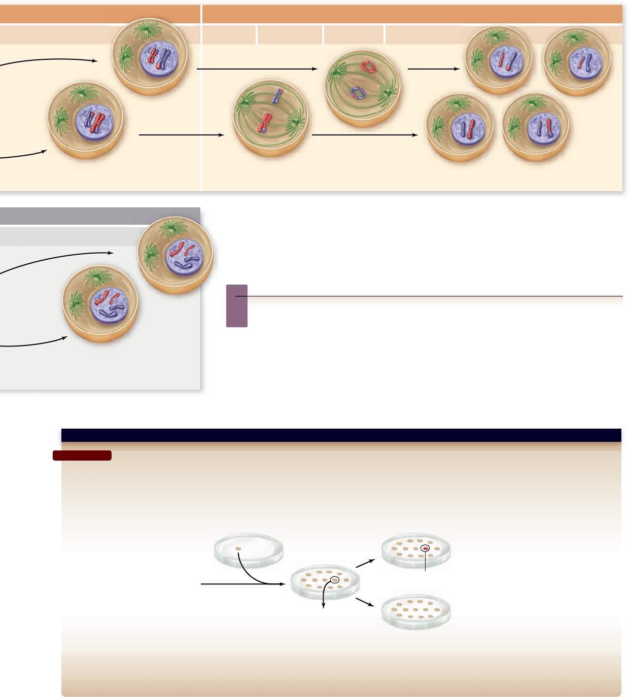
In prophase I of meiosis I, the
chromosomes begin to
condense, and the spindle of
microtubules begins to form.
The DNA has been replicated,
and each chromosome consists
of two sister chromatids
attached at the centromere. In
the cell illustrated here, there
are four chromosomes, or two
pairs of homologues.
Homologous chromosomes pair
up and become closely
associated during synapsis.
Crossing over occurs, forming
chiasmata, which hold
homologous chromosomes
together.
In metaphase I, the pairs of
homologous chromosomes
align along the metaphase
plate. Chiasmata help keep the
pairs together and produce
tension when microtubules from
opposite poles attach to sister
kinetochores of each
homologue. A kinetochore
microtubule from one pole of
the cell attaches to one
homologue of a chromosome,
while a kinetochore microtubule
from the other cell pole
attaches to the other
homologue of a pair.
In anaphase I, kinetochore
microtubules shorten, and
homologous pairs are pulled
apart. One duplicated homologue
goes to one pole of the cell, while
the other duplicated homologue
goes to the other pole. Sister
chromatids do not separate.This
is in contrast to mitosis, where
duplicated homologues line up
individually on the metaphase
plate, kinetochore microtubules
from opposite poles of the cell
attach to opposite sides of one
homologue's centromere, and
sister chromatids are pulled apart
in anaphase.
MEIOSIS I
Prophase I Metaphase I Anaphase I Telophase I
In telophase I, the separated
homologues form a cluster at
each pole of the cell, and the
nuclear envelope re-forms
around each daughter cell
nucleus. Cytokinesis may occur.
The resulting two cells have
half the number of
chromosomes as the original
cell: In this example, each
nucleus contains two
chromosomes (versus four in
the original cell). Each
chromosome is still in the
duplicated state and consists of
two sister chromatids, but sister
chromatids are not identical
because crossing over has
occurred.
Chromosome (replicated)
Sister
chromatids
Spindle
Paired homologous
chromosomes
Chiasmata
Kinetochore microtubule
Homologue pair
on metaphase plate
Homologous chromosomes
Sister chromatids
Homologous
chromosomes
Chromosome
Nonidentical sister chromatids
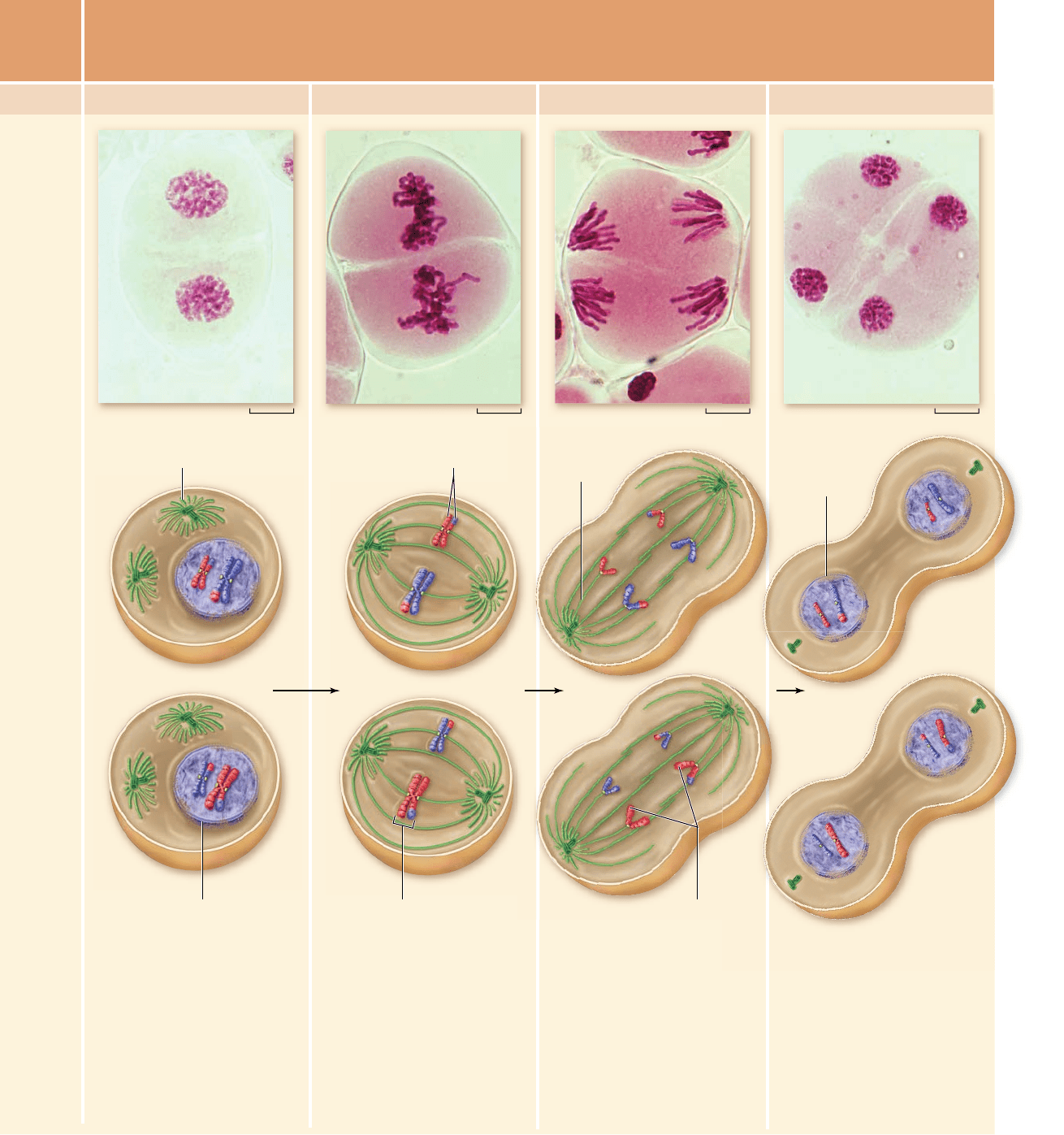
Figure 11.7
The stages of meiosis. Meiosis in plant cells (photos) and animal cells (drawings) is shown.
212
part
III
Genetic and Molecular Biology
rav32223_ch11_207-220.indd 212rav32223_ch11_207-220.indd 212 11/9/09 12:12:39 PM11/9/09 12:12:39 PM
Apago PDF Enhancer
40 µm 40 µm 40 µm 40 µm
Following a typically brief
interphase, with no S phase,
meiosis II begins. During
prophase II, a new spindle
apparatus forms in each cell,
and the nuclear envelope
breaks down. In some species
the nuclear envelope does not
re-form in telophase I removing
the need for nuclear envelope
breakdown.
In metaphase II, a completed
spindle apparatus is in place
in each cell. Chromosomes
consisting of sister chromatids
joined at the centromere align
along the metaphase plate in
each cell. Now, kinetochore
microtubules from opposite
poles attach to kinetochores of
sister chromatids.
When microtubules shorten in
anaphase II, the centromeres
split, and sister chromatids
are pulled to opposite poles
of the cells.
MEIOSIS II
Prophase II Metaphase II Anaphase II Telophase II
In telophase II, the nuclear
membranes re-form around
four different clusters of
chromosomes. After
cytokinesis, four haploid cells
result. No two cells are alike
due to the random alignment of
homologous pairs at
metaphase I and crossing over
during prophase I.
Sister chromatids
Sister chromatids Spindle
Nuclear membrane breaking down
Nuclear
membrane
re-forming
Kinetochore
microtubule
Chromosome
chapter
11
Sexual Reproduction and Meiosis
213www.ravenbiology.com
rav32223_ch11_207-220.indd 213rav32223_ch11_207-220.indd 213 11/9/09 12:12:47 PM11/9/09 12:12:47 PM
Apago PDF Enhancer
Anaphase I results from the di erential loss
of sister chromatid cohesion along the arms
In anaphase I, the microtubules of the spindle fibers begin to
shorten. As they shorten, they break the chiasmata and pull the
centromeres toward the poles, dragging the chromosomes
along with them.
Anaphase I comes about by the release of sister chromatid
cohesion along the chromosome arms, but not at the cen tro-
meres. This release is thought to be the result of the destruc-
tion of meiosis-specific cohesin in a process analogous to
anaphase in mitosis. The difference is that the destruction is
inhibited at the centromeres by a mechanism that is only re-
cently becoming clear.
As a result of this release, the homologues are pulled
apart, but not the sister chromatids. Each homologue moves to
one pole, taking both sister chromatids with it. When the spin-
dle fibers have fully contracted, each pole has a complete hap-
loid set of chromosomes consisting of one member of each
homologous pair.
Because of the random orientation of homologous chro-
mosomes on the metaphase plate, a pole may receive either the
maternal or the paternal homologue from each chromosome
pair. As a result, the genes on different chromosomes assort in-
dependently; that is, meiosis I results in the independent as-
sortment of maternal and paternal chromosomes into the
gametes (see chapter 12).
Telophase I completes meiosis I
By the beginning of telophase I, the chromosomes have segre-
gated into two clusters, one at each pole of the cell. Now the
nuclear membrane re-forms around each daughter nucleus.
Because each chromosome within a daughter nucleus
had replicated before meiosis I began, each now contains two
sister chromatids attached by a common centromere. Note
that the sister chromatids are no longer identical because of the
crossing over that occurred in prophase I (see figure 11.7); as
you will see, this change has important implications for ge-
netic variability.
Cytokinesis, the division of the cytoplasm and its contents,
may or may not occur after telophase I. The second meiotic di-
vision, meiosis II, occurs after an interval of variable length.
Achiasmate segregation
of homologues is possible
The preceding description of meiosis I relies on the observa-
tion that homologues are held together by chiasmata and by
sister chromatid cohesion. This connection produces the criti-
cal behavior of chromosomes during metaphase I and anaphase
I, when homologues move to the metaphase plate and then
move to opposite poles.
Although this connection of homologues is the rule, there
are exceptions. In fruit fly ( Drosophila) males for example, there
is no recombination, and yet meiosis proceeds accurately, a pro-
cess called achiasmate segregation (“without chiasmata”).
This seems to involve an alternative mechanism for joining
homologues and then allowing their segregation during ana-
phase I. Telomeres and other heterochromatic sequences have
been implicated, but the details remain unclear.
Despite these exceptions, the vast majority of species that
have been examined use the formation of chiasmata and sister
chromatid cohesion to hold homologues together for segrega-
tion during anaphase I.
Meiosis II is like a mitotic division
without DNA replication
Typically, interphase between meiosis I and meiosis II is brief
and does not include an S phase: Meiosis II resembles a normal
mitotic division. Prophase II, metaphase II, anaphase II, and
telophase II follow in quick succession (see figure 11.7).
Prophase II. At the two poles of the cell, the clusters of
chromosomes enter a brief prophase II, each nuclear
envelope breaking down as a new spindle forms.
Metaphase II. In metaphase II, spindle bers from opposite
poles bind to kinetochores of each sister chromatid,
allowing each chromosome to migrate to the metaphase
plate as a result of tension on the chromosomes from
polar microtubules pulling on sister centromeres. This
process is the same as metaphase during a mitotic
division.
Anaphase II. The spindle bers contract, and the cohesin
complex joining the centromeres of sister chromatids is
destroyed, splitting the centromeres and pulling the
sister chromatids to opposite poles. This process is also
the same as anaphase during a mitotic division.
Telophase II. Finally, the nuclear envelope re-forms around
the four sets of daughter chromosomes. Cytokinesis
then follows.
The final result of this division is four cells containing
haploid sets of chromosomes. The cells that contain these
haploid nuclei may develop directly into gametes, as they do
in animals. Alternatively, they may themselves divide mitoti-
cally, as they do in plants, fungi, and many protists, eventually
producing greater numbers of gametes or, as in some plants
and insects, adult individuals with varying numbers of chro-
mosome sets.
Errors in meiosis produce
aneuploid gametes
It is critical that the process of meiosis be accurate because any
failure produces gametes without the correct number of chro-
mosomes. Failure of chromosomes to move to opposite poles
during either meiotic division is called nondisjunction, and it
produces one gamete that lacks a chromosome and one that has
two copies. Gametes with an improper number of chromo-
somes are called aneuploid gametes. In humans, this condi-
tion is the most common cause of spontaneous abortion. The
implications of aneuploid gametes are explored in more detail
in chapter 13.
214
part
III
Genetic and Molecular Biology
rav32223_ch11_207-220.indd 214rav32223_ch11_207-220.indd 214 11/9/09 12:12:50 PM11/9/09 12:12:50 PM
Apago PDF Enhancer
Learning Outcomes
1. Understand the distinct features of meiosis.
2. Describe the differences in chromatid cohesion in meiosis
and mitosis.
Explain the importance of the suppression of replication 3.
between meiotic divisions.
The key to meiosis is understanding the differences between
meiosis and mitosis. The basic machinery in both processes is
the same, but the behavior of chromosomes is distinctly differ-
ent during the first meiotic division (figure 11.8).
Meiosis is characterized by four distinct features:
Homologous pairing and crossing over joins maternal and 1.
paternal homologues during meiosis I.
Sister chromatids remain connected at the centromere 2.
and segregate together during anaphase I.
Kinetochores of sister chromatids are attached to the 3.
same pole in meiosis I and to opposite poles in mitosis.
DNA replication is suppressed between the two 4.
meiotic divisions.
Although the underlying molecular mechanisms are un-
clear, we will consider what we know of each of these features
in the following sections.
Homologous pairing is speci c
to meiosis
The pairing of homologues during prophase I of meiosis is the
first deviation from mitosis and sets the stage for all of the sub-
sequent differences (see figure 11.8). How homologues find
each other and become aligned is one of the great mysteries of
meiosis. Some cytological evidence implicates telomeres and
11.4
Summing Up: Meiosis
Versus Mitosis
Learning Outcomes Review 11.3
Sister chromatid cohesion, combined with crossing over, connects
homologous chromosomes during meiosis I. The centromere of each
homologue shows monopolar attachment, leading to the alignment of
homologous pairs at metaphase I. Loss of cohesion on the arms but not
the centromere leads to homologues moving to opposite poles during
anaphase I. During anaphase II, cohesin proteins holding sister chromatids
together at the centromere are removed, allowing them to move to
opposite poles.
■ What would be the result of improper disjunction at
anaphase I? At anaphase II?
other specific sites as being necessary for pairing, but this find-
ing does little to clarify the essential process.
Some light has been shed on the mechanisms with the dis-
covery of meiosis-specific cohesin proteins. In yeast, the protein
Rec8 replaces the mitotic Scc1 protein as part of the cohesin
complex. You saw in chapter 10 that Scc1 is destroyed during
anaphase of mitosis to allow sister chromatids to be pulled to
opposite poles. The replacement of this critical cohesin compo-
nent with a meiosis-specific version seems to be a common fea-
ture in systems analyzed to date.
Synaptonemal complex proteins have been identified in
diverse species, but these proteins show little sequence conser-
vation. This is despite the similarity of structures observed
cytologically. The transverse elements, while showing no se-
quence conservation, do share the feature of coiled-coil do-
mains that promote protein–protein interactions.
The molecular details of the recombination process that
produces crossing over are complex, but many of the proteins
involved have been identified. The process is initiated with the
introduction of a double-strand break in one homologue. This
explains the similarity in the machinery necessary for meiotic
recombination and the machinery involved in the repair of
double-strand breaks in DNA. Recombination probably first
evolved as a repair mechanism and was later co-opted for use in
disjoining chromosomes. The importance of recombination for
proper disjunction is clear from the observation in many organ-
isms that loss of function for recombination proteins also re-
sults in higher levels of nondisjunction.
Sister chromatid cohesion is maintained
through meiosis I but released in meiosis II
Meiosis I is characterized by the segregation of homologues,
not sister chromatids, during anaphase. For this separation to
occur, the centromeres of sister chromatids must move to the
same pole, or cosegregate, during anaphase I. This means that
meiosis-specific cohesin proteins must first be removed from
the chromosome arms, then later from sister centromeres.
Homologues are joined by chiasmata, and sister chroma-
tid cohesion around the site of exchange then holds homo-
logues together. The destruction of Rec8 protein on the
chromosome arms appears to be what allows homologues to be
pulled apart at anaphase I.
This leaves the key distinction between meiosis and mito-
sis being the maintenance of sister chromatid cohesion at the
centromere during all of meiosis I, but the loss of cohesion
from the chromosome arms during anaphase I (see figure 11.8).
Recently, some light was shed on this problem with the identi-
fication of conserved proteins, called Shugoshin (a Japanese
term meaning “guardian spirit”) required for cohesin protec-
tion from separase-mediated cleavage during meiosis I
( figure 11.9 ). Mice have two Shugoshins: Sgo-1 and Sgo-2.
Depletion of Sgo-2 results in early sister chromatid separation.
This leaves the problem of why Sgo-2 acts only at anaphase I
and not anaphase II. It has been suggested that the tension pro-
duced by anaphase II causes Sgo-2 to migrate from the cen-
tromere to the kinetochore.
chapter
11
Sexual Reproduction and Meiosis
215www.ravenbiology.com
rav32223_ch11_207-220.indd 215rav32223_ch11_207-220.indd 215 11/9/09 12:12:50 PM11/9/09 12:12:50 PM
Apago PDF Enhancer
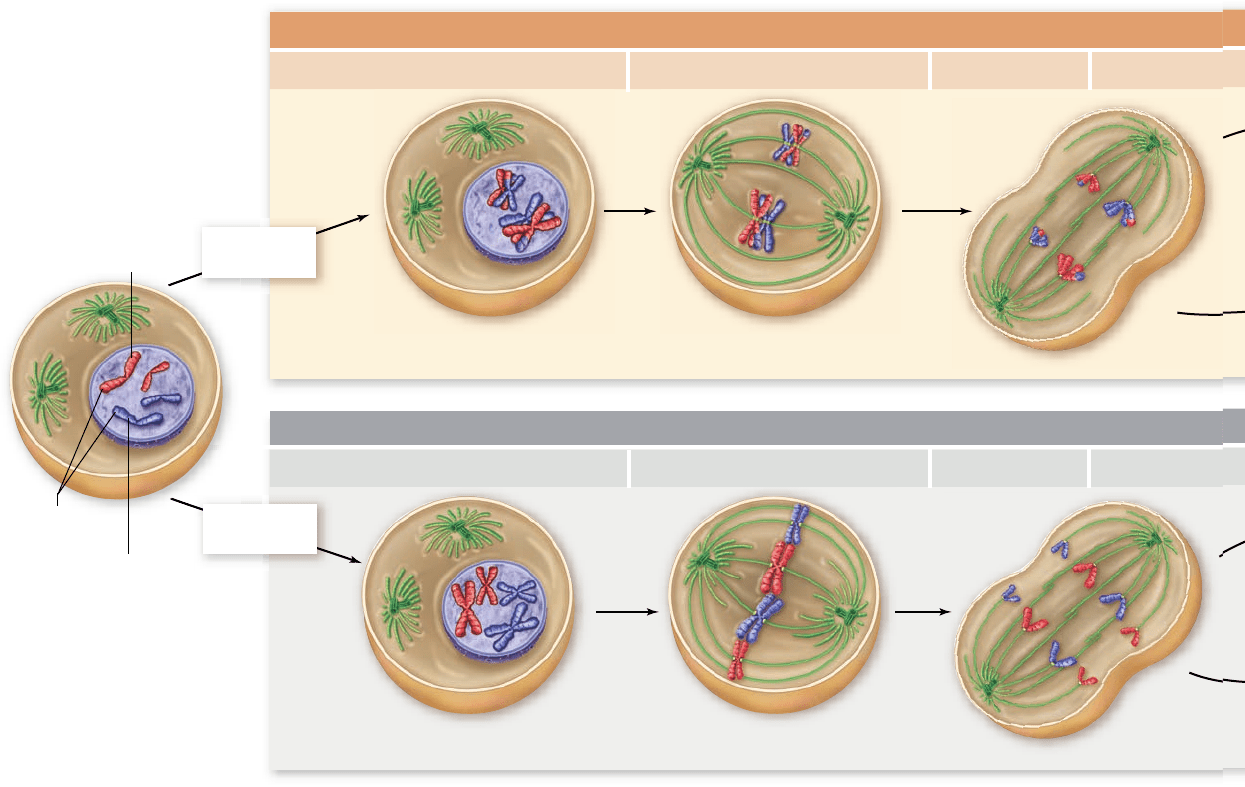
MITOSIS
Parent cell (2n)
Homologous chromosomes
do not pair
.
Maternal
homologue
Paternal
homologue
Homologous
chromosomes
Homologous chromosomes pair;
synapsis and crossing over occur.
Paired homologous chromosomes
align on metaphase plate.
Individual homologues align
on metaphase plate.
MEIOSIS I
Prophase I Metaphase I Anaphase I Telophase I
Prophase Metaphase Anaphase Telophase
Chromosome
replication
Chromosome
replication
Replication is suppressed
between meiotic divisions
After a mitotic division, a new round of DNA replication
must occur before the next division. For meiosis to succeed
in halving the number of chromosomes, this replication
must be suppressed between the two divisions. The detailed
mechanism of suppression of replication between meiotic
division is unknown. One clue is the observation that the
level of one of the cyclins, cyclin B, is reduced between mei-
otic divisions, but is not lost completely, as it is between
mitotic divisions.
During mitosis, the destruction of mitotic cyclin is nec-
essary for a cell to enter another division cycle. The result of
this maintenance of cyclin B between meiotic divisions in
germ-line cells is the failure to form initiation complexes nec-
essary for DNA replication to proceed. This failure to form
initiation complexes appears to be critical to suppressing
DNA replication.
Sister kinetochores are attached
to the same pole during meiosis I
The cosegregation of sister centromeres requires that the kine-
tochores of sister chromatids are attached to the same pole dur-
ing meiosis I. This attachment is in contrast to both mitosis (see
figure 11.8 ) and meiosis II, in which sister kinetochores must
become attached to opposite poles.
The underlying basis of this monopolar attachment of
sister kinetochores is unclear, but it seems to be based on struc-
tural differences between centromere–kinetochore complexes
in meiosis I and in mitosis. Mitotic kinetochores visualized with
the electron microscope appear to be recessed, making bipolar
attachment more likely. Meiosis I kinetochores protrude more,
making monopolar attachment easier.
It is clear that both the maintenance of sister chromatid
cohesion at the centromere and monopolar attachment are re-
quired for the segregation of homologues that distinguishes
meiosis I from mitosis.
216
part
III
Genetic and Molecular Biology
rav32223_ch11_207-220.indd 216rav32223_ch11_207-220.indd 216 11/9/09 12:12:51 PM11/9/09 12:12:51 PM
Apago PDF Enhancer
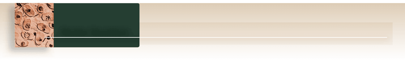
Question: Why are cohesin proteins at the centromeres of sister chromatids not destroyed at anaphase I of meiosis?
Hypothesis: Meiosis-specific cohesin component Rec8 is protected by another protein at centromeres.
Prediction: If Rec8 and the centromere protecting protein are both expressed in mitotic cells, chromosome separation will be prevented. This is lethal to a dividing cell.
Test: Fission yeast strain is designed to produce Rec8 instead of normal mitotic cohesin. These cells are transformed with a cDNA library that expresses all cellular
proteins. Transformed cells are duplicated onto media containing dye for dead cells (allows expression of Rec8 and cDNA), and media that will result in loss of
plasmid cDNA (expresses only Rec8). Cells containing cDNA for protecting protein will be dead in presence of Rec8.
Result: Transformed cells that die on the plates where Rec8 is coexpressed with cDNA identify the protecting protein. When the cDNA is extracted and analyzed, the
encoded protein localizes to the centromeres of meiotic cells.
Conclusion: This screen identifies a protein with Rec8 protecting activity.
Further Experiments: If the gene encoding the protecting protein is deleted from cells, what would be the expected phenotype? In mitotic cells? In meiotic cells?
SCIENTIFIC THINKING
Strain that expresses
Rec8 in mitosis
cDNA library that
expresses all proteins
Extract plasmid
containing cDNA
Red colony = dead cells
Expresses cDNA + Rec8
Expresses Rec8 alone
Homologous chromosomes separate;
sister chromatids remain together.
MEIOSIS II
Prophase II Metaphase II Anaphase II Telophase II
Chromosomes align, sister chromatids separate, and four haploid cells result,
each containing half the original number of homologues.
Four
daughter
cells
(each n)
Sister chromatids separate,
cytokinesis occurs, and two cells
result, each containing the
original number of homologues.
Two
daughter
cells
(each 2n)
Figure 11.9
Identi cation of meiosis-speci c cohesin protector.
Figure 11.8
A comparison of meiosis and mitosis. Meiosis involves two nuclear divisions
with no DNA replication between them. It thus produces four daughter cells, each with half the
original number of chromosomes. Crossing over occurs in prophase I of meiosis. Mitosis involves a
single nuclear division after DNA replication. It thus produces two daughter cells, each containing
the original number of chromosomes.
Inquiry question
?
If the chromosomes of a mitotic cell behaved the same as chromosomes in meiosis I, would the
resulting cells have the proper chromosomal constitution?
chapter
11
Sexual Reproduction and Meiosis
217www.ravenbiology.com
rav32223_ch11_207-220.indd 217rav32223_ch11_207-220.indd 217 11/9/09 12:12:56 PM11/9/09 12:12:56 PM
Apago PDF Enhancer
11.3 The Process of Meiosis (see gures 11.7 and 11.8)
Prophase I sets the stage for the reductive division.
Meiotic cells have an interphase period similar to mitosis with G
1
, S,
and G
2
phases. This is followed by prophase I in which homologous
chromosomes align along their entire length. The sister chromatids
are held together by cohesin proteins. Homologues exchange
chromosomal material by crossing over, which assists in holding
the homologues together during meiosis I. The nuclear envelope
disperses and the spindle apparatus forms.
During metaphase I, paired homologues align.
Spindle bers attach to the kinetochores of the homologues; the
kinetochores of sister chromatids behave as a single unit. Homologues
of each pair become attached by kinetochore microtubules to opposite
poles, and homologous pairs move to the metaphase plate as a unit. The
orientation of each homologous pair on the equator is random; either the
maternal or paternal homologue may be oriented toward a given pole.
Anaphase I results from the di erential loss of sister chromatid
cohesion along the arms.
During anaphase I the homologues of each pair are pulled to
opposite poles as kinetochore microtubules shorten. Loss of sister
chromatid cohesion on the arms but not at the centromeres allows
homologues to separate, but sister chromatids to stay together.
This is due to the loss of cohesin proteins on the arms but not at
the centromere. At the end of anaphase I each pole has a complete
set of haploid chromosomes, consisting of one member of each
homologous pair. Because of the random orientation of homologous
pairs at metaphase I, meiosis I results in the independent assortment
of maternal and paternal chromosomes in gametes.
Telophase I completes meiosis I.
During telophase I the nuclear envelope re-forms around each
daughter nucleus. This phase does not occur in all species.
Cytokinesis may or may not occur after telophase I.
11.1 Sexual Reproduction Requires Meiosis
( gure 11.2)
Meiosis reduces the number of chromosomes.
Eggs and sperm are haploid (1n) cells,
which contain one set of all
chromosomes, and products of meiotic division.
Sexual life cycles have both haploid and diploid stages.
During fertilization, or syngamy, the fusion of two haploid
gametes results in a diploid (2n) zygote, which contains two sets of
chromosomes. Meiosis and fertilization constitute a reproductive
cycle in sexual organisms as they alternate between diploid and
haploid chromosome numbers. Somatic cells divide by mitosis and
form the body of an organism.
Germ-line cells are set aside early in animal development.
Cells that eventually will form haploid gametes by meiosis are called
germ-line cells. These are set aside early in development in animals.
11.2 Features of Meiosis
Homologous chromosomes pair during meiosis.
The pairing of homologous chromosomes, called synapsis, occurs
during early prophase I. Paired homologues are often joined by the
synaptonemal complex (see gure 11.3). During synapsis, crossing
over occurs between homologous chromosomes, exchanging
chromosomal material (see gure 11.4). Because the homologues
are paired, they move as a unit to the metaphase plate during
metaphase I. During anaphase I, homologues of each pair are pulled
to opposite poles, producing two cells that each have one complete
set of chromosomes.
Meiosis features two divisions with one round of DNA replication.
Meiosis II is like mitosis but without replication of DNA. Sister
chromatids are pulled to opposite poles to yield four haploid cells.
Chapter Review
of heredity. The different cells produced by meiosis form the
basis for understanding the behavior of observable traits in ge-
netic crosses. In the next two chapters we will follow the behav-
ior of traits in genetic crosses and see how this correlates with
the behavior of chromosomes in meiosis.
Learning Outcomes Review 11.4
Meiosis is characterized by homologue pairing and crossing over ; by loss of
sister chromatid cohesion in the arms, but not at the centromere at the fi rst
division; and by the suppression of DNA replication between the two meiotic
divisions. Sister chromatid cohesion would be disastrous in mitosis, but in
meiosis I it allows the reduction of chromosome number. If replication were
not suppressed between meiosis I and meiosis II, gametes would be diploid,
and zygotes would be tetraploid.
■ What features of meiosis lead to genetic variation in the
products?
Meiosis produces cells that
are not identical
The daughter cells produced by mitosis are identical to the pa-
rental cell, at least in terms of their chromosomal constitution.
This exact copying is critical to producing new cells for growth,
for development, and for wound healing. Meiosis, because of
the random orientation of different chromosomes at the first
meiotic division and because of crossing over, rarely produces
cells that are identical . The gametes from meiosis all carry an
entire haploid set of chromosomes, but these chromosomes are
a mixture of maternal and paternal homologues; furthermore,
the homologues themselves have exchanged material by cross-
ing over. The resulting variation is essential for evolution and is
the reason that sexually reproducing populations have much
greater variation than asexually reproducing ones.
Meiosis is not only critical for the process of sexual repro-
duction, but is also the foundation for understanding the basis
218
part
III
Genetic and Molecular Biology
rav32223_ch11_207-220.indd 218rav32223_ch11_207-220.indd 218 11/9/09 12:13:02 PM11/9/09 12:13:02 PM
Apago PDF Enhancer
Achiasmate segregation of homologues is possible.
Although homologues are usually held together by chiasmata, some
systems are able to segregate chromosomes without this.
Meiosis II is like a mitotic division without DNA replication.
A brief interphase with no DNA replication occurs after meiosis I.
During meiosis II, cohesin proteins at the centromeres that hold
sister chromatids together are destroyed, allowing each to migrate to
opposite poles of the cell. The result of meiosis I and II is four cells,
each containing haploid sets of chromosomes that are not identical.
Once completed, the haploid cells may produce gametes or divide
mitotically to produce even more gametes or haploid adults.
Errors in meiosis produce aneuploid gametes.
Errors occur during meiosis because of nondisjunction, the failure of
chromosomes to move to opposite poles. It may result in aneuploid
gametes: one gamete with no chromosome, and another gamete with
two copies of a chromosome.
11.4 Summing Up: Meiosis Versus Mitosis
Four distinct features of meiosis I are not found in mitosis: Maternal
and paternal homologues pair, and exchange genetic information
by crossing over; the kinetochores of sister chromatids function as
a unit during meiosis I, allowing sister chromatids to cosegregate
during anaphase I; kinetochores of sister chromatids are connected to
a single pole in meiosis I and to opposite poles in mitosis; and DNA
replication is suppressed between meiosis I and meiosis II.
Homologous pairing is speci c to meiosis.
How homologues nd each other during meiosis is not known. The
proteins of the synaptonemal complex do not seem to be conserved in
different species, but there are meiosis-speci c cohesin proteins. These
are involved in the differential destruction of cohesins on the arms versus
the centromere during meiosis I. The recombination process that occurs
between paired homologues is better known. This process uses proteins
involved in DNA repair and starts with a double-stranded break in DNA.
Sister chromatid cohesion is maintained through meiosis I
but released in meiosis II.
Shugoshin protein protects centromeric cohesin in anaphase I, so
that sister chromatids remain connected. Cohesins on the arms are
not protected and are thus degraded during anaphase I, allowing
homologues to move to opposite poles.
Sister kinetochores are attached to the same pole during meiosis I.
Kinetochores of sister chromatids must be attached to the same spindle
bers (monopolar attachment) to segregate together.
Replication is suppressed between meiotic divisions.
Suppression of replication may be related to the maintenance of
some cyclin proteins that are degraded at the end of mitosis.
Meiosis produces cells that are not identical.
Because of the independent assortment of homologues and the process
of crossing over, gametes show great variation.
5. During anaphase I
a. sister chromatids separate and move to the poles.
b. homologous chromosomes move to opposite poles.
c. homologous chromosomes align at the middle of the cell.
d. all the chromosomes align independently at the middle of
the cell.
6. At metaphase I the kinetochores of sister chromatids are
a. attached to microtubules from the same pole.
b. attached to microtubules from opposite poles.
c. held together with cohesin proteins.
d. not attached to any microtubules.
7. What occurs during anaphase of meiosis II?
a. The homologous chromosomes align.
b. Sister chromatids are pulled to opposite poles.
c. Homologous chromosomes are pulled to opposite poles.
d. The haploid chromosomes line up.
APPLY
1. Which of the following does not contribute to genetic diversity?
a. Independent assortment
b. Recombination
c. Metaphase of meiosis II
d. Metaphase of meiosis I
UNDERSTAND
1. In comparing somatic cells and gametes, somatic cells are
a. diploid with half the number of chromosomes.
b. haploid with half the number of chromosomes.
c. diploid with twice the number of chromosomes.
d. haploid with twice the number of chromosomes.
2. What are homologous chromosomes?
a. The two halves of a replicated chromosome
b. Two identical chromosomes from one parent
c. Two genetically identical chromosomes, one from
each parent
d. Two genetically similar chromosomes, one from
each parent
3. Chiasmata form
a. between homologous chromosomes.
b. sister chromatids.
c. between replicated copies of the same chromosomes.
d. sex chromosomes but not autosomes.
4. Crossing over involves each of the following with the
exception of
a. the transfer of DNA between two nonsister chromatids.
b. the transfer of DNA between two sister chromatids.
c. the formation of a synaptonemal complex.
d. the alignment of homologous chromosomes.
Review Questions
chapter
11
Sexual Reproduction and Meiosis
219www.ravenbiology.com
rav32223_ch11_207-220.indd 219rav32223_ch11_207-220.indd 219 11/9/09 12:13:03 PM11/9/09 12:13:03 PM

Apago PDF Enhancer
2. How does DNA replication differ between mitosis and meiosis?
a. DNA replication takes less time in meiosis because the
cells are haploid.
b. During meiosis, there is only one round of replication for
two divisions.
c. During mitosis, there is only one round of replication
every other division.
d. DNA replication is exactly the same in mitosis and meiosis.
3. Which of the following is not a distinct feature of meiosis?
a. Pairing and exchange of genetic material between
homologous chromosomes
b. Attachment of sister kinetochores to spindle microtubules
c. Movement of sister chromatids to the same pole
d. Suppression of DNA replication
4. Which phase of meiosis I is most similar to the comparable
phase in mitosis?
a. Prophase I c. Anaphase I
b. Metaphase I d. Telophase I
5. Structurally, meiotic cohesins have different components than
mitotic cohesins. This leads to the following functional difference:
a. During metaphase I, the sister kinetochores become
attached to the same pole.
b. Centromeres remain attached during anaphase I of meiosis.
c. Centromeres remain attached through both divisions.
d. Centromeric cohesins are destroyed at anaphase I, and
cohesins along the arms are destroyed at anaphase II.
6. Mutations that affect DNA repair often also affect the accuracy
of meiosis. This is because
a. the proteins involved in the repair of double-strand breaks
are also involved in crossing over.
b. the proteins involved in DNA repair are also involved in
sister chromatid cohesion.
c. DNA repair only occurs on condensed chromosomes such
as those found in meiosis.
d. cohesin proteins are also necessary for DNA repair.
SYNTHESIZE
1. Diagram the process of meiosis for an imaginary cell with six
chromosomes in a diploid cell.
a. How many homologous pairs are present in this cell? Create
a drawing that distinguishes between homologous pairs.
b. Label each homologue to indicate whether it is maternal
(M) or paternal (P).
c. Draw a new cell showing how these chromosomes would
arrange themselves during metaphase of meiosis I. Do all
the maternal homologues have to line up on the same side
of the cell?
d. How would this picture differ if you were diagramming
anaphase of meiosis II?
2. Mules are the offspring of the mating of a horse and a donkey.
Mules are unable to reproduce. A horse has a total of 64
chromosomes, whereas donkeys have 62 chromosomes. Use
your knowledge of meiosis to predict the diploid chromosome
number of a mule. Propose a possible explanation for the
inability of mules to reproduce.
3. Compare the processes of independent assortment and crossing over.
Which process has the greatest in uence on genetic diversity?
4. Aneuploid gametes are cells that contain the wrong number of
chromosomes. Aneuploidy occurs as a result of nondisjunction,
or lack of separation of the chromosomes during either phase
of meiosis.
a. At what point in meiotic cell division would nondisjunction
occur?
b. Imagine a cell had a diploid chromosome number of four.
Create a diagram to illustrate the effects of nondisjunction
of one pair of homologous chromosomes in meiosis I
versus meiosis II.
ONLINE RESOURCE
www.ravenbiology.com
Understand, Apply, and Synthesize—enhance your study with
animations that bring concepts to life and practice tests to assess
your understanding. Your instructor may also recommend the
interactive eBook, individualized learning tools, and more.
220
part
III
Genetic and Molecular Biology
rav32223_ch11_207-220.indd 220rav32223_ch11_207-220.indd 220 11/9/09 12:13:03 PM11/9/09 12:13:03 PM
Apago PDF Enhancer
E
Chapter
12
Patterns of Inheritance
Chapter Outline
12.1 The Mystery of Heredity
12.2 Monohybrid Crosses: The Principle
of Segregation
12.3 Dihybrid Crosses: The Principle
of Independent Assortment
12.4 Probability: Predicting the Results
of Crosses
12.5 The Testcross: Revealing Unknown Genotypes
12.6 Extensions to Mendel
Introduction
Every living creature is a product of the long evolutionary history of life on Earth. All organisms share this history, but as far
as we know, only humans wonder about the processes that led to their origin and investigate the possibilities. We are far
from understanding everything about our origins, but we have learned a great deal. Like a partially completed jigsaw puzzle,
the boundaries of this elaborate question have fallen into place, and much of the internal structure is becoming apparent. In
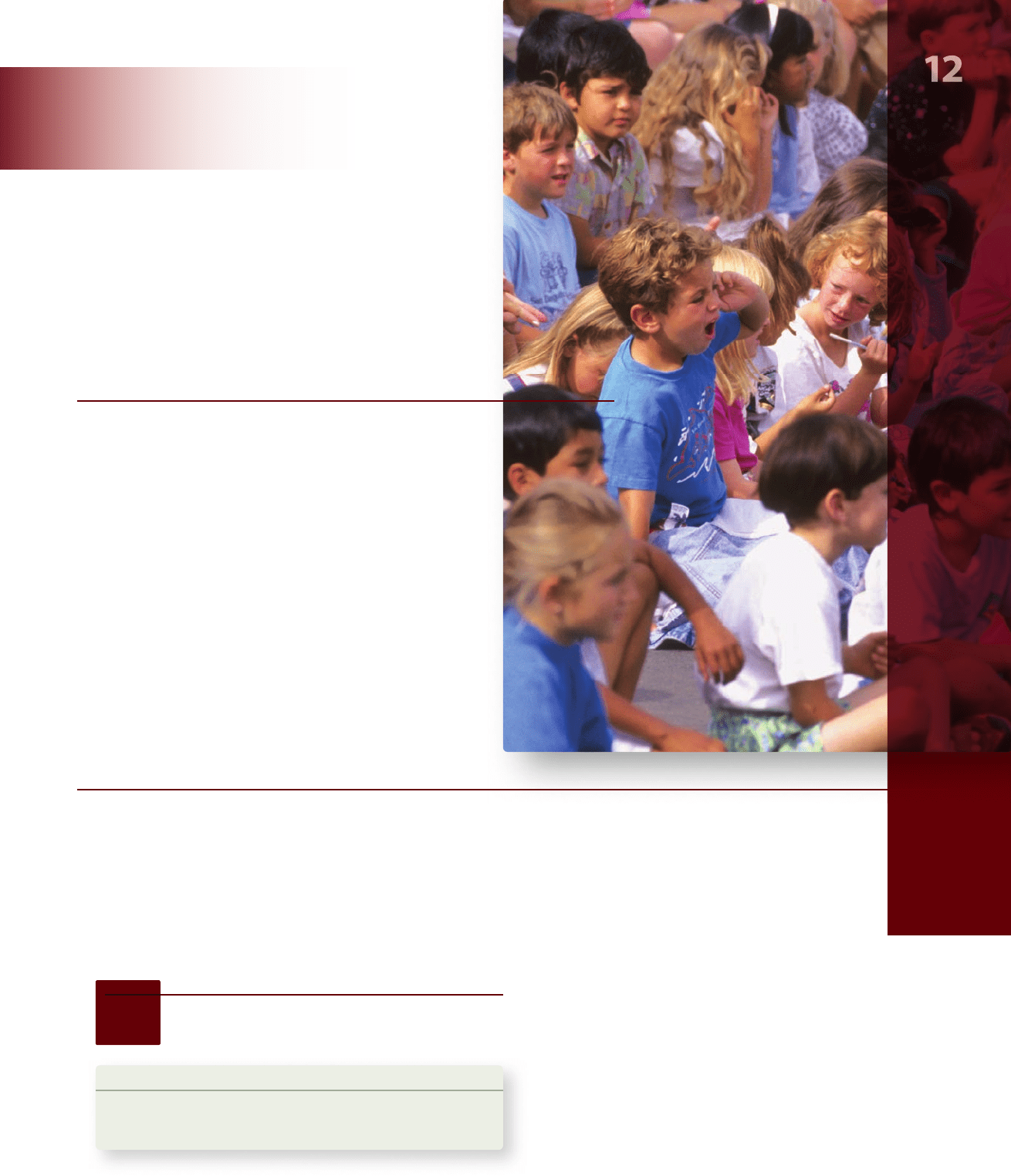
this chapter, we discuss one piece of the puzzle—the enigma of heredity. Why do individuals, like the children in this picture,
differ so much in appearance despite the fact that we are all members of the same species? And, why do members of a single
family tend to resemble one another more than they resemble members of other families?
CHAPTER
As far back as written records go, patterns of resemblance
among the members of particular families have been noted and
commented on (figure 12.1), but there was no coherent model
to explain these patterns. Before the 20th century, two concepts
provided the basis for most thinking about heredity. The first
was that heredity occurs within species. The second was that
traits are transmitted directly from parents to offspring. Taken
together, these ideas led to a view of inheritance as resulting
from a blending of traits within fixed, unchanging species.
12.1
The Mystery of Heredity
Learning Outcomes
Describe explanations for inheritance prior to Mendel.1.
Explain the advantages of Mendel’s experimental system.2.
rav32223_ch12_221-238.indd 221rav32223_ch12_221-238.indd 221 11/6/09 2:44:53 PM11/6/09 2:44:53 PM