Raven P.H., Johnson G.B., Mason K.A. Biology (Ninth Edition)
Подождите немного. Документ загружается.

Apago PDF Enhancer
5„ cap
Eukaryotic DNA template
exons
introns
1 2 3 4 1 2 3 4
Transcription
Primary RNA transcript
Introns are cut out,
and coding regions are
spliced together.
5„ cap
Mature RNA transcript
3„ poly-A tail
Degraded
mRNA
3„ poly-A tail
mRNA–cDNA hybrid
Isolation of mRNA
Addition of reverse
transcriptase
Addition of mRNA-
degrading enzymes
DNA polymerase
Double-stranded cDNA
with no introns
Reverse
transcriptase
Reverse
transcriptase
utilizes mRNA
to create cDNA.
use a known, specific DNA molecule to find its partner in a
complex mixture.
Any single-stranded nucleic acid (DNA or RNA) can be
tagged with a radioactive label or with another detectable label,
such as a fluorescent dye. This can then be used as a probe to
identify its complement in a complex mixture of DNA or RNA.
This renaturing is termed hybridization because the combina-
tion of labeled probe and unlabeled DNA form a hybrid mol-
ecule through base-pairing.
Probes have been made historically by a variety of tech-
niques. One technique involved isolating a protein of interest
books, which is organized and catalogued, a DNA library is a
random collection of overlapping DNA fragments. We explore
how to find a sequence of interest in this random collection
later in the chapter.
Reverse transcriptase can make
a DNA copy of RNA
In addition to genomic libraries, investigators often wish to iso-
late only the expressed part of genes. The structure of eukaryotic
genes is such that the mRNA may be much smaller than the
gene itself due to the presence of introns in the gene. After
transcription by RNA polymerase II, the primary transcript is
spliced to produce the mRNA (chapter 15 ). Because of this,
genomic libraries are crucial to understanding the structure of
the gene, but are not of much use if we want to express the gene
in a bacterial species, whose genes do not contain introns and
which has no mechanism for splicing.
A library of only expressed sequences represents a much
smaller amount of DNA than the entire genome. The starting
point for a cDNA library is isolated mRNA representing the
genes expressed in a specific tissue at a specific developmental
stage. Such a library of expressed sequences is made possible by
the use of another enzyme: reverse transcriptase.
Reverse transcriptase was isolated from a class of viruses
called retroviruses. The life cycle of a retrovirus requires mak-
ing a DNA copy from its RNA genome. We can take advantage
of the activity of the retrovirus enzyme to make DNA copies
from isolated mRNA. DNA copies of mRNA are called
complementary DNA (cDNA) (figure 17.5). A cDNA library
is made by first isolating mRNA from genes being expressed
and then using the reverse transcriptase enzyme to make cDNA
from the mRNA. The cDNA is then used to make a library, as
mentioned earlier. These cDNA libraries are extremely useful
and are commonly made to represent the genes expressed in
many different tissues or cells. While all genomic libraries made
from an individual will be identical, cDNA libraries from the
same cells at different developmental stages or different tissues
will each be distinct.
Inquiry question
?
Suppose you wanted a copy of a section of a eukaryotic
genome that included the introns and exons. Would the
creation of cDNA be a good way to go about this?
Hybridization allows identi cation of speci c
DNAs in complex mixtures
The technique of molecular hybridization is commonly used
to identify specific DNAs in complex mixtures such as librar-
ies. Hybridization, also called annealing, takes advantage of
the specificity of base-pairing between the two strands of
DNA. If a DNA molecule is denatured, that is, the two strands
are separated, the strands can only reassociate with partners
that have the correct complementary sequence. Molecular
biologists can take advantage of this feature experimentally to
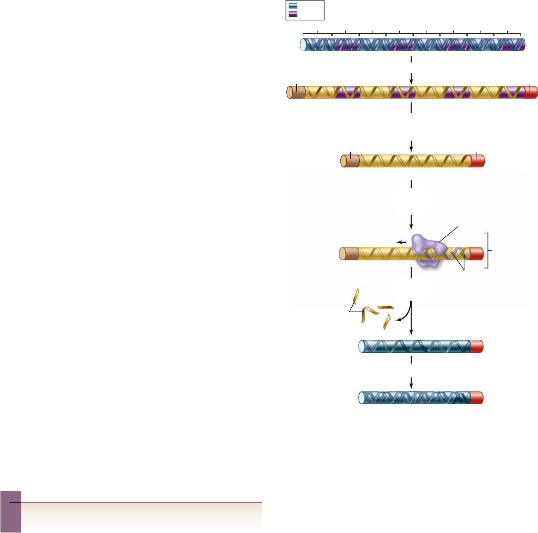
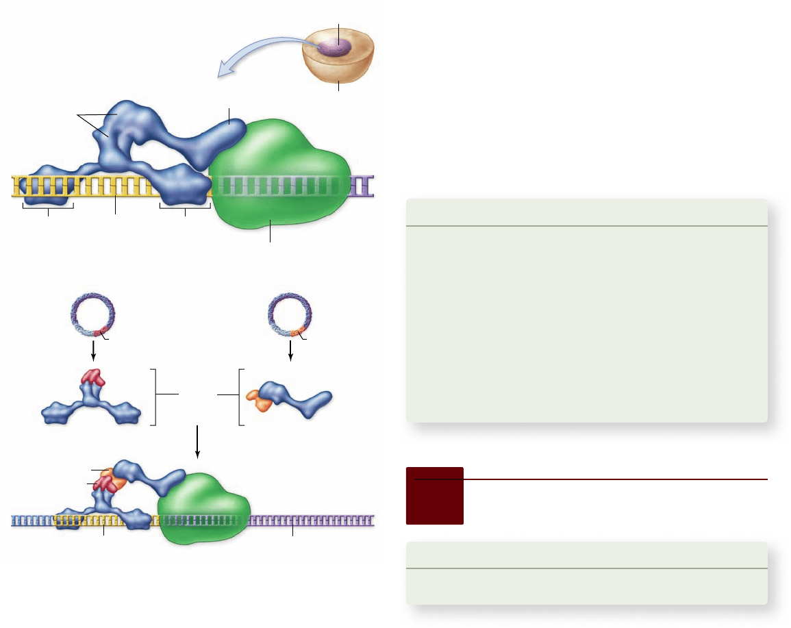
Figure 17.5
The formation of cDNA.
A mature mRNA
transcript is usually much smaller than the gene due to the loss of
intron sequences by splicing. mRNA is isolated from the cytoplasm
of a cell, which the enzyme reverse transcriptase uses as a template
to make a DNA strand complementary to the mRNA . That newly
made strand of DNA is the template for the enzyme DNA
polymerase, which assembles a complementary DNA strand along
it, producing cDNA—a double-stranded DNA version of the
intron-free mRNA.
332
part
III
Genetic and Molecular Biology
rav32223_ch17_327-351.indd 332rav32223_ch17_327-351.indd 332 11/10/09 2:43:16 PM11/10/09 2:43:16 PM
Apago PDF Enhancer
Film
Filter paper
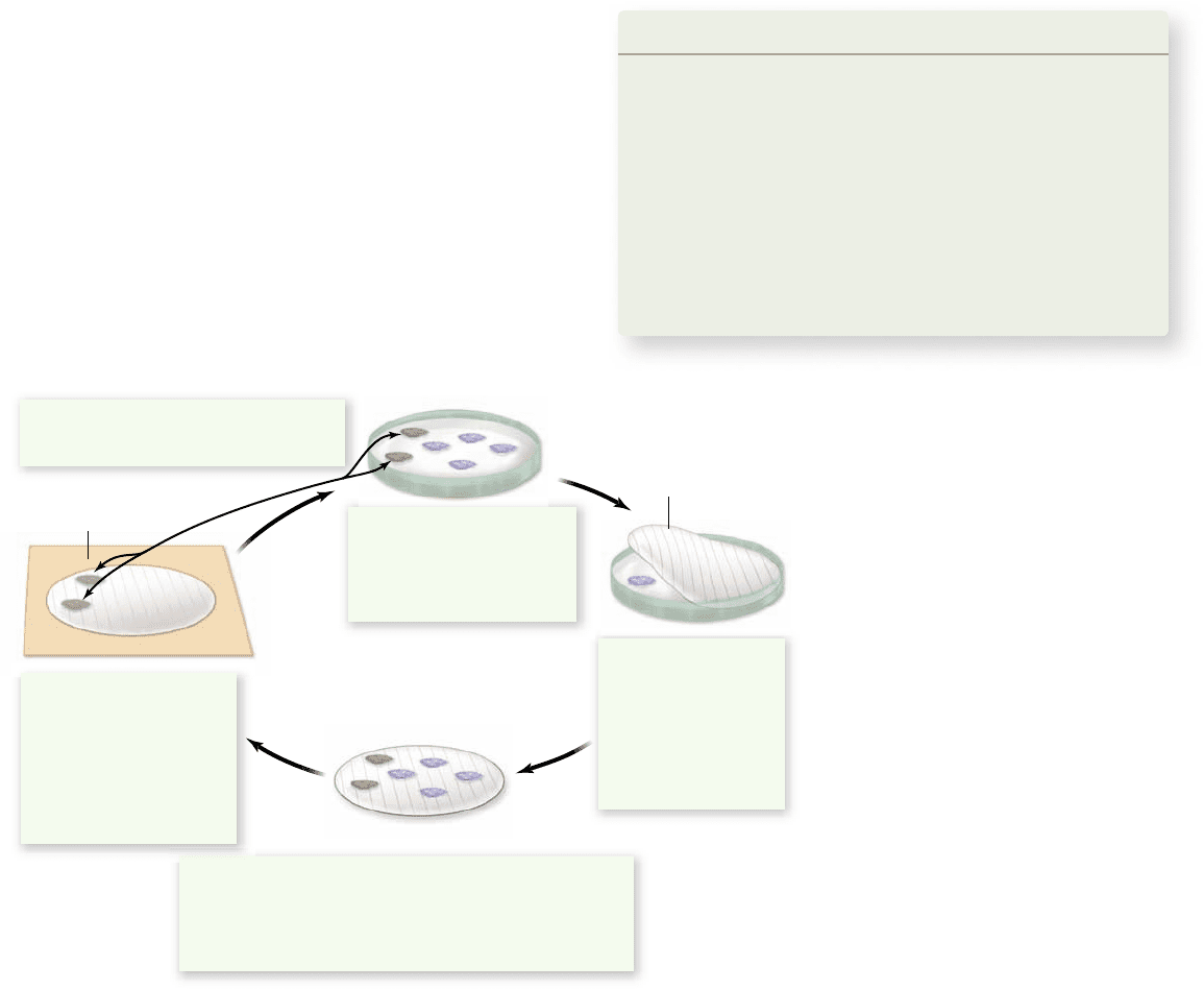
1. Colonies of plasmid
containing bacteria, each
containing a single DNA
from the library, are grown
on agar.
3. The filter is washed with a solution to break the cells open
and denature the DNA, which sticks to the filter at the site
of each colony. The filter is incubated with a radioactively
labeled probe that can form hybrids with complementary
DNA in the gene of interest.
4. The only sites on the
filter that will retain
probe DNA will contain
DNA complementary to
the probe. These
represent the sites of
colonies containing the
gene of interest.
5. A comparison with the original plate
identifies the colony containing the gene.
2. A replica of the plate
is made by pressing
a piece of filter paper
against the agar and
bacterial colonies.
Some cells from
each colony adhere
to the filter.
Stage 2: Replicating the library
Once the library has been grown on plates, a replica can be
made by laying a piece of filter paper on the plate; some of
the viruses or cells in each colony will stick to the filter, and
some will be left on the plate. The result is a copy of the li-
brary on a piece of filter paper. The DNA can be affixed to
the filter paper by baking or by cross-linking it to the filter
using UV light.
Stage 3: Screening the library
Once a replica of the library has been formed on a filter, a spe-
cific clone can be identified by hybridization. The probe, which
represents the specific sequence of interest, is labeled with a
radioactive nucleotide. The probe is then added to the filters
with the library replicated on them. Film sensitive to radio-
active emissions is then placed in contact with the filters; where
radioactivity is present, a dark spot appears on the film. When
the film is aligned with the original plate, the clone of interest
can be identified (see figure 17.6).
Learning Outcomes Review 17.2
Molecular cloning is the isolation and amplifi cation of a specifi c DNA
sequence. A vector is a carrier into which a sequence of interest may be
introduced. The most common vectors are plasmids and phages. The
vector takes the sequence into a cell, which then multiplies, copying its own
DNA along with that of the vector. DNA libraries are representations of
complex mixtures of DNA, such as an entire genome, stored in a host–vector
system. DNA libraries are often screened for specifi c clones using
molecular hybridization.
■ How does a gene’s sequence in a cDNA library compare
with the sequence of the gene itself?
and then chemically sequencing the protein. With the protein
sequence in hand, the DNA sequence could be predicted using
the genetic code. This information can then be used to make a
synthetic DNA for use as a probe.
Speci c clones can be isolated from a library
The isolation of a specific clone from the random collection
that is a DNA library is akin to finding the proverbial needle in
a haystack. It requires some information about the gene of in-
terest. For example, many of the first genes isolated were those
that are highly expressed in a specific cell type, such as the
globin genes that encode the proteins found in the oxygen car-
rier hemoglobin.
Hybridization is the most common way of identifying a
clone within a DNA library. This procedure is outlined for a
DNA library in a plasmid vector in figure 17.6.
In the early days of molecular biology, individual investi-
gators made their own DNA libraries, as is shown earlier in
figure 17.4. Now, genomic and cDNA libraries are commer-
cially available for a large number of organisms. Screening
such a library involves growing the library on agar plates, mak-
ing a replica of the library, and screening for the cloned se-
quence of interest.
Stage 1: Plating the library
Physically, the library is either a collection of bacterial viruses
that each contain an inserted DNA, or bacterial cells that each
harbor a plasmid or artificial chromosome with inserted DNA.
To find a specific clone, the library needs to be represented in
an organized fashion. Figure 17.6 shows this representation for
a plasmid vector. The library of bacteria containing plasmids is
grown on agar plates at a high density, but not so high that in-
dividual colonies cannot be distinguished.
Figure 17.6
Screening a library
using hybridization.
This technique
takes advantage of DNA’s ability to be
denatured and renatured, with
complementary strands nding each other.
Cells containing the library are plated on
agar gel. A replica of the plates is made
using special lter paper, nitrocellulose or
nylon, which binds to single-stranded
DNA. The lter paper with replica
colonies is treated to lyse the cells and
denature the DNA, producing a pattern of
DNA bound to the lter that corresponds
to the pattern of colonies. When a
radioactive probe is added, it nds
complementary DNA and forms hybrids at
the site of colonies that contained the gene
of interest.
chapter
17
Biotechnology
333www.ravenbiology.com
rav32223_ch17_327-351.indd 333rav32223_ch17_327-351.indd 333 11/10/09 2:43:17 PM11/10/09 2:43:17 PM
Apago PDF Enhancer
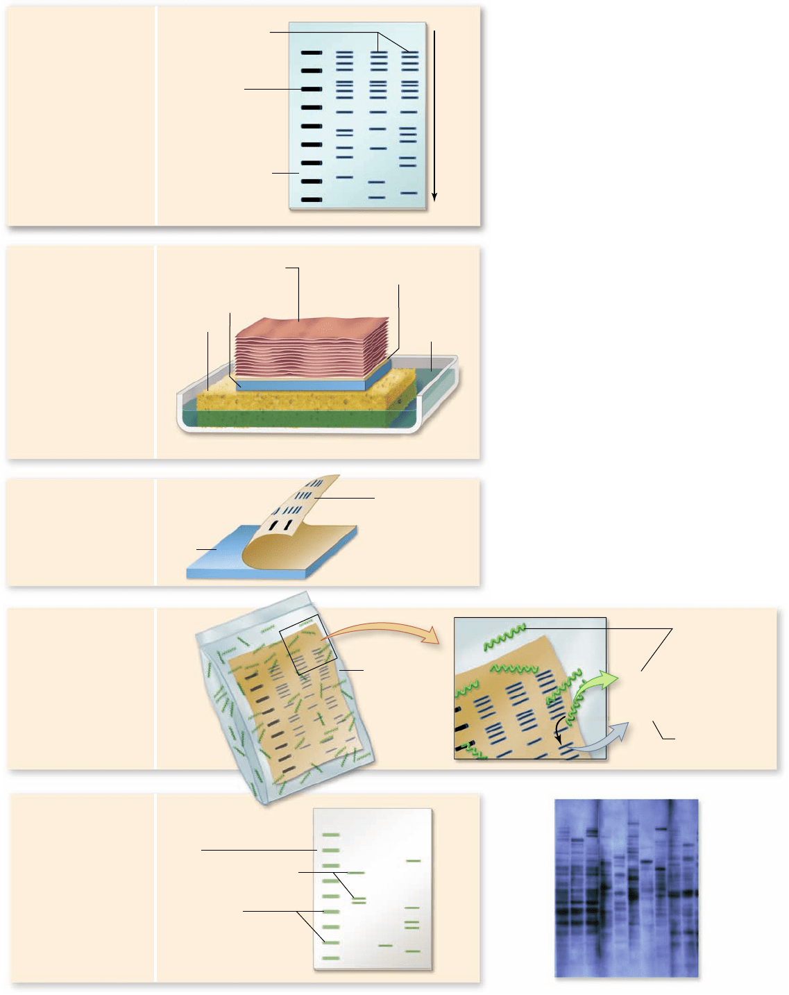
3. DNA in the gel is
transferred, or
“blotted,” onto the
nitrocellulose.
4. Nitrocellulose with
bound DNA is
incubated with
radioactively labeled
nucleic acids and is
then rinsed.
5. Photographic film is laid
over the filter and is
exposed only in areas
that contain radioactivity
(autoradiography).
Bands on the film
represent DNA in the
gel that is
complementary to the
probe sequence.
Size markers
Hybridized nucleic acids
Film
Gel
Buffer
Sponge
Nitrocellulose filter
Gel
1. Electrophoresis is
performed, using
radioactively labeled
markers as a size
guide in the first lane.
Test nucleic acids
Radioactively
labeled markers
with specific sizes
Electrophoretic gel
Electrophoresis
DNA fragments
within bands
Radioactive
probe (single-
stranded DNA)
2. The gel is covered
with a sheet of
nitrocellulose and
placed in a tray of
buffer on top of a
sponge. Alkaline
chemicals in the buffer
denature the DNA into
single strands. The
buffer wicks its way up
through the gel and
nitrocellulose into a
stack of paper towels
placed on top of the
nitrocellulose.
Stack of paper towels
—TTACC—
—AATGG—
Sealed
container
Nitrocellulose
paper now
contains nucleic
acid “print”
Figure 17.7
The Southern blot
procedure.
Edwin M. Southern developed
this procedure in 1975 to enable DNA
fragments of interest to be visualized in a
complex sample containing many other
fragments of similar size. In steps 1–3, the DNA
is separated on a gel, and then transferred
(“blotted”) onto a solid support medium such as
nitrocellulose paper or a nylon membrane.
Sequences of interest can be detected by using a
radioactively labeled probe. This probe (usually
several hundred nucleotides in length) of
single-stranded DNA (or an mRNA
complementary to the gene of interest) is
incubated with the lter containing the DNA
fragments. All DNA fragments that contain
nucleotide sequences complementary to the
probe will form hybrids with the probe. Only a
short segment of the probe and the
complementary sequence are shown in panel 4.
The fragments differ in size, with the smallest
moving the farthest in the gel. The fragments
of interest are then detected using photographic
lm. A representative image is shown in panel 5.
The use of lm for detection is being replaced
by phosphor imagers, computer-controlled
devices that have electronic sensors for light or
radioactive emissions.
334
part
III
Genetic and Molecular Biology
rav32223_ch17_327-351.indd 334rav32223_ch17_327-351.indd 334 11/10/09 2:43:17 PM11/10/09 2:43:17 PM
Apago PDF Enhancer
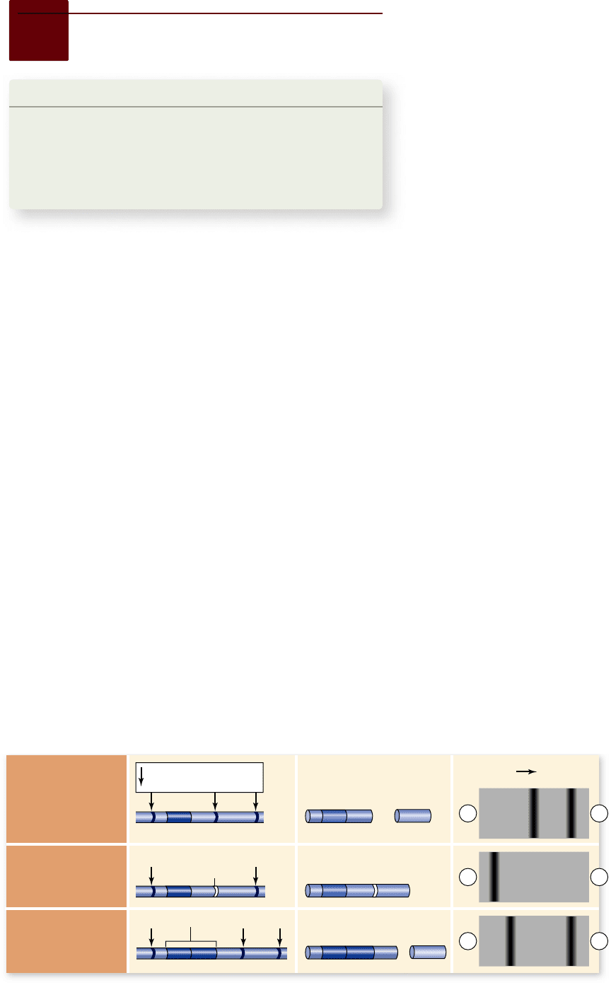
a. Three different
DNA duplexes
b. Cut DNA c. Gel electrophoresis of
restriction fragments
Original Sequence
of Restriction Sites
(no mutations)
Point Mutations
Change the
Sequence of
Restriction Sites
Sequence
Repetitions Can
Occur Between
Restriction Sites
Larger
fragments
Smaller
fragments
restriction endonuclease
cutting sites
Sequence duplication
Single base-pair
change
+
+
+
+
+
-
-
-
Learning Outcomes
Explain the Southern blotting method of identifying genes.1.
Compare endogenous DNA replication with sequencing 2.
and with the polymerase chain reaction.
Explain how the yeast system is used to study protein–3.
protein interactions.
Molecular cloning provides specific DNA for further manipulation
and analysis. The number of ways that DNA can be manipulated
could fill the rest of this book, but for our purposes, we will highlight
a few important methods of analysis and uses of molecular clones.
Restriction maps provide molecular “landmarks”
If you are new to a city, the easiest way to find your way around
is to obtain a map and compare that map with your surround-
ings. In a similar fashion, molecular biologists need maps to
analyze and compare cloned DNAs.
The first kind of physical maps were restriction maps that
included the location and order of sites cut by the battery of
restriction enzymes available. Initially, these maps were created
by cutting the DNA with different enzymes, separating the
fragments by gel electrophoresis, and analyzing the resulting
patterns. Although this method is still in use, many restriction
maps are now generated by computer searching of known DNA
sequences for the sites cut by restriction enzymes.
Southern blotting reveals DNA di erences
Once a gene has been cloned, it may be used as a probe to iden-
tify the same or a similar gene in DNA isolated from a cell or
tissue (figure 17.7). In this procedure, called a Southern blot,
DNA from the sample is cleaved into fragments with a restric-
tion endonuclease, and the fragments are separated by gel elec-
trophoresis. The double-stranded helix of each DNA fragment
is then denatured into single strands by making the pH of the
gel basic. Then the gel is “blotted” with a sheet of filter paper,
transferring some of the DNA strands to the sheet.
17.3
DNA Analysis
Next, the filter is incubated with a labeled probe consist-
ing of purified, single-stranded DNA corresponding to a spe-
cific gene (or mRNA transcribed from that gene). Any fragment
that has a nucleotide sequence complementary to the probe’s
sequence hybridizes with the probe (see figure 17.7).
This kind of blotting technique has also been adapted for
use with RNA and proteins. When mRNA is separated by electro-
phoresis, the technique is called a Northern blot. The methodol-
ogy is the same except for the starting material (mRNA instead of
DNA) and that no denaturation step is required. Proteins can also
be separated by electrophoresis and blotted by a procedure called
a Western blot. In this case both the electrophoresis and the de-
tection step are different from Southern blotting. The detection,
in this case, requires an antibody that can bind to one protein.
The names of these techniques all go back to the original
investigator, the British biologist Edwin M. Southern; the
Northern and Western blotting names were word play on
Southern’s name using the cardinal points of the compass.
RFLP analysis
In some cases, an investigator wants to do more than find a
specific gene, but instead is looking for variation in the genes of
different individuals. One powerful way to do this is by analyz-
ing restriction fragment length polymorphisms, or RFLPs,
using Southern blotting (figure 17.8).
Point mutations that change the sequence of DNA can elim-
inate sequences recognized by restriction enzymes or create new
recognition sequences, changing the pattern of fragments seen in a
Southern blot. Sequence repetitions may also occur between the
restriction endonuclease sites, and differences in repeat number be-
tween individuals can also alter the length of the DNA fragments.
These differences can all be detected with Southern blotting.
When a genetic disease has an associated RFLP, the
RFLP can be used to diagnose the disease. Huntington disease,
cystic fibrosis, and sickle cell anemia all have associated RFLPs
that have been used as molecular markers for diagnosis.
DNA fingerprinting
RFLP analysis has been used in DNA fingerprinting. When a
probe is made for DNA that is repetitive, it often detects a large
number of fragments. These fragments are often not identical in
different individuals. We say that the population is polymorphic
for these molecular markers. These markers can be used as
Figure 17.8
Restriction
fragment length
polymorphism (RFLP)
analysis.
a. Three samples of
DNA differ in their restriction
sites due to a single base-pair
substitution in one case and a
sequence duplication in another
case. b. When the samples are cut
with a restriction endonuclease,
different numbers and sizes of
fragments are produced. c. Gel
electrophoresis separates the
fragments, and different banding
patterns result.
chapter
17
Biotechnology
335www.ravenbiology.com
rav32223_ch17_327-351.indd 335rav32223_ch17_327-351.indd 335 11/10/09 2:43:18 PM11/10/09 2:43:18 PM
Apago PDF Enhancer
Suspect’s blood
Rapist’s semen
Rapist’s semen
Victim
Victim
Suspect’s blood
NH
2
O
J
J
P
J
O
-
-
O
J
O
J
CH
2
J
5„
4„
3„ 2„
1„
H
H
N N
N
O
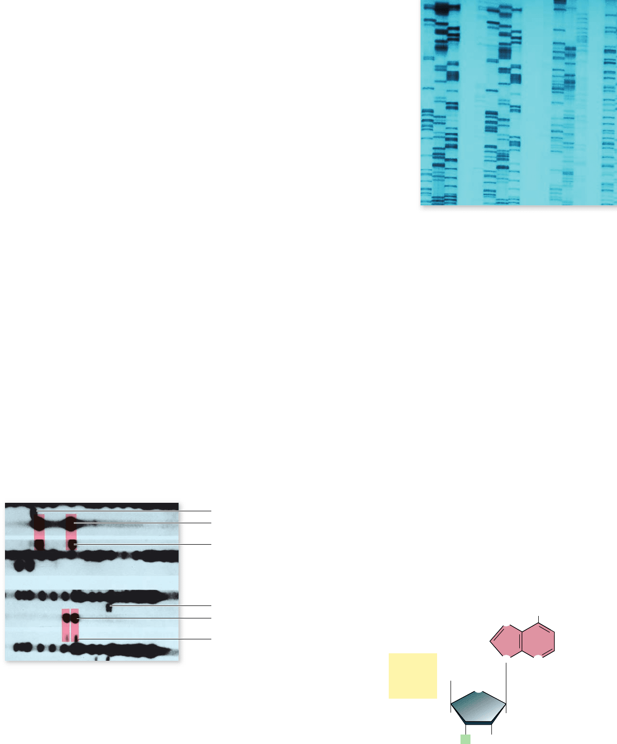
Figure 17.9
Two of the DNA pro les that led to the
conviction of Tommie Lee Andrews for rape in 1987.
The two
DNA probes seen here were used to characterize DNA isolated from
the victim, the semen left by the rapist, and the suspect. The dark
channels are multiband controls. There is a clear match between the
suspect’s DNA and the DNA of the rapist’s semen in these two pro les.
DNA “fingerprints” in criminal investigations and other iden-
tification applications.
Figure 17.9 shows the DNA fingerprints a prosecuting
attorney presented in a rape trial in 1987. They consist of auto-
radiographs, parallel bars on X-ray film. These bars can be
thought of as being similar to the product price codes on con-
sumer goods in that they may provide unique identification.
Each bar represents the position of a DNA restriction endonu-
clease fragment produced by techniques similar to those de-
scribed in figures 17.7 and 17.8. The long dark lane with many
bars in figure 17.9 represents a standardized control.
Two different probes were used to identify the restriction
fragments. A vaginal swab had been taken from the victim with-
in hours of her attack; from it, semen was collected and its DNA
analyzed for restriction endonuclease patterns.
Compare the restriction endonuclease patterns of the se-
men to that of blood from the suspect. You can see that the sus-
pect’s two patterns match that of the rapist (and are not at all like
those of the victim). The suspect was Tommie Lee Andrews, and
on November 6, 1987, the jury returned a verdict of guilty. An-
drews became the first person in the United States to be con-
victed of a crime based on DNA evidence.
Since the Andrews verdict, DNA fingerprinting evidence is
now a determining factor in at least forty percent of the criminal
cases in the United States. Although some probes highlight profiles
shared by many people, others are quite rare. Using several probes,
the probability of identity can be calculated or identity can be ruled
out. Laboratory analyses of DNA samples, however, must be carried
out properly—sloppy procedures could lead to a wrongful con-
viction. After widely publicized instances of questionable lab
procedures, national standards are being developed.
DNA fingerprinting is also used to identify human re-
mains. After the September 11, 2001 attacks on the World
Trade Centers in New York, DNA fingerprinting was the only
option for identifying some of the victims of the attack. by
2005, 1585 of the 2792 people who were missing had been
identified using DNA fingerprinting. Advances in forensic
technology, including improved DNA isolation from very small
amounts of tissue, have made it possible to identify additional
individuals since 2005.
DNA sequencing provides information
about genes and genomes
The ultimate level of analysis is determination of the actual
sequence of bases in a DNA molecule. The development of
sequencing technology has paralleled the advancement of molecu-
lar biology. As it became possible to determine the sequence of an
entire genome relatively rapidly, the field of genomics emerged.
The basic idea used in DNA sequencing is to generate a set
of nested fragments that each begin with the same sequence and
end in a specific base. When this set of fragments is separated by
high-resolution gel electrophoresis, the result is a “ladder” of
fragments (figure 17.10) in which each band consists of fragments
that end in a specific base. By starting with the shortest fragment,
one can then read the sequence by moving up the ladder.
The problem then became how to generate the sets of
fragments that end in specific bases. In the early days of sequenc-
ing, both a chemical method and an enzymatic method were
utilized. The chemical method involved organic reactions spe-
cific for the different bases that made breaks in the DNA chains
at specific bases. The enzymatic method used DNA polymerase
to synthesize chains, but it also included in the reaction modi-
fied nucleotides that could be incorporated but not extended:
so-called chain terminators. The enzymatic method has proved
more versatile, and it is easier to adapt to different uses.
Enzymatic sequencing
The enzymatic method of sequencing was devel-
oped by Fredrick Sanger, who also was the first to
determine the complete sequence of a protein.
This method uses
dideoxynucleotides
as chain terminators
in DNA synthesis re-
actions. A dideoxy-
nucleotide has H in
place of OH at both
the 2' position and at
the 3
' position.
Figure 17.10
Ladder of
fragments used in DNA
sequencing.
The photo
shows the autoradiograph of
the fragments generated by
DNA-sequencing reactions.
These fragments are generated
by either organic reactions that
cleave at speci c bases or
enzymatic reactions that
terminate in speci c bases.
The gel can separate fragments
that differ by a single base.
336
part
III
Genetic and Molecular Biology
rav32223_ch17_327-351.indd 336rav32223_ch17_327-351.indd 336 11/10/09 2:43:19 PM11/10/09 2:43:19 PM
Apago PDF Enhancer
G
5„ 3„
Primer
Template
DNA polymerase
G C C C A A A T T
Manual Enzymatic DNA Sequencing Automated Enzymatic DNA Sequencing
a. b.
G
G
C T A
G G C T A
G G G C C T T A A
C T A
G G C C T T A A
A
G G C T T A A
T A
G G C T T A
G
T
G
T
G
A
C
C
C
C
T
A
G C A T
T
G
T
G
A
G
A
T
5„ 3„
5„
5„
3„
3„
3„
5„
5„
Reaction
for ddG
Primer
Template
DNA polymerase
Reaction
for ddC
Reaction
for ddA
Reaction
for ddT
Longer
segments
Shorter
segments
G G G C C T
T
T
T
T T A A
G G G C C T T T A A
G
G
G
G
G G C C T T A A
G G C
C
C
C T T A A
G G C T T A
A
A
A
G G C T T A
G G C T A
G C T A
C T A
T A
A
Laser
Photo detector
reads colors
+
-
G C
C C A A A T T
5„
5„
5„
5„
5„
5„
5„
5„
5„
5„
5„
5„
5„
5„
5„
5„
5„
5„
5„
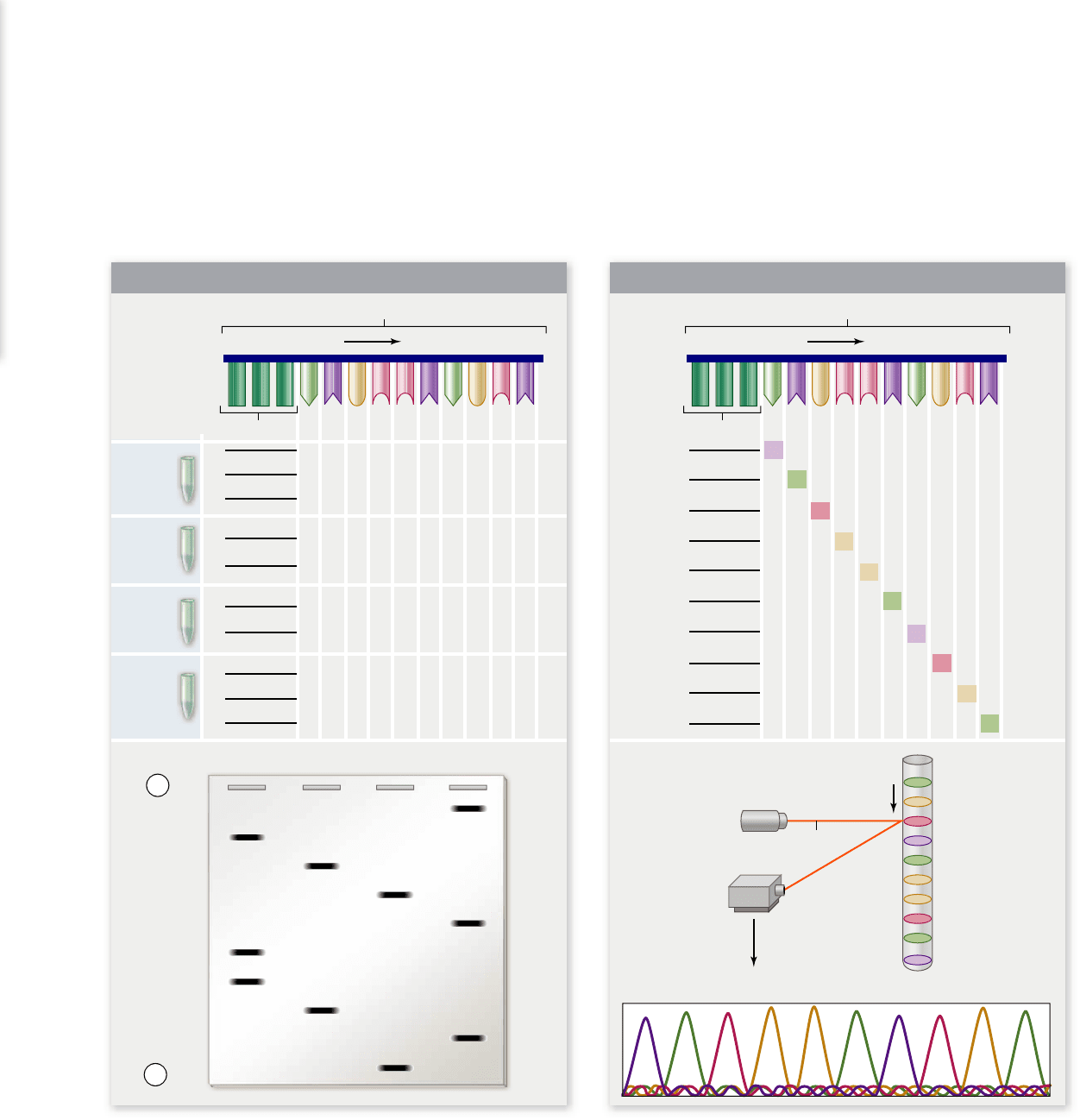
Figure 17.11
Manual and automated enzymatic DNA sequencing.
The sequence to be determined is shown at the top as a
template strand for DNA polymerase with a primer attached. a. In the manual method, four reactions were done, one for each nucleotide. For
example, the A tube would contain dATP, dGTP, dCTP, dTTP, and ddATP. This leads to fragments that end in A due to the dideoxy
terminator. The fragments generated in each reaction are shown along with the results of gel electrophoresis. b. In automated sequencing,
each ddNTP is labeled with a different color uorescent dye, which allows the reaction to be done in a single tube. The fragments generated
by the reactions are shown. When these are electrophoresed in a capillary tube, a laser at the bottom of the tube excites the dyes, and each will
emit a different color that is detected by a photodetector.
All DNA nucleotides lack
–
OH at the 2' carbon of the sugar,
but dideoxynucleotides have no 3'
–
OH at which the enzyme
can add new nucleotides. Thus the chain is terminated.
The experimenter must perform four separate reac-
tions, each with a single dideoxynucleotide, to generate a set
of fragments that terminate in specific bases. Thus all of the
fragments produced in the A reaction incorporate dideoxy-
adenosine and must end in A, and the same for the other
three reactions with different terminators. When these frag-
ments are separated by high-resolution gel electrophoresis,
each reaction is run in a different track, or lane, to generate a
pattern of nested fragments that can be read from the small-
est fragment to fragments that are each longer by one base
(figure 17.11a).
chapter
17
Biotechnology
337www.ravenbiology.com
rav32223_ch17_327-351.indd 337rav32223_ch17_327-351.indd 337 11/10/09 2:43:20 PM11/10/09 2:43:20 PM
Apago PDF Enhancer
NH
2
O
J
J
P
J
O
-
-
O
J
O
J
CH
2
J
5„
4„
3„ 2„
1„
OH
NN
N
O
A
Adapter
Adapter
Bridge
amplification
with unlabeled
dNTPs
Free end
binds to
primer
First round of
synthesis with
labeled dNTPs
Reversible terminator
A
A
T
T
G
G
C
A
T
G
C
C
Image capture for each
round of synthesis
35 cycles
of bridge
amplification
Fragments
become
double-
stranded
Denature
double-
stranded
molecules
Attached Attached Clusters
Free
terminus
DNA fragment
Dense primer lawn
in flow cell
DNA
Adapters
a. b.
c. d. e. f.
g
.h.
Flow cell
1 cm
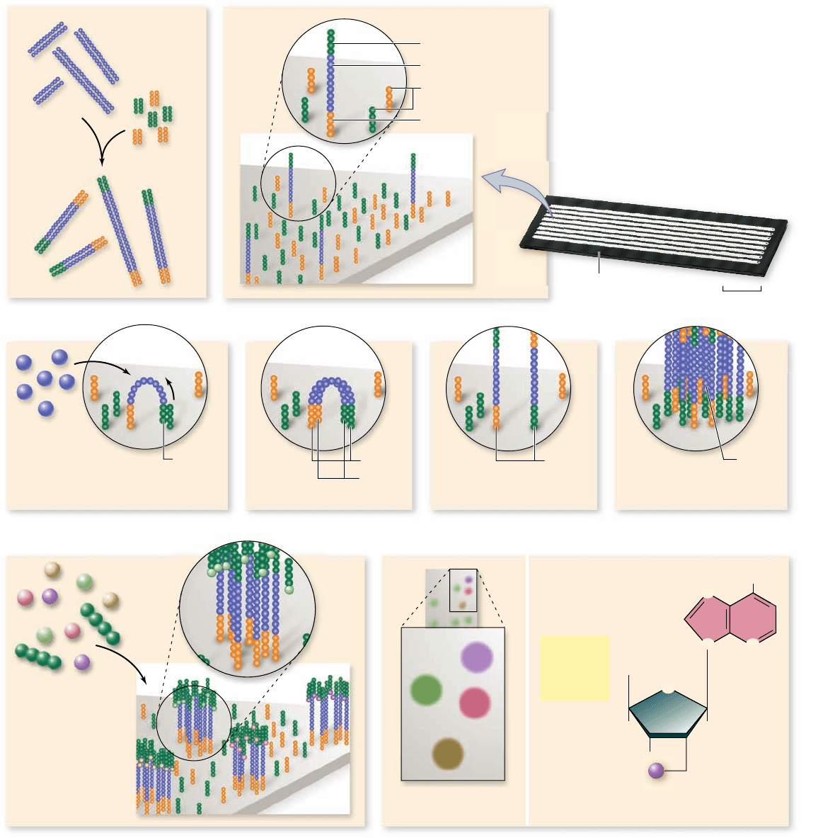
Figure 17.12
New approach to sequencing.
DNA is cleaved into short fragments that will be sequenced. a. Adapters are added to
the end of the DNA. b. DNA is denatured and the adapters bind to complementary primers in the ow cell. c–f. Individual fragments are
ampli ed using dNTPs and polymerase. g. Fluorescently labeled dNTPs with cleavable dye that blocks the formation of additional
phosphodiester bonds are added, and the rst uorescently labeled base is added. h. A CCD camera records the uorescence pattern before the
uorescent dye is removed, and the next base is added to each DNA sequence.
338
part
III
Genetic and Molecular Biology
rav32223_ch17_327-351.indd 338rav32223_ch17_327-351.indd 338 11/10/09 2:43:22 PM11/10/09 2:43:22 PM
Apago PDF Enhancer
clusters of fragments. Amplification works like DNA replica-
tion where a polymerase is added that recognizes the primer
and starts copying. The fragments are again denatured to yield
single-stranded molecules. They are now ready for sequencing.
As with Sanger sequencing, deoxyribonucleotide triphosphates
(dNTPs) have a fluorescent tag, but it can be removed. Four
colors are used to distinguish each base. The fluorescent tag is
reversibly attached to the 2' position on the deoxyribose sugar
and it blocks the 3' OH so that only a single phosphodiester
bond forms, but the blocking group can be removed after each
round of DNA extension so the DNA strands continue to elon-
gate. Very powerful charge-coupled device (CCD) cameras,
once used exclusively by astronomers, record the pattern of
fluorescence in the flow cell after each round of elongation.
The technology works because a solid material holds the DNA
fragments in place while they are being synthesized so that the
repeated CCD images can be compiled and provide informa-
tion about the sequence of each cluster of fragments. The
amount of data generated each time another round of base pairs
is added is enormous, so digital storage space and computa-
tional power to make sense of the data are the limiting factors.
The polymerase chain reaction accelerates
the process of analysis
The next revolution in molecular biology was the development
of the polymerase chain reaction (PCR). Kary Mullis devel-
oped PCR in 1983 while he was a staff chemist at the Cetus
Corporation; in 1993, he was awarded the Nobel Prize in
chemistry for his discovery.
The idea of the polymerase chain reaction is simple: Two
primers are used that are complementary to the opposite strands
of a DNA sequence, oriented toward each other. When DNA
polymerase acts on these primers and the sequence of interest,
the primers produce complementary strands, each containing
the other primer. If this procedure is done cyclically, the result
is a large quantity of a sequence corresponding to the DNA
that lies between the two primers (figure 17.13).
The PCR procedure
Two developments turned this simple concept into a power-
ful technique. First, each cycle requires denaturing the DNA
after each round of synthesis, which is easily done by raising
the temperature; however, this destroys most polymerase
enzymes. The solution was to isolate a DNA polymerase
from a thermophilic, or heat-loving bacteria, Thermus aquat-
icus. This enzyme, called Taq polymerase, allows the reac-
tion mixture to be repeatedly heated without destroying
enzyme activity.
The second innovation was the development of machines
with heating blocks that can be rapidly cycled over large tem-
perature ranges with very accurate temperature control.
Thus each cycle of PCR involves three steps:
Denaturation (high temperature)1.
Annealing of primers (low temperature)2.
Synthesis (intermediate temperature)3.
Steps 1 to 3 are now repeated, and the two copies become
four. It is not necessary to add any more polymerase, because
Notice that since this is a DNA polymerase reaction, it
requires a primer to begin synthesis. The vectors used for DNA
sequencing have known regions next to the site where DNA is
inserted. Short DNAs that are complementary to these regions
are then synthesized and can be used as primers. This serves the
dual purposes of providing a primer and ensuring that the first
few bases sequenced are known because they are known in the
vector itself. This allows the investigator to determine where
the sequence of interest begins. As the sequence is generated,
new primers can be designed near the end of the known se-
quence and DNA synthesized to use as a primer to extend the
region sequenced in the next set of reactions.
Automated sequencing
The technique of enzymatic sequencing is very powerful, but it
is also labor-intensive and takes a significant amount of time. It
requires a series of enzymatic manipulations, time for electro-
phoresis, then time to expose the gel to film. At the end of this,
a skilled researcher can read around 300 bases of sequence reli-
ably. The development of automated techniques made sequenc-
ing a much more practical and less human-intensive procedure.
Automated sequencing machines use fluorescent dyes in-
stead of a radioactive label and separate the products of the se-
quencing reactions using gels in thin capillary tubes instead of
the large slab gels. The tubes run in front of a laser that excites
the dyes, causing them to fluoresce. With a different colored
dye for each base, a photodetector can determine the identity
of each base by its color.
The data are assembled by a computer that generates a
visual image consisting of different colored peaks; these are
converted into the raw sequence data (figure 17.11b). The se-
quence data come directly from the electrophoresis, eliminat-
ing the time needed for exposing gels to film and for manual
reading of the sequences. The use of different colored dyes also
reduces handling and allows more sequence to be produced at
one time.
With increases in the number of samples per run and the
length of sequences able to be read, along with decreases in
handling time, the amount of sequence information that can be
generated is limited mainly by the number of machines that can
be run at once.
New sequencing technology
For over 30 years, the basic chemistry of DNA sequencing did
not change. Automation increased the speed of sequencing to
the point that sequencing large eukaryotic genomes became
possible. In the last few years, however, fundamentally new
methods for sequencing have vastly accelerated the rate of se-
quence generation. Here we explore one new approach, which
can generate 20 billion base pairs of sequence in a single run
(figure 17.12). DNA is cleaved into smaller pieces, a few hun-
dred base pairs, using a nebulizer—a device that converts the
liquid to a very fine spray. Both ends are ligated to adapters that
are complementary to specific primers. These DNA fragments
are injected into a flow cell, which is like a microscope slide
with seven channels, each containing a solid substrate with
primers that complement the ligated ends of the DNA frag-
ments. Millions of DNA fragments are placed in these chan-
nels, made single-stranded, and then amplified so there are
chapter
17
Biotechnology
339www.ravenbiology.com
rav32223_ch17_327-351.indd 339rav32223_ch17_327-351.indd 339 11/10/09 2:43:23 PM11/10/09 2:43:23 PM
Apago PDF Enhancer
DNA is denatured
into single strands
DNA segment
to be amplified
5„
3„
5„ 3„
3„
5„
3„ 5„
Primers anneal to DNA
5„ 3„
3„ 5„
Taq DNA polymerase
5„ 3„
5„ 3„
3„ 5„
3„ 5„
5„ 3„
5„ 3„
5„ 3„
5„ 3„
5„ 3„
5„ 3„
5„ 3„
5„ 3„
3„ 5„
3„ 5„
3„ 5„
3„ 5„
3„ 5„
3„ 5„
3„ 5„
3„ 5„
5„ 3„
5„ 3„
5„ 3„
5„ 3„
3„ 5„
3„ 5„
3„ 5„
3„ 5„
PCR
machine
Cycle 2:
4 copies
Cycle 3:
8 copies
1. Sample is first heated
to denature DNA.
2. DNA is cooled to a
lower temperature
to allow annealing
of primers.
3. DNA is heated to
72°C, the optimal
temperature for Taq
DNA polymerase to
extend primers.
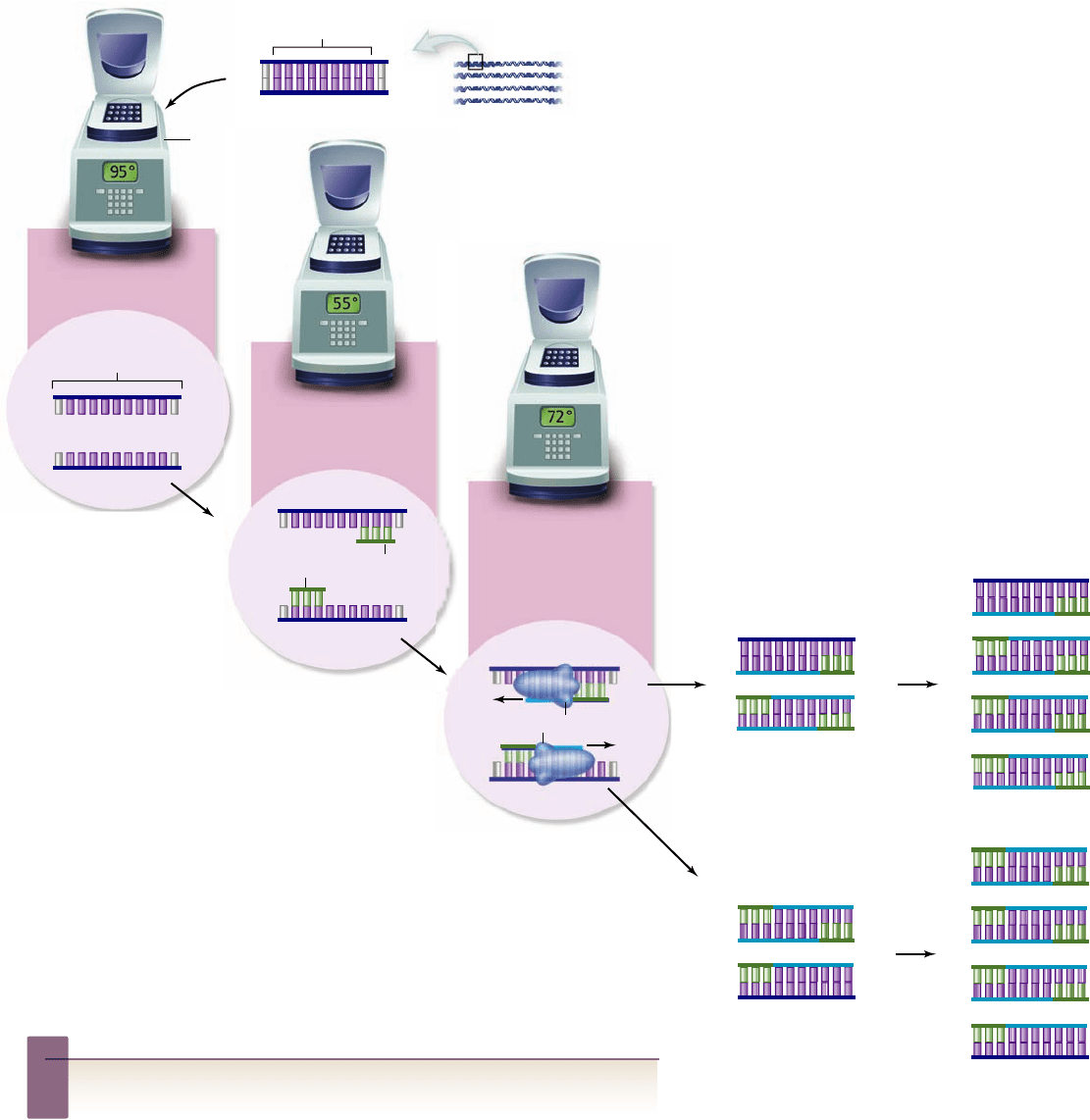
Figure 17.13
The polymerase chain reaction.
The
polymerase chain reaction (PCR) allows a single
sequence in a complex mixture to be ampli ed for
analysis. The process involves using short primers
for DNA synthesis that ank the region to be
ampli ed and (1) repeated rounds of denaturation,
(2) annealing of primers, and (3) synthesis of DNA. The
enzyme used for synthesis is a thermostable polymerase that can survive the high
temperatures needed for denaturation of template DNA. The reaction is
performed in a thermocycler machine that can be programmed to change
temperatures quickly and accurately. The annealing temperature used depends on
the length and base composition of the primers. Details of the synthesis process
have been simpli ed to illustrate the ampli cation process. Newly synthesized
strands are shown in light blue with primers in green.
Inquiry question
?
Could PCR be used to amplify mRNA?
manipulations, or directly sequenced. There are limitations on
the size of the fragment that can be synthesized in this way, but
it has been adapted for an amazing number of uses.
Applications of PCR
PCR, now fully automated, has revolutionized many aspects of
science and medicine because it allows the investigation of
minute samples of DNA. In criminal investigations, DNA
finger prints can now be prepared from the cells in a tiny speck
of dried blood or from the tissue at the base of a single human
hair. In medicine, physicians can detect genetic defects in very
early embryos by collecting a single cell and amplifying its
DNA. Due to its sensitivity, speed, and ease of use, technicians
now routinely use PCR methods for these applications.
PCR has even been used to analyze mitochondrial DNA
from the early human species Homo neanderthalensis. This appli-
cation provides the first glimpse of data from extinct related spe-
cies. The amplification of ancient DNA has been a controversial
field because contamination with modern DNA is difficult to
avoid. But it remains an active area of genetic research.
Protein interactions can be detected
with the two-hybrid system
Protein–protein interactions form the basis of many
biological structures. Just as human society is ulti-
mately dependent on interactions between people,
cells are dependent on interactions between proteins.
This observation has led to the large-scale goal of de-
termining all interactions among proteins in differ-
ent cells. This goal once would have been a dream,
but it is now becoming a reality. The yeast two-
hybrid system is one of the workhorses of this
kind of analysis (figure 17.14).
the heating step does not harm Taq polymerase. Each complete
cycle, which takes only 1–2 min, doubles the number of DNA
molecules. After 20 cycles, a single fragment produces more
than one million (2
20
) copies!
In this way, the process of PCR allows the amplification
of a single DNA fragment from a small amount of a complex
mixture of DNA. This result is similar to what is isolated using
molecular cloning, but in the case of PCR, the DNA cannot be
reintroduced directly into a cell. The PCR product can be ana-
lyzed using electrophoresis, cloned into a vector for other
340
part
III
Genetic and Molecular Biology
rav32223_ch17_327-351.indd 340rav32223_ch17_327-351.indd 340 11/10/09 2:43:23 PM11/10/09 2:43:23 PM
Apago PDF Enhancer
Fusion
proteins
Bait vector
Bait protein
Prey vector
Prey protein
Reporter gene
Yeast nucleus
Yeast cell
Inserted DNA Inserted DNA
Transcription-
activating domain
Gal4 protein
RNA polymerase
DNA DNA-
binding
domain
DNA-
binding
domain
Figure 17.14
The yeast two-hybrid system detects
interacting proteins. The Gal4 protein is a transcriptional
activator (top). The Gal4 gene has been split and engineered into two
different vectors such that one will encode only the DNA-binding
domain (bait vector) and the other the transcription-activating
domain (prey vector). When other genes are spliced into these
vectors, they produce fusion proteins containing part of Gal4 and
the proteins to be tested. If the proteins being tested interact, this
will restore Gal4 function and activate expression of a reporter gene.
When cDNAs are inserted into each of these two vectors
in the proper reading frame, they are expressed as a single pro-
tein consisting of the protein of interest and part of the Gal4
activator protein (see figure 17.13). These hybrid proteins are
called fusion proteins since they are literally fused in the same
polypeptide chain. The DNA-binding hybrid is called the bait,
and the activating domain hybrid is called the prey.
These vectors are inserted into cells of different mating
types that can be crossed. One of these vectors also contains a
so-called reporter gene encoding a protein that can be assayed
for enzymatic activity. The reporter gene is under control of a
Gal4-responsive regulatory region, so that when active Gal4 is
present, the reporter gene is expressed and can be detected by
an enzymatic assay.
The DNA-binding hybrid binds to DNA adjacent to the
reporter gene. When the two proteins in bait and prey interact,
the prey hybrid brings the activating domain into position to turn
on gene expression from the reporter gene (see figure 17.13).
The beauty of this system is that it is both simple and
flexible. It can be used with two known proteins or with a
known protein in the bait vector and entire cDNA libraries in
the prey vector. In the latter case, all of the possible interactions
in a cell type can be mapped.
It is already clear that even more protein interactions oc-
cur in cells than anticipated. In the future these data will form
the basis for understanding the networks of protein interac-
tions that make up the normal activities of a cell.
Learning Outcomes Review 17.3
The Southern blotting technique allows identifi cation of a target DNA by
separating single-stranded DNA fragments and hybridizing fragments
of interest with a labeled probe. In living cells, DNA polymerase is a key
enzyme in replication. DNA sequencing uses a modifi ed DNA polymerase
reaction that contains chain terminators, allowing fragments to be ordered
in sequence. The polymerase chain reaction (PCR) produces a large amount
of a specifi c DNA from a small amount of starting material. The yeast system
for detecting protein–protein interactions involves a bait protein, a prey
protein, and a reporter gene.
■ What key component of PCR allows the rapid
amplification of a sample?
The yeast two-hybrid system integrates much of the tech-
nology discussed in this chapter. It takes advantage of one fea-
ture of eukaryotic gene regulation, namely that the structure of
proteins that turn on eukaryotic gene expression, transcription
factors, have a modular structure.
The Gal4 gene of yeast encodes a transcriptional activa-
tor with modular structure consisting of a DNA-binding do-
main that binds sequences in Gal4-responsive promoters, and
an activation domain that interacts with the transcription ap-
paratus to turn on transcription. The system uses two vectors:
one containing a fragment of the Gal4 gene that encodes the
DNA-binding domain, and another containing a fragment of
the Gal4 gene that encodes the transcription activation domain.
Neither of these alone can activate transcription.
Learning Outcome
Describe three applications of cloning technology.1.
The ability to clone individual genes for analysis ushered in an
era of unprecedented advancement in research. At the time,
these advancements were not accompanied by grand announce-
ments of potential medical breakthroughs and other applica-
tions. The ability to truly genetically engineer any kind of cell
17.4
Genetic Engineering
chapter
17
Biotechnology
341www.ravenbiology.com
rav32223_ch17_327-351.indd 341rav32223_ch17_327-351.indd 341 11/10/09 2:43:48 PM11/10/09 2:43:48 PM