Raven P.H., Johnson G.B., Mason K.A. Biology (Ninth Edition)
Подождите немного. Документ загружается.


Apago PDF Enhancer
3. Determine which of the following is correct regarding the yeast
Saccharomyces cerevisiae.
a. It reproduces asexually by a process called budding.
b. It produces an ascocarp during reproduction.
c. It belongs in the group Zygomycota.
d. All of the above are correct.
4. Appraise the fungal relationship between a forest tree and a
basidiomycetes and determine the most suitable classi cation
for the symbiosis.
a. Parasitism only
b. An arbuscular mycorrhizae
c. Ectomycorrhizae
d. A lichen
5. Choose which of the following best re ects the symbiotic
relationships between animals and fungi.
a. Protection from bacteria
b. Colonization of land
c. Protection from desiccation
d. Exchange of nutrients
S Y N THES IZE
1. Historically fungi have been classi ed as being more plantlike
despite their lack of photosynthetic ability. Although we now
know that fungi are more closely related to the animals than the
plants, review characteristics that initially led scientists to place
them closer to the plants.
2. The importance of fungi in the evolution of terrestrial life is
typically understated. Evaluate the importance of fungi in the
colonization of land.
3. Based on your understanding of fungi, hypothesize why
antibiotics won’t work in the treatment of a fungal infection.
U N DERS TAN D
1. Which of the following is not a characteristic of a fungus?
a. Cell walls made of chitin
b. A form of mitosis different from plants and animals
c. Ability to conduct photosynthesis
d. Filamentous structure
2. A fungal cell that contains two genetically different nuclei
would be classi ed as
a. monokaryotic. c. homokaryotic.
b. bikaryotic. d. heterokaryotic.
3. Which of the following groups of fungi is not monophyletic?
a. Zygomycota c. Glomeromycota
b. Basidiomycota d. Ascomycota
4. Based on physical characteristics, the __________ represent
the most ancient phylum of fungi.
a. Basidiomycota c. Ascomycota
b. Zygomycota d. Chytridiomycota
5. The early evolution of terrestrial plants was made possible
by mycorrhizal relationships with the
a. Zygomycetes. c. Ascomycota.
b. Glomeromycota. d. Basidiomycota.
6. Symbiotic relationships occur between the fungi and
a. plants. c. animals.
b. bacteria. d. all of the above.
7. Which of the following species of fungi is not associated
with diseases in humans?
a. Pneumocystis jiroveci
b. Aspergillus avus
c. Candida albicans
d. Batrachochytrium dendrobatidis
APPLY
1. In a culture of hyphae of unknown origin you notice that the
hyphae lack septa and that the fungi reproduce asexually by using
clumps of erect stalks. However, at times sexual reproduction can
be observed. To what group of fungi would you assign it?
a. Chytridiomycota c. Ascomycota
b. Basidiomycota d. Zygomycota
2. Examine the life cycle of a typical basidiomycetes and determine
where you would expect to nd a dikaryotic cell.
a. Primary mycelium c. In the basidiospores
b. Secondary mycelium d. In the zygote
Review Questions
O N LIN E RES OURCE
www.ravenbiology.com
Understand, Apply, and Synthesize—enhance your study with
animations that bring concepts to life and practice tests to assess
your understanding. Your instructor may also recommend the
interactive eBook, individualized learning tools, and more.
632
part
V
Diversity of Life on Earth
rav32223_ch31_614-632.indd 632rav32223_ch31_614-632.indd 632 11/13/09 1:18:37 PM11/13/09 1:18:37 PM
Apago PDF Enhancer
W
Chapter
32
Overview of Animal
Diversity
Chapter Outline
32.1 Some General Features of Animals
32.2 Evolution of the Animal Body Plan
32.3 The Classi cation of Animals
32.4 The Roots of the Animal Tree of Life
Introduction
We now explore the great diversity of modern animals, the result of a long evolutionary history. Animals are among the
most abundant living organisms. Found in almost every habitat, they bewilder us with their diversity in form, habitat,
behavior, and lifestyle. About a million and a half species have been described, and several million more are thought to
await discovery. Despite their great diversity, animals have much in common. For example, locomotion is a distinctive
characteristic, although not all animals can move about. Early naturalists thought that sponges and corals were plants
because the adults are attached to the surface on which they live.
CHAPTER
rav32223_ch32_633-648.indd 633rav32223_ch32_633-648.indd 633 11/16/09 12:57:53 PM11/16/09 12:57:53 PM
Apago PDF Enhancer
TABLE 32.1
General Features of Animals
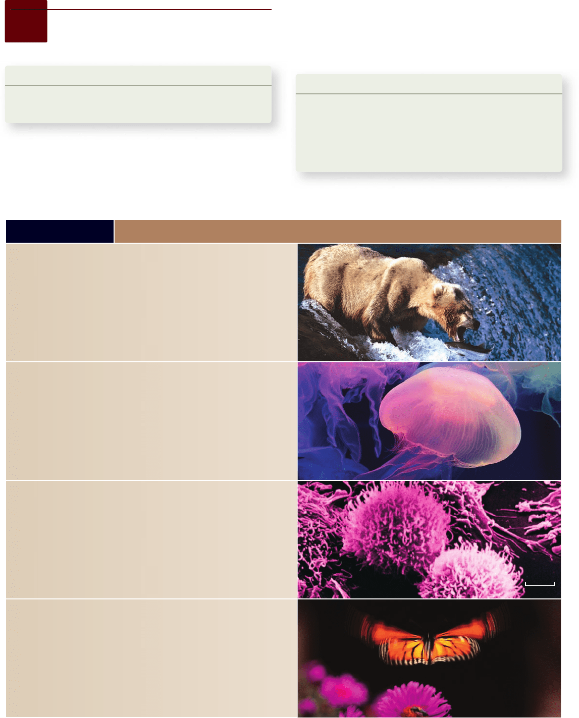
Heterotrophy. All animals are heterotrophs—that is, they obtain energy and
organic molecules by ingesting other organisms. Unlike autotrophic plants and
algae, animals cannot construct organic molecules from inorganic chemicals.
Some animals (herbivores) consume autotrophs; other animals (carnivores)
consume heterotrophs; some animals (omnivores) consume both autotrophs and
heterotrophs; and still others (detritivores) consume decomposing organisms.
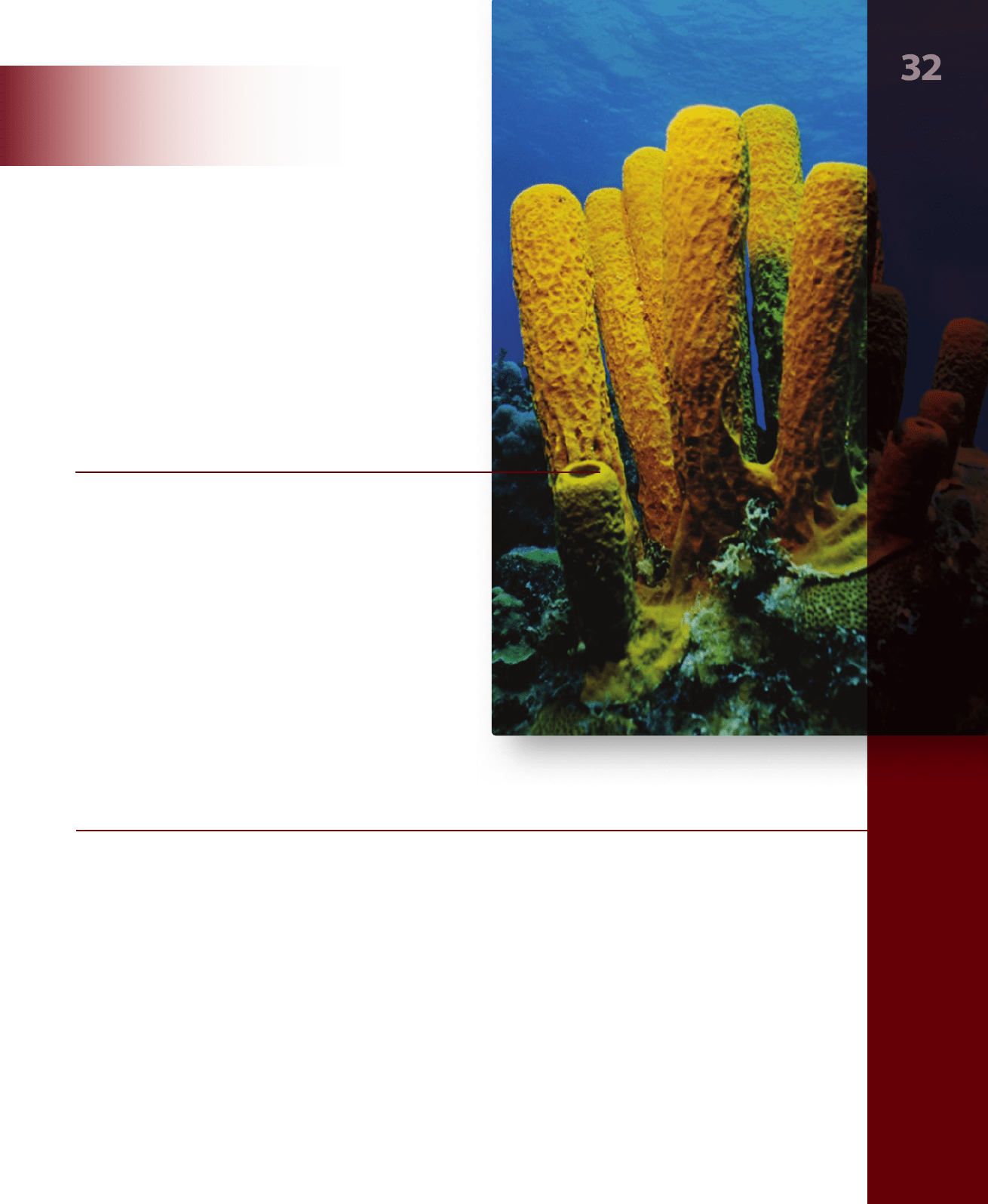
Multicellularity. All animals are multicellular; many have complex bodies like that
of this jelly sh (phylum Cnidaria). The unicellular heterotrophic organisms called
Protozoa, which were at one time regarded as simple animals, are now considered
members of the large and diverse kingdom Protista, discussed in chapter 29.
No Cell Walls. Animal cells di er from those of other multicellular organisms: they
lack rigid cell walls and are usually quite exible. The many cells of animal bodies
are held together by extracellular frames of structural proteins such as collagen.
Other proteins form unique intercellular junctions between animal cells.
Active Movement. Animals move more rapidly and in more complex ways
than members of other kingdoms—this ability is perhaps their most striking
characteristic, one directly related to the exibility of their cells and the evolution
of nerve and muscle tissues. A remarkable form of movement unique to animals
is ying, a capability that is well developed among vertebrates and insects such
as this buttery (phylum Arthropoda). Many animals cannot move from place to
place (they are sessile) or do so rarely or slowly (they are sedentary) although they
have muscles or muscle bers that allow parts of their bodies to move. Sponges,
however, have little capacity for movement.
2.2 µm
tions. Taken together, the universal characteristics and other
features of major importance that have exceptions are convinc-
ing evidence that animals are monophyletic—that they de-
scended from a common ancestor. Table 32.1 describes the
general features of animals.
Learning Outcome Review 32.1
All animals are multicellular and heterotrophic, and their cells lack cell walls.
Most animals can move from place to place, can reproduce sexually, and
possess unique tissues. Animals can be found in almost all habitats.
■ What evidence is there that animals could not have
been the first type of life to have evolved?
32.1
Some General Features
of Animals
Learning Outcome
Identify three features that characterize all animals and 1.
three that characterize only some types of animals.
Animals are so diverse that few criteria fit them all. But some,
such as animals being eaters, or consumers, apply to all. Others,
such as their being mobile (they can move about) have excep-
634
part
V
Diversity of Life on Earth
rav32223_ch32_633-648.indd 634rav32223_ch32_633-648.indd 634 11/13/09 1:35:19 PM11/13/09 1:35:19 PM
Apago PDF Enhancer
TABLE 32.1
General Features of Animals, continued
Diversity in Form. Animals vary greatly in form, ranging in size from organisms
too small to see with the unaided eye to enormous whales and giant squids.
Almost all animals, like this millipede (phylum Arthropoda), lack a backbone—
they are therefore called invertebrates. Of the million known living animal species,
only 42,500 have a backbone—they are therefore referred to as vertebrates.
Probably most of the many millions of animal species awaiting discovery
are invertebrates.
Diversity in Habitat. The animal kingdom is divided into 35–40 phyla, most
of which have members that occur only in the sea like this brittlestar (phylum
Echinodermata). Members of fewer phyla occur in fresh water, and members of
fewer still occur on land. Members of three phyla that are successful in the marine
environment—Arthropoda, Mollusca, and Chordata—also dominate animal
life on land. Only one animal phylum, Onychophora (velvet worms) is
entirely terrestrial.
Sexual Reproduction. Most animals reproduce sexually; these tortoises
(phylum Chordata) are engaging in the rst step of that process. Animal eggs,
which are nonmobile, are much larger than the small, usually agellated sperm.
In animals, cells formed in meiosis function as gametes. These haploid cells do
not divide by mitosis rst, as they do in plants and fungi, but rather fuse directly
with each other to form the zygote. Consequently, there is no counterpart among
animals to the alternation of haploid (gametophyte) and diploid (sporophyte)
generations characteristic of plants. Some individuals of some species and all
individuals of a very few animal species are incapable of sexual reproduction.
Embryonic Development. An animal zygote rst undergoes a series of mitotic
divisions, called cleavage, and like this dividing frog’s egg, that produces a ball
of cells, the blastula. In most animals, the blastula folds inward at one point to
form a hollow sac with an opening at one end called the blastopore. An embryo
at this stage is called a gastrula. The subsequent growth and movement of the
cells of the gastrula di er from one group of animals to another, re ecting the
evolutionary history of the group. Embryos of most kinds of animals develop into
a larva, which looks unlike the adult of the species, lives in a di erent habitat,
and eats di erent sorts of food; in most groups, it is very small. A larva undergoes
metamorphosis, a radical reorganization, to transform into the adult body form.
Tissues. The cells of all animals except sponges are organized into structural
and functional units called tissues, collections of cells that together are
specialized to perform specic tasks. Animals are unique in having two tissues
associated with movement: (1) muscle tissue, which contracts, and (2) nervous
tissue, which conducts signals among cells. Neuromuscular junctions, where
nerves connect with muscle tissue, are shown here.
chapter
32
Overview of Animal Diversity
635www.ravenbiology.com
rav32223_ch32_633-648.indd 635rav32223_ch32_633-648.indd 635 11/13/09 1:35:23 PM11/13/09 1:35:23 PM
Apago PDF Enhancer
a.
Radial Symmetry
b.
Bilateral Symmetry
Ventral
Dorsal
T
ransverse
plane
Frontal plane
Sagittal plane
Anterior
Posterior
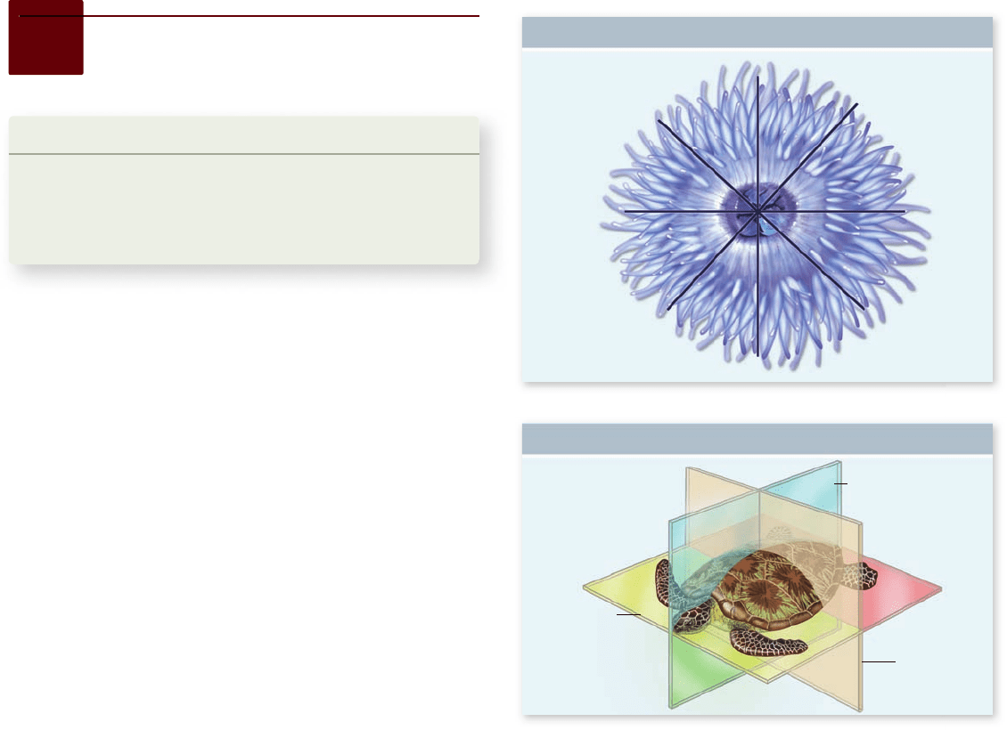
Figure 32.1
A comparison of radial and bilateral
symmetry. a. Radially symmetrical animals, such as this sea
anemone (phylum Cnidaria), can be bisected into equal halves by any
longitudinal plane that passes through the central axis. b. Bilaterally
symmetrical animals, such as this turtle (phylum Chordata), can only
be bisected into equal halves in one plane (the sagittal plane).
image halves, one along the mouth and one perpendicular to it;
these animals are actually biradially symmetrical (figure 32.1b).
Bilateral symmetry
The bodies of most animals other than sponges and cnidarians
exhibit bilateral symmetry , in which the body has right and left
halves that are mirror images of each other. Animals with this
body plan are collectively termed the Bilateria. The sagittal plane
defines these halves. A bilaterally symmetrical body has, in addi-
tion to left and right halves, dorsal and ventral portions, which
are divided by the frontal plane, and anterior (front) and poste-
rior (rear) ends, which are divided by the transverse plane (in an
animal that walks on all fours, dorsal is the top side). In echino-
derms (sea stars and their relatives), adults are radially symmetri-
cal (actually pentaradially symmetrical, because the body has five
clear sections), but the larvae are bilaterally symmetrical.
32.2
Evolution of the Animal
Body Plan
Learning Outcomes
Differentiate between a pseudocoelom and a coelom.1.
Explain the difference between protostomes and 2.
deuterostomes.
Describe the advantages of segmentation.3.
The features described in the preceding section evolved over
the course of millions of years. We can understand how the his-
tory of life has proceeded by examining the types of animal
bodies and body plans present in fossils and in existence today.
Five key innovations can be noted in animal evolution:
The evolution of symmetry1.
The evolution of tissues, allowing specialized structures 2.
and functions
The evolution of a body cavity3.
The evolution of various patterns of embryonic 4.
development
The evolution of segmentation, or repeated body units5.
These innovations are explained in the sections that follow.
Some innovations appear to have evolved only once, some twice
or more. Scientists use an innovation that evolved once as evi-
dence that all the animals possessing it are more closely related
to one another than they are to any animal lacking the innova-
tion. The animals with the innovation and their ancestor in
which the innovation arose are said to constitute a clade—an
evolutionarily coherent group (see chapter 23). On the other
hand, some innovations evolve more than once in different
clades. This is the phenomenon of convergent evolution (see
chapter 23). Although not indicative of close evolutionary rela-
tionship, convergently evolved innovations may be important
to how species have adapted to their environments.
Most animals exhibit radial
or bilateral symmetry
A typical sponge lacks definite symmetry, growing as an irregu-
lar mass. Virtually all other animals have a definite shape and
symmetry that can be defined along an imaginary axis drawn
through the animal’s body. The two main types of symmetry are
radial and bilateral.
Radial symmetry
The body of a member of phylum Cnidaria (jellyfish, sea anemo-
nes, and corals: the C of Cnidaria is silent; see chapter 34) exhibits
radial symmetry . Its parts are arranged in such a way that any
longitudinal plane passing through the central axis divides the
organism into halves that are approximate mirror images
(figure 32.1a) . A pie, for example, is radially symmetrical. In cni-
darians such as corals and sea anemones, the mouth is not circular,
but oval, because it opens into a sort of throat that is like a flattened
sleeve. Thus there are two planes that divide the body into mirror-
636
part
V
Diversity of Life on Earth
rav32223_ch32_633-648.indd 636rav32223_ch32_633-648.indd 636 11/13/09 1:35:28 PM11/13/09 1:35:28 PM
Apago PDF Enhancer
Flatworm
Ectodermally
derived tissue
Mesodermally
derived tissue
Ectodermally
derived tissue
Ectodermally
derived tissue
Mesodermally
derived tissue
Mesodermally
derived tissue
Endodermally
derived tissue
Acoelomate
Annelid
Coelomate
Coelom
Roundworm
Endodermally
derived tissue
Endodermally
derived tissue
Pseudocoelomate
Pseudocoelom
Figure 32.2
Three body plans for bilaterally
symmetrical animals. Acoelomates, such as atworms, have no
body cavity between the digestive tract (derived from the
endoderm) and the musculature layer (derived from the mesoderm).
Pseudocoelomates have a body cavity, the pseudocoelom, between
tissues derived from the endoderm and those derived from the
mesoderm. Coelomates have a body cavity, the coelom, that
develops entirely within tissues derived from the mesoderm, and so
is lined on both sides by tissue derived from the mesoderm.
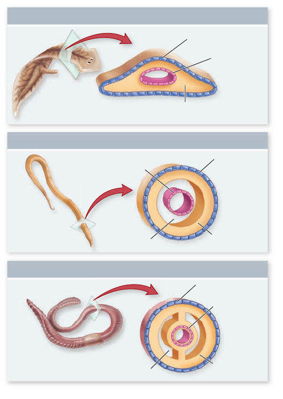
A key innovation in the body plan of some bilaterians was a
body cavity isolated from the exterior of the animal. This is differ-
ent from the digestive cavity, which is open to the exterior at least
through the mouth, and in most animals at the opposite end as
well, via the anus. The evolution of efficient organ systems within
the animal body was not possible until a body cavity evolved for
accommodating and supporting organs (such as our heart and
lungs), distributing materials, and fostering complex develop-
mental interactions. The cavity is filled with fluid: in most ani-
mals, the fluid is liquid, but in vertebrates, it is gas—the body
cavity of humans filling with liquid is a life-threatening condition.
A very few types of bilaterians have no body cavity, the space be-
tween tissues that develop from the mesoderm and those that de-
velop from the endoderm being filled with cells and connective
tissue. These are the so-called acoelomate animals (figure 32.2).
Bilateral symmetry constitutes a major evolutionary advance
in the animal body plan. Bilaterally symmetrical animals have the
ability to move through the environment in a consistent direction
(typically with the anterior end leading)—a feat that is difficult for
radially symmetrical animals. Associated with directional move-
ment is the grouping of nerve cells into a brain, and sensory struc-
tures, such as eyes and ears, at the anterior end of the body. This
concentration of nervous tissue at the anterior end, which appears
to have occurred early in evolution, is called cephalization. Much
of the layout of the nervous system in bilaterally symmetrical ani-
mals is centered on one or more major longitudinal nerve cords
that transmit information from the anterior sense organs and
brain to the rest of the body. Cephalization is often considered a
consequence of the development of bilateral symmetry.
The evolution of tissues allowed for
specialized structures and functions
The zygote (a fertilized egg), has the capability to give rise to all
the kinds of cells in an animal’s body. That is, it is totipotent (all
powerful). During embryonic development, cells specialize to
carry out particular functions. In all animals except sponges, the
process is irreversible: once a cell differentiates to serve a func-
tion, it and its descendants can never serve any other.
A sponge cell that had specialized to serve one function (such
as lining the cavity where feeding occurs) can lose the special at-
tributes that serve that function and change to serve another func-
tion (such as being a gamete). Thus a sponge cell can dedifferentiate
and redifferentiate. Cells of all other animals are organized into
tissues, each of which is characterized by cells of particular mor-
phology and capability. But their competence to dedifferentiate
prevents sponge cells from forming clearly defined tissues (and
therefore, of course, organs, which are composed of tissues).
Because cells differentiate irreversibly in all animals ex-
cept sponges, scientists infer that bodies containing cells spe-
cialized to serve particular functions have an advantage
compared to those with cells that potentially have multiple
functions. Judging by the relative diversity of animals with spe-
cialized tissues and those lacking them, tissues are a favorable
adaptation. Presumably the advantage to the animal is embod-
ied in the old adage “Jack of all trades, master of none.”
A body cavity made possible the development
of advanced organ systems
In the process of embryonic development, the cells of animals of
most groups organize into three layers (called germ layers): an
outer ectoderm, an inner endoderm, and an intermediate
mesoderm. Animals with three embryonic cell layers are said to be
triploblastic. Part of the maturation from the embryo is that certain
organs and organ systems develop from each germ layer. The ecto-
derm gives rise to the outer covering of the body and the nervous
system; the endoderm gives rise to the digestive system, including
the intestine; and the skeleton and muscles develop from the meso-
derm. Cnidarians have only two layers (thus they are diplobastic),
the endoderm and the ectoderm, and lack organs. Sponges lack
germ layers altogether; they, of course, have no tissues or organs.
All triploblastic animals are members of the Bilateria.
chapter
32
Overview of Animal Diversity
637www.ravenbiology.com
rav32223_ch32_633-648.indd 637rav32223_ch32_633-648.indd 637 11/13/09 1:35:29 PM11/13/09 1:35:29 PM
Apago PDF Enhancer
the “primitive gut”); it communicates with the outside by
a blastopore.
In a protostome, the mouth of the adult animal develops
from the blastopore or from an opening near the blastopore
(protostome means “first mouth”—the first opening becomes
the mouth). Protostomes include most bilaterians, including
flatworms, nematodes, mollusks, annelids, and arthropods. In
some protostomes, both mouth and anus form from the embry-
onic blastopore; in other protostomes, the anus forms later in
another region of the embryo. Two outwardly dissimilar groups,
the echinoderms and the chordates, together with a few other
small phyla, constitute the deuterostomes, in which the mouth
of the adult animal does not develop from the blastopore. The
deuterostome blastopore gives rise to the organism’s anus, and
the mouth develops from a second pore that arises later in de-
velopment (deuterostome means “second mouth”). Proto-
stomes and deuterostomes differ in several other aspects of
embryology too, as discussed later.
Cleavage patterns
The cleavage pattern relative to the embryo’s polar axis deter-
mines how the resulting cells lie with respect to one another. In
some protostomes, each new cell cleaves off at an angle oblique
to the polar axis. As a result, a new cell nestles into the space
between the older ones in a closely packed array. This pattern is
called spiral cleavage because a line drawn through a sequence
of dividing cells spirals outward from the polar axis (figure 32.3
top) . Spiral cleavage is characteristic of annelids, mollusks,
nemerteans, and related phyla; the clade of animals with this
cleavage pattern is therefore known as the Spiralia.
In all deuterostomes, by contrast, the cells divide parallel
to and at right angles to the polar axis. As a result, the pairs of
cells from each division are positioned directly above and be-
low one another, a process that gives rise to a loosely packed
ball. This pattern is called radial cleavage because a line drawn
through a sequence of dividing cells describes a radius outward
from the polar axis (figure 32.3 bottom).
Determinate versus indeterminate development
Many protostomes exhibit determinate development, in
which the type of tissue each embryonic cell will form in the
adult is determined early, in many lineages even before cleav-
age begins, when the molecules that act as developmental
signals are localized in different regions of the egg. Conse-
quently, the cell divisions that occur after fertilization segre-
gate molecular signals into different daughter cells, specifying
the fate of even the very earliest embryonic cells. Each em-
bryonic cell is destined to occur only in particular parts of
the adult body, so if the cells are separated, development
cannot proceed.
Deuterostomes, on the other hand, display indeterminate
development. The first few cell divisions of the zygote pro-
duce identical daughter cells. If the cells are separated, any one
can develop into a complete organism because the molecules
that signal the embryonic cells to develop differently are not
segregated in different cells until later in the embryo’s devel-
opment. (This is how identical twins are formed.) Thus, each
cell remains totipotent and its fate is not determined for sev-
eral cleavages.
Body cavities
Body cavities appear to have evolved multiple times in the Bila-
teria (see figure 32.2). A body cavity called the pseudocoelom
develops embryologically between mesoderm and endoderm,
so occurs in the adult between tissues derived from the meso-
derm and those derived from endoderm; animals with this type
of body cavity are termed pseudocoelomates. Although the
word pseudocoelom means “false coelom,” this is a true body
space and characterizes many successful groups of animals. A
coelom is a cavity that develops entirely within the mesoderm.
The coelom is surrounded by a layer of epithelial cells derived
from the mesoderm and termed the peritoneum.
Zoologists previously inferred that the first animals were
acoelomate, that some of their descendants evolved a pseudo-
coelom, and that some pseudocoelomate descendants evolved
the coelom. However, as you saw in chapter 21, evolution rarely
occurs in such a linear and directional way. Rather, pseudo-
coeloms seem to have evolved several times, and some animals
have lost the body space, becoming acoelomate secondarily.
However, a coelom appears to have evolved just once. Thus,
species possessing a coelom form a clade, but those with a
pseudocoelom do not.
The circulatory system
In many small animals, nutrients and oxygen are distributed
and wastes are removed by fluid in the body cavity. Most
larger animals, in contrast, have a circulatory system, a net-
work of vessels that carry fluids to and from the parts of the
body distant from the sites of digestion (gut) and gas ex-
change (gills or lungs). The circulating fluid carries nutrients
and oxygen to the tissues and removes wastes, including car-
bon dioxide, by diffusion between the circulatory fluid and
the other cells of the body.
In an open circulatory system, the blood passes from
vessels into sinuses, mixes with body fluid that bathes the cells
of tissues, then reenters vessels in another location. In a closed
circulatory system, the blood is entirely confined to blood
vessels, so is physically separated from other body fluids. Blood
moves through a closed circulatory system faster and more ef-
ficiently than it does through an open system; open systems are
typical of animals that are relatively inactive and so do not have
a high demand for oxygen. In small animals, blood can be
pushed through a closed circulatory system by movement of
the animal. In larger animals, the body musculature does not
provide enough force, so the blood must be propelled by con-
traction of one or more hearts, which are specialized, muscular
parts of the blood vessels.
Bilaterians have two main types
of development
The processes of embryonic development in animals is dis-
cussed fully in chapter 54 . Briefly, development of a bilater-
ally symmetrical animal begins with mitotic cell divisions
(called cleavages) of the egg that lead to the formation of a
hollow ball of cells, which subsequently indents to form a
two-layered ball. The internal space that is created through
such indentation (figure 54.11) is the archenteron (literally
638
part
V
Diversity of Life on Earth
rav32223_ch32_633-648.indd 638rav32223_ch32_633-648.indd 638 11/13/09 1:35:29 PM11/13/09 1:35:29 PM
Apago PDF Enhancer
Spiralian Protostomes
Radial cleavage
Blastopore
becomes mouth
Blastopore
becomes anus
Archenteron
Mesoderm
Archenteron
Mesoderm
Development
arrested
Determinate
development
Indeterminate
development
Normal
embryos
Spiral cleavage
Side view Top view
Axis
Cleavage
Fate of
Embryonic Cells
Fate of
Blastopore
Formation of
Coelom
Four-cell
embryo
Four-cell
embryo
Deuterostomes
Coelom
Mouth
Anus
Side view Top view
Axis
Cell
excised
Cell
excised
Figure 32.3
Embryonic development in protostomes and deuterostomes. In spiralian protostomes, embryonic cells cleave in
a spiral pattern and exhibit determinate development; the blastopore becomes the animal’s mouth, and the coelom originates from a split
among endodermal cells. In deuterostomes, embryonic cells cleave radially and exhibit indeterminate development; the blastopore becomes
the animal’s anus, and the coelom originates from an invagination of the archenteron.
early in development, but that may have specialized functions.
Development of segmentation is mediated at the molecular
level by Hox genes (see chapters 19 and 25 , and in section 32.4).
During early development, segments first are obvious in
the mesoderm but later are reflected in the ectoderm and
endoderm. Two advantages result from early embryonic
segmentation:
In highly segmental animals, such as earthworms (phylum 1.
Annelida), each segment may develop a more or less
complete set of adult organ systems. Because these are
redundant systems, damage to any one segment need not
be fatal because other segments duplicate the damaged
segment’s functions.
Locomotion is more ef cient when individual segments 2.
can move semi-independently. Because partitions isolate
the segments, each can contract or expand autonomously.
Therefore, a long body can move in ways that are often
quite complex.
Segmentation underlies the organization of body plans of
the most morphologically complex animals. In some adult
Formation of the coelom
The coelom arises within the mesoderm. In protostomes, cells
simply move apart from one another to create an expanding
coelomic cavity within the mass of mesodermal cells. In deu-
terostomes, groups of cells pouch off the end of the archenteron,
which you will recall is the primitive gut—the hollow in the
center of the developing embryo that is lined with endoderm.
The consistency of deuterostome development and its
distinctiveness from that of the protostomes suggest that it
evolved once, in the ancestor of the deuterostome phyla. The
mode of development in protostomes is more diverse, but be-
cause of the distinctiveness of spiral development, scientists
infer it also evolved once, in the common ancestor to all
spiralian phyla.
Segmentation allowed for redundant systems
and improved locomotion
Segmented animals consist of a series of linearly arrayed com-
partments that typically look alike (see figure 34.14 ), at least
chapter
32
Overview of Animal Diversity
639www.ravenbiology.com
rav32223_ch32_633-648.indd 639rav32223_ch32_633-648.indd 639 11/13/09 1:35:30 PM11/13/09 1:35:30 PM

Apago PDF Enhancer
arthropods, the segments are fused, but segmentation is usu-
ally apparent in embryological development. In vertebrates,
the backbone and muscle blocks are segmented, although seg-
mentation is often disguised in the adult form.
Previously, zoologists considered that true segmentation
was found only in annelids, arthropods, and chordates, but seg-
mentation is now recognized to be more widespread. Animals
such as onychophorans (velvet worms), tardigrades (water
bears), and kinorhynchs (mud dragons) are also segmented.
Learning Outcomes Review 32.2
Animals are distinguished on the basis of symmetry, tissues, type of
body cavity, sequence of embryonic development, and segmentation.
A pseudocoelom is a space that develops between the mesoderm and
endoderm; a coelom develops entirely within mesoderm. In bilaterians,
protostomes develop the mouth prior to the anus; deuterostomes develop
the mouth after the anus has formed. Segmentation allows redundant
systems and more effi cient locomotion.
■ How is cephalization related to body symmetry?
32.3
The Classi cation of Animals
Learning Outcomes
List the major criteria scientists have used to distinguish 1.
animal phyla.
Distinguish between spiralian and ecdysozoan 2.
organisms.
Identify the placement of humans among the 3.
animal phyla.
Multicellular animals, or metazoans, are traditionally divided
into 35 to 40 phyla (singular, phylum). There is little disagree-
ment among biologists about the placement of most animals in
phyla, although zoologists disagree on the status of some, par-
ticularly those with few members or recently discovered ones.
The diversity of animals is obvious in tables 32.2 and 32.3 ,
which describe key characteristics of 20 of the phyla.
Traditionally, the phylogeny of animals has been in-
ferred using features of anatomy and aspects of embryological
development, as discussed earlier, from which a broad consen-
sus emerged over the last century concerning the main
branches of the animal tree of life. In the past 30 years, data
derived from molecular features have been added, leading to
some rethinking of classification schemes. Depending on the
features compared, biologists may draw quite different family
trees—although, of course, there is only one way that evolu-
tion actually occurred, and the goal of phylogeny is to detect
that history.
Whether morphological or molecular characters (or
both) are used, the underlying principle is the same: system-
atists use features they assume to have evolved only once, so the
animals sharing such a feature are inferred to be more closely
related to one another than they are to animals not sharing the
feature. The shared derived characters unique to a group and
its ancestors define a monophyletic assemblage termed a clade
(see chapter 23). The animal phylogenetic tree viewed in these
terms is a hierarchy of clades nested within larger clades, and
containing smaller clades.
Tissues and symmetry separate
the Parazoa and Eumetazoa
Systematists traditionally divided the kingdom Animalia (also
termed Metazoa) into two main branches. Parazoa (“near ani-
mals”) comprises animals that, for the most part, lack definite
symmetry, and that do not possess tissues. These are the
sponges, phylum Porifera. Because they are so different in so
many ways from other animals, some scientists inferred that
sponges were not closely related to other animals, which would
mean that what we consider animals had two separate origins.
Eumetazoa (“true animals”) are animals that have a definite
shape and symmetry. All have tissues, and most have organs and
organ systems. Now most systematists agree that Parazoa and
Eumetazoa are descended from a common ancestor, so animal
life had a single origin. And although most trees constructed
including molecular data consider Parazoa to be at the base of
the animal tree of life, some do not.
Further divisions are based on other key features, as dis-
cussed previously. Bilaterally symmetrical animals (which are
also triploblastic) are divided into the groups Protostomia and
Deuterostomia depending on whether the embryonic blasto-
pore (see figure 32.3) becomes the mouth or the anus (or both),
respectively, in the adult animal.
Animals are traditionally classified into 35 to 40 phyla.
The evolutionary relationships among the animal phyla are
based on the inference that phyla sharing certain fundamental
morphological and molecular characters are more closely re-
lated to one another than they are to phyla not sharing those
characters. Phylogenetically informative characters are inferred
to have arisen only once.
Molecular data help reveal
evolutionary relationships
Gene sequence data are accumulating at an accelerating pace
for all animal groups. Phylogenies developed from different
molecules sometimes suggest quite different evolutionary rela-
tionships among the same groups of animals. However, com-
bining data from multiple genes has resolved the relationships
of most phyla. Current studies are using sequences from hun-
dreds of genes to try to fully resolve the animal tree of life.
Molecular data are helping to resolve some problems
with the traditional phylogeny, such as puzzling groups that did
not fit well into the widely accepted phylogeny. These data may
be especially helpful in clarifying relationships that conven-
tional data cannot, as, for example, in animals such as parasites.
Through dependence on their host, the anatomy, physiology,
and behavior of parasites tends to be greatly altered, so features
that may reveal the phylogenetic affinities of free-living ani-
mals can be highly modified or lost.
640
part
V
Diversity of Life on Earth
rav32223_ch32_633-648.indd 640rav32223_ch32_633-648.indd 640 11/13/09 1:35:30 PM11/13/09 1:35:30 PM
Apago PDF Enhancer
TABLE 32.2
Animal Phyla with the Most Species
Phylum
Typical
Examples Key Characteristics
Approximate
Number of
Named Species
Arthropoda (arthropods) Beetles, other insects,
crabs, spiders,
krill, scorpions,
centipedes,
millipedes
Chitinous exoskeleton covers segmented, coelomate body. With
paired, jointed appendages; many types of insects have wings.
Occupy marine, terrestrial, and freshwater habitats. Most arthropods
are insects (as are most animals!).
1,000,000
Mollusca
(mollusks)
Snails, oysters, clams,
octopuses, slugs
Coelomate body of many mollusks is covered by one or more shells
secreted by a part of the body termed the mantle. Many kinds
possess a unique rasping tongue, a radula. Members occupy marine,
terrestrial, and freshwater habitats (35,000 species are terrestrial).
110,000
Chordata
(chordates)
Mammals, sh,
reptiles, amphibians
Each coelomate individual possesses a notochord, a dorsal nerve
cord, pharyngeal slits, and a postanal tail at some stage of life. In
vertebrates, the notochord is replaced during development by the
spinal column. Members occupy marine, terrestrial, and freshwater
habitats (20,000 species are terrestrial).
56,000
Platyhelminthes
( atworms)
Planarians,
tapeworms, liver and
blood ukes
Unsegmented, acoelomate, bilaterally symmetrical worms. Digestive
cavity has only one opening; tapeworms lack a gut. Many species are
parasites of medical and veterinary importance. Members occupy
marine, terrestrial, and freshwater habitats (as well as the bodies of
other animals)
20,000
Nematoda (roundworms) Ascaris, pinworms,
hookworms, larial
worms
Pseudocoelomate, unsegmented, bilaterally symmetrical worms;
tubular digestive tract has mouth and anus. Members occupy marine,
terrestrial, and freshwater habitats; some are important parasites of
plants and animals, including humans.
25,000 (but it is thought
by some that the
number of nematode
species may be
much greater)
Annelida
(segmented worms)
Earthworms,
polychaetes, tube
worms, leeches
Segmented, bilaterally symmetrical, coelomate worms with a
complete digestive tract; most have bristles (chaetae) on each
segment that anchor them in tubes or aid in crawling. Occupy marine,
terrestrial, and freshwater habitats.
16,000
Cnidaria
(cnidarians)
Jellysh, Hydra,
corals, sea anemones,
sea fans
Radially symmetrical, acoelomate body has tissues but no organs.
Mouth opens into a simple digestive sac and is surrounded by
tentacles armed with stinging capsules (nematocysts). In some
groups, individuals are joined into colonies; some can secrete a hard
exoskeleton. The very few nonmarine species live in fresh water.
10,000
Echinodermata
(echinoderms)
Sea stars, sea urchins,
sand dollars, sea
cucumbers
Adult body pentaradial ( vefold) in symmetry. Water-vascular system
is a coelomic space; endoskeleton of calcium carbonate plates. Many
can regenerate lost body parts. Fossils are more diverse in body plan
than extant species. Exclusively marine.
7000
Porifera
(sponges)
Barrel sponges,
boring sponges,
basket sponges, bath
sponges
Bodies of most asymmetrical: de ning “an individual” is di cult. Body
lacks tissues or organs, being a meshwork of cells surrounding channels
that open to the outside through pores, and that expand into internal
cavities lined with food-ltering agellated cells (choanocytes). Most
species are marine (150 species live in fresh water).
7000
Bryozoa (moss animals)
(also called Polyzoa and
Ectoprocta)
Sea mats, sea moss The only exclusively colonial phylum; each colony comprises small,
coelomate individuals (zooids) connected by an exoskeleton
(calcareous in marine species, organic in most freshwater ones). A
ring of ciliated tentacles (lophophore) surrounds the mouth of each
zooid; the anus lies beyond the lophophore.
4500
chapter
32
Overview of Animal Diversity
641www.ravenbiology.com
rav32223_ch32_633-648.indd 641rav32223_ch32_633-648.indd 641 11/13/09 1:35:31 PM11/13/09 1:35:31 PM