Raven P.H., Johnson G.B., Mason K.A. Biology (Ninth Edition)
Подождите немного. Документ загружается.

Apago PDF Enhancer
Olfactory
bulb
Cerebrum Thalamus
Optic
tectum
Cerebellum
Spinal
cord
Medulla
oblongata
Pituitary
Hypothalamus
Optic chiasm
Forebrain
(Prosencephalon)
Midbrain
(Mesencephalon)
Hindbrain
(Rhombencephalon)
TABLE 44.3
Subdivisions of the Central
Nervous System
Major Subdivision Function
SPINAL CORD Spinal re exes; relays sensory and motor information
BRAIN
Hindbrain
(Rhombencephalon)
Medulla oblongata Sensory nuclei; reticular-activating system; autonomic
functions
Pons Reticular-activating system; autonomic functions
Cerebellum Coordination of movements; balance
Midbrain (Mesencephalon) Re exes involving eyes and ears
Forebrain (Prosencephalon)
Diencephalon
Thalamus Relay station for ascending sensory and descending
motor tracts; autonomic functions
Hypothalamus Autonomic functions; neuroendocrine control
Telencephalon (cerebrum)
Basal ganglia Motor control
Corpus callosum Connects and relays information between the two
hemispheres
Hippocampus (limbic system) Memory; emotion
Cerebral cortex Higher cognitive functions; integrates and interprets
sensory information; organizes motor output
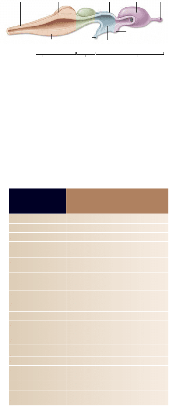
Figure 44.20
The basic organization of the vertebrate
brain can be seen in the brains of primitive shes.
The brain is divided into three regions that are found in differing
proportions in all vertebrates: the hindbrain, which is the largest
portion of the brain in shes; the midbrain, which in shes is
devoted primarily to processing visual information; and the
forebrain, which is concerned mainly with olfaction (the sense of
smell) in shes. In terrestrial vertebrates, the forebrain plays a far
more dominant role in neural processing than it does in shes.
in which all neurons are similar and linked to one another in a
web, or nerve net. There is no associative activity, no control
of complex actions, and little coordination.
The simplest animals with associative activity in the ner-
vous system are the free-living flatworms, phylum Platy hel-
minthes. Running down the bodies of these flatworms are two
nerve cords, from which peripheral nerves extend outward to
the muscles of the body. The two nerve cords converge at the
front end of the body, forming an enlarged mass of nervous tis-
sue that also contains interneurons with synapses connecting
neurons to one another. This primitive “brain” is a rudimentary
central nervous system and permits a far more complex control
of muscular responses than is possible in cnidarians.
All of the subsequent evolutionary changes in nervous
systems can be viewed as a series of elaborations on the charac-
teristics already present in flatworms. For example, among coe-
lomate invertebrates (see chapter 34), earthworms exhibit a
central nervous system that is connected to all other parts of
the body by peripheral nerves. And in arthropods, the central
coordination of complex responses is increasingly localized in
the front end of the nerve cord. As this region evolved, it came
to contain a progressively larger number of interneurons and to
develop tracts, which are major information highways within
the brain.
Vertebrate brains have three basic divisions
Casts of the interior braincases of fossil agnathans, fishes that
swam 500 mya (see chapter 35), have revealed much about the
early evolutionary stages of the vertebrate brain. Although
small, these brains already had the three divisions that charac-
terize the brains of all contemporary vertebrates:
the 1. hindbrain, or rhombencephalon;
the 2. midbrain, or mesencephalon; and
the 3. forebrain, or prosencephalon ( gure 44.20 and
table 44.3).
The hindbrain in fishes
The hindbrain was the major component of these early brains,
as it still is in fishes today. Composed of the cerebellum,
pons, and medulla oblongata, the hindbrain may be consid-
ered an extension of the spinal cord devoted primarily to co-
ordinating motor reflexes. Tracts containing large numbers of
axons run like cables up and down the spinal cord to the hind-
brain. The hindbrain, in turn, integrates the many sensory
signals coming from the muscles and coordinates the pattern
of motor responses.
Much of this coordination is carried on within a small
extension of the hindbrain called the cerebellum (“little cere-
brum”). In more advanced vertebrates, the cerebellum plays an
increasingly important role as a coordinating center for move-
ment and it is correspondingly larger than it is in the fishes. In
all vertebrates, the cerebellum processes data on the current
position and movement of each limb, the state of relaxation or
contraction of the muscles involved, and the general position of
the body and its relation to the outside world.
902
part
VII
Animal Form and Function
rav32223_ch44_887-914.indd 902rav32223_ch44_887-914.indd 902 11/17/09 3:32:25 PM11/17/09 3:32:25 PM
Apago PDF Enhancer
Chordate Ancestor
Lancelets
Tunicates
Jawless Fish
Bony Fish
Mammals
Birds
Reptiles
Amphibians
Cartilaginous Fish
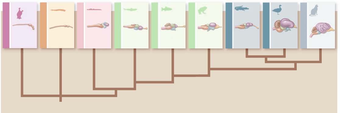
Figure 44.21
Evolution of the vertebrate brain. The relative sizes of different brain regions have changed as vertebrates have
evolved. In sharks and other shes, the hindbrain is predominant, and the rest of the brain serves primarily to process sensory information. In
amphibians and reptiles, the forebrain is far larger, and it contains a larger cerebrum devoted to associative activity. In birds, which evolved
from reptiles, the cerebrum is even more pronounced. In mammals, the cerebrum covers the optic tectum and is the largest portion of the
brain. The dominance of the cerebrum is greatest in humans, in whom it envelops much of the rest of the brain.
The midbrain and forebrain of fishes
In fishes, the remainder of the brain is devoted to the reception
and processing of sensory information. The midbrain is com-
posed primarily of the optic tectum, which receives and processes
visual information, whereas the forebrain is devoted to the pro-
cessing of olfactory (smell) information.
The brains of fishes continue growing throughout their
lives. This continued growth is in marked contrast to the
brains of other classes of vertebrates, which generally com-
plete their development by infancy. The human brain con-
tinues to develop through early childhood, but few new
neurons are produced once development has ceased. One ex-
ception is the hippocampus, which has control over which
experiences are filed away into long-term memory and which
are forgotten. The extent of neurogenesis (production of
new neurons) in adult brains is controversial, and one area of
active current research.
The dominant forebrain in more recent vertebrates
Starting with the amphibians and continuing more prominently
in the reptiles, processing of sensory information is increasingly
centered in the forebrain. This pattern was the dominant evo-
lutionary trend in the further development of the vertebrate
brain (figure 44.21).
The forebrain in reptiles, amphibians, birds, and mam-
mals is composed of two elements that have distinct functions.
The diencephalon consists of the thalamus and hypothalamus.
The thalamus is an integration and relay center between in-
coming sensory information and the cerebrum. The hypo-
thalamus participates in basic drives and emotions and controls
the secretions of the pituitary gland. The telencephalon, or
“end brain,” is located at the front of the forebrain and is de-
voted largely to associative activity. In mammals, the tel-
encephalon is called the cerebrum. The telencephalon also
includes structures we discuss later on when describing the
human brain.
The expansion of the cerebrum
In examining the relationship between brain mass and body
mass among the vertebrates, a remarkable difference is ob-
served between fishes and reptiles on the one hand, and birds
and mammals on the other. Mammals have brains that are par-
ticularly large relative to their body mass. This is especially true
of porpoises and humans.
The increase in brain size in mammals largely reflects the
great enlargement of the cerebrum, the dominant part of the
mammalian brain. The cerebrum is the center for correlation,
association, and learning in the mammalian brain. It receives
sensory data from the thalamus and issues motor commands to
the spinal cord via descending tracts of axons.
In vertebrates, the central nervous system is composed
of the brain and the spinal cord (see table 44.3). These
two structures are responsible for most of the information
processing within the nervous system and they consist pri-
marily of interneurons and neuroglia. Ascending tracts carry
sensory information to the brain. Descending tracts
carry impulses from the brain to the motor neurons and
inter neurons in the spinal cord that control the muscles of
the body.
The human forebrain exhibits exceptional
information-processing ability
The human cerebrum is so large that it appears to envelop
the rest of the brain. It is split into right and left cerebral
hemispheres, which are connected by a tract called the corpus
chapter
44
The Nervous System
903www.ravenbiology.com
rav32223_ch44_887-914.indd 903rav32223_ch44_887-914.indd 903 11/17/09 3:32:25 PM11/17/09 3:32:25 PM
Apago PDF Enhancer
Parietal lobe
Occipital
lobe
Cerebellum
Frontal lobe
Temporal
lobe
Motor areas involved
with the control of
voluntary muscles
Motor speech area
(Broca's area)
Lateral
sulcus
Auditory
area
Interpretation of sensory
experiences, memory of
visual and auditory patterns
Combining
visual images,
visual recognition
of objects
General interpretative
area (Wernicke's area)
Sensory areas involved
with cutaneous
and other senses
Central
sulcus
Corpus
callosum
Parietal lobe of
cerebral cortex
Pineal gland
Occipital
lobe of
cerebral
cortex
Cerebellum
Medulla
oblongata
Pons
Hypothalamus
Pituitary
gland
Thalamus
Frontal lobe
of cerebral
cortex
Lateral
ventricle
Optic chiasm
Optic
recess
Temporal
lobe of cerebral
cortex
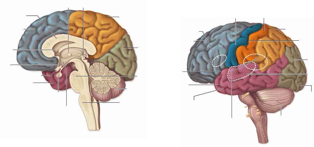
Figure 44.23
The cerebrum. This diagram shows the
lobes of the cerebrum and indicates some of the known regions
of specialization.
lips, and tongue because of the need for manual dexterity and
speech. The auditory cortex lies within the temporal lobe, and
different regions of this cortex deal with different sound fre-
quencies. The visual cortex lies on the occipital lobe, with dif-
ferent sites processing information from different positions
on the retina, equivalent to particular points in the visual
fields of the eyes.
The portion of the cerebral cortex that is not occupied by
these motor and sensory cortices is referred to as the association
cortex. The site of higher mental activities, the association cor-
tex reaches its greatest extent in primates, especially humans,
where it makes up 95% of the surface of the cerebral cortex.
Basal ganglia
Buried deep within the white matter of the cerebrum are sev-
eral collections of cell bodies and dendrites that produce islands
of gray matter. These aggregates of neuron cell bodies, which
are collectively termed the basal ganglia, receive sensory infor-
mation from ascending nerve tracts and motor commands from
the cerebral cortex and cerebellum.
Outputs from the basal ganglia are sent down the spinal
cord, where they participate in the control of body move-
ments. Damage to specific regions of the basal ganglia can
produce the resting tremor of muscles that is characteristic of
Parkinson disease.
Thalamus and hypothalamus
The thalamus is a primary site of sensory integration in the
brain. Visual, auditory, and somatosensory information is sent
to the thalamus, where the sensory tracts synapse with associa-
tion neurons. The sensory information is then relayed via the
thalamus to the occipital, temporal, and parietal lobes of the
cerebral cortex, respectively. The transfer of each of these types
callosum (figure 44.22) . The hemispheres are further divided
into the frontal, parietal, temporal, and occipital lobes.
Each hemisphere primarily receives sensory input from
the opposite, or contralateral, side of the body and exerts motor
control primarily over that side. Therefore, a touch on the right
hand is relayed primarily to the left hemisphere, which may
then initiate movement of the right hand in response to the
touch. Damage to one hemisphere due to a stroke often results
in a loss of sensation and paralysis on the contralateral side of
the body.
The cerebral cortex
Much of the neural activity of the cerebrum occurs within a
layer of gray matter only a few millimeters thick on its outer
surface. This layer, called the cerebral cortex, is densely packed
with nerve cells. In humans, it contains over 10 billion nerve
cells, amounting to roughly 10% of all the neurons in the brain.
The surface of the cerebral cortex is highly convoluted; this is
particularly true in the human brain, where the convolutions
increase the surface area of the cortex threefold.
The activities of the cerebral cortex fall into one of three
general categories: motor, sensory, and associative. Each of its
regions correlates with a specific function (figure 44.23) . The
primary motor cortex lies along the gyrus (convolution) on
the posterior border of the frontal lobe, just in front of the cen-
tral sulcus (crease). Each point on the surface of the motor cor-
tex is associated with the movement of a different part of the
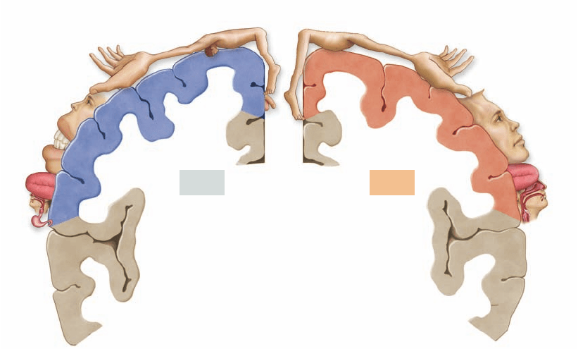
body (figure 44.24, right).
Just behind the central sulcus, on the anterior edge of
the parietal lobe, lies the primary somatosensory cortex.
Each point in this area receives input from sensory neurons
serving skin and muscle senses in a particular part of the body
(figure 44.24, left). Large areas of the primary motor cortex
and primary somatosensory cortex are devoted to the fingers,
Figure 44.22
A section through the human brain. In
this sagittal section showing one cerebral hemisphere, the corpus
callosum, a ber tract connecting the two cerebral hemispheres,
can be clearly seen.
904
part
VII
Animal Form and Function
rav32223_ch44_887-914.indd 904rav32223_ch44_887-914.indd 904 11/17/09 3:32:26 PM11/17/09 3:32:26 PM
Apago PDF Enhancer
Tongue
Lips
Jaw
Face
Eye
Tongue
Jaw
Lips
Teeth
Gums
Face
Eye
Nose
Brow
Neck
Thumb
Fingers
Fingers
Forefinger
Hand
Hand
Forearm
Wrist
Elbow
Elbow
Arm
Arm
Shoulder
Trunk
Trunk
Hip
Hip
Genitals
Knee
Toes
Pharynx
Leg
Motor
Sensory
Figure 44.24
The
primary somatosensory
cortex (left) and the
primary motor cortex
(right). Each of these
regions of the cerebral cortex
is associated with a different
region of the body, as
indicated in this stylized
map. The areas of the body
are drawn in relative
proportion to the amount of
cortex dedicated to their
sensation or control. For
example, the hands have
large areas of sensory and
motor control, and the
pharynx has a considerable
area of motor control but
little area devoted to the
sensations of the pharynx.
tors the information coming into the brain and identifies im-
portant stimuli. When the reticular-activating system has been
stimulated to arousal, it increases the level of activity in many
parts of the brain. Neural pathways from the reticular forma-
tion to the cortex and other brain regions are depressed by an-
esthetics and barbiturates.
The reticular-activating system controls both sleep and
the waking state. It is easier to sleep in a dark room than in a
lighted one because there are fewer visual stimuli to stimulate
the reticular-activating system. In addition, activity in this sys-
tem is reduced by serotonin, a neurotransmitter discussed ear-
lier. Serotonin causes the level of brain activity to fall, bringing
on sleep.
Brain state can be monitored by means of an electro-
encephalogram (EEG), a recording of electrical activity. Awake
but relaxed individuals with eyes closed exhibit a brain pattern
of large, slow waves termed alpha waves. In an alert individual
with eyes open, the waves are more rapid (beta waves) and more
desynchronized as sensory input is being received. Theta waves
and delta waves are very slow waves seen during sleep. When an
individual is in REM sleep—characterized by rapid eye move-
ments with the eyes closed—the EEG is more like that of an
awake, relaxed individual.
Language
Although the two cerebral hemispheres seem structurally
similar, they are responsible for different activities. The most
thoroughly investigated example of this lateralization of func-
tion is language.
The left hemisphere is the “dominant” hemisphere for
language in 90% of right-handed people and nearly two-thirds
of left-handed people. (By dominant, we mean it is the hemi-
sphere in which most neural processing related to language is
performed.) Different brain regions control language in the
of sensory information is handled by specific aggregations of
neuron cell bodies within the thalamus.
The hypothalamus integrates the visceral activities. It
helps regulate body temperature, hunger and satiety, thirst,
and—along with the limbic system—various emotional states.
The hypothalamus also controls the pituitary gland, which in
turn regulates many of the other endocrine glands of the body.
By means of its interconnections with the cerebral cortex and
with control centers in the brainstem (a term used to refer col-
lectively to the midbrain, pons, and medulla oblongata), the
hypothalamus helps coordinate the neural and hormonal re-
sponses to many internal stimuli and emotions.
The hippocampus and amygdala, along with the hypothal-
amus, are the major components of the limbic system—an
evolutionarily ancient group of linked structures deep within
the cerebrum that are responsible for emotional responses, as
described earlier. The hippocampus is also believed to be im-
portant in the formation and recall of memories.
Complex functions of the human brain may be
controlled in speci c areas
Although studying brain function is difficult, it has long fasci-
nated researchers. The distinction between sleep and waking,
the use and acquisition of language, spatial recognition, and
memory are all areas of active research. Although far from
understood, one generalization that emerged was the region-
alization of function.
Sleep and arousal
The brainstem contains a diffuse collection of neurons referred
to as the reticular formation. One part of this formation, the
reticular-activating system, controls consciousness and alertness.
All of the sensory pathways feed into this system, which moni-
chapter
44
The Nervous System
905www.ravenbiology.com
rav32223_ch44_887-914.indd 905rav32223_ch44_887-914.indd 905 11/17/09 3:32:32 PM11/17/09 3:32:32 PM
Apago PDF Enhancer
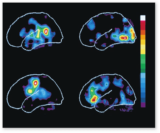
Hearing Words
Speaking Words
Seeing Words
Generating Words
Min
Max
F igure 44.25
Di erent brain regions control various
language activities. This illustration shows how the brain reacts
in human subjects asked to listen to a spoken word, to read that
same word silently, to repeat the word out loud, and then to speak a
word related to the rst. Regions of white, red, and yellow show the
greatest activity. Compare this with gure 44.24 to see how regions
of the brain are mapped.
Memory and learning
One of the great mysteries of the brain is the basis of memory
and learning. Memory appears dispersed across the brain.
Specific cortical sites cannot be identified for particular mem-
ories because relatively extensive cortical damage does not
selectively remove memories. Although memory is impaired if
portions of the brain, particularly the temporal lobes, are re-
moved, it is not lost entirely. Many memories persist in spite
of the damage, and the ability to access them is gradually re-
covered with time.
Fundamental differences appear to exist between short-
term and long-term memory. Short-term memory is transient,
lasting only a few moments. Such memories can readily be
erased by the application of an electrical shock, leaving previ-
ously stored long-term memories intact. This result suggests
that short-term memories are stored in the form of a transient
neural excitation. Long-term memory, in contrast, appears to
involve structural changes in certain neural connections with-
in the brain.
Two parts of the temporal lobes, the hippocampus and
the amygdala, are involved in both short-term memory and its
consolidation into long-term memory. Damage to these struc-
tures impairs the ability to process recent events into long-
term memories.
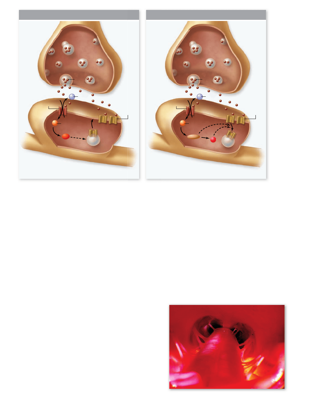
Synaptic plasticity
Part of the basis of learning and memory are changes to the
function of a synapse over time. Two examples of this synaptic
plasticity are long-term potentiation (LTP), and long-term de-
pression (LTD). The mechanism of LTP is complex and not
completely understood. One well-studied form involves syn-
apses that release the neurotransmitter glutamate, and have N-
methyl-d-aspartic acid (NMDA) type of receptors. When either
the same synapse is stimulated repeatedly, or neighboring syn-
apses are stimulated, the postsynaptic membrane becomes sig-
nificantly depolarized. This releases a block of the NMDA
receptor by Mg
2+
such that glutamate binding causes an influx
of Ca
2+
that stimulates a signal transduction pathway involving
calcium/calmodulin-dependent protein kinase II. This pathway
leads to the insertion of another receptor type, the α-amino-3-
hydroxyl-5-methyl-4-isoxazole-propionate (AMPA) receptor,
into the postsynaptic membrane, making the synapse more sen-
sitive to future stimulation (figure 44.26) .
If the stimulation of an NMDA receptor is less, and the
postsynaptic membrane is less depolarized, LTD can result. In
this case, a different Ca
2+
-dependent signaling pathway results
in the loss of AMPA receptors from the membrane. Taken to-
gether, these two mechanisms can make a synapse more or less
sensitive to future stimulation.
Alzheimer disease: Degeneration of brain neurons
In the past, little was known about Alzheimer disease, a condition
in which the memory and thought processes of the brain be-
come dysfunctional. Scientists disagree about the biological
nature of the disease and its cause. Two hypotheses have been
proposed: One suggests that nerve cells in the brain are killed
from the outside in, and the other that the cells are killed from
the inside out.
dominant hemisphere (figure 44.25) . Wernicke’s area, located
in the parietal lobe between the primary auditory and visual
areas, is important for language comprehension and the for-
mulation of thoughts into speech (see figure 44.23). Broca’s
area, found near the part of the motor cortex controlling the
face, is responsible for the generation of motor output needed
for language communication.
Damage to these brain areas can cause language disorders
known as aphasias. For example, if Wernicke’s area is damaged,
the person’s speech is rapid and fluid but lacks meaning; words
are tossed together as in a “word salad.”
Spatial recognition
Whereas the dominant hemisphere for language is adept at se-
quential reasoning, like that needed to formulate a sentence,
the nondominant hemisphere (the right hemisphere in most
people) is adept at spatial reasoning, the type of reasoning
needed to assemble a puzzle or draw a picture. It is also the
hemisphere primarily involved in musical ability—a person
with damage to Broca’s speech area in the left hemisphere may
not be able to speak but may retain the ability to sing.
Damage to the nondominant hemisphere may lead to an
inability to appreciate spatial relationships and may impair
musical activities such as singing. Even more specifically,
damage to the inferior temporal cortex in that hemisphere
eliminates the capacity to recall faces, a condition known as
prosopagnosia. Reading, writing, and oral comprehension re-
main normal, and patients with this disability can still recog-
nize acquaintances by their voices. The nondominant
hemisphere is also important for the consolidation of memo-
ries of nonverbal experiences.
906
part
VII
Animal Form and Function
rav32223_ch44_887-914.indd 906rav32223_ch44_887-914.indd 906 11/17/09 3:32:33 PM11/17/09 3:32:33 PM
Apago PDF Enhancer
Ca
2
+
PPI
Mg
2
+
CaMKII
Calcineurin
Ca
2
+
Mg
2
+
LTP LTD
Presynaptic
terminal
Postsynaptic
dendrite
Dendritic
spine
NMDAR
AMPAR
Expression: postsynaptic insertion of AMPARs Expression: internalization of postsynaptic AMPARs
Glu
NMDAR
AMPAR
Glu
ventral columns. These nerve tracts may also contain the
dendrites of other nerve cells. Messages from the body and
the brain run up and down the spinal cord, the body’s “infor-
mation highway.”
In addition to relaying messages, the spinal cord also
functions in reflexes, the sudden, involuntary movement of
muscles. A reflex produces a rapid motor response to a stimulus
because the sensory neuron passes its information to a motor
In the first hypothesis, external proteins called β-amyloid
exist in an abnormal form, which then forms aggregates, or
plaques. The plaques begin to fill in the brain and then dam-
age and kill nerve cells. However, these amyloid plaques have
been found in autopsies of people who did not exhibit Alz hei-
mer disease.
The second hypothesis maintains that the nerve cells are
killed by an abnormal form of an internal protein called tau (τ),
which normally functions to maintain protein transport micro-
tubules. Abnormal forms of τ-protein assemble into helical
segments that form tangles, which interfere with the normal
functioning of the nerve cells. At this point, the association of
tangles with actual neuronal death is stronger.
The spinal cord conveys messages
and controls some responses directly
The spinal cord is a cable of neurons extending from the brain
down through the backbone (figure 44.27) . It is enclosed and
protected by the vertebral column and layers of membranes
called meninges, which also cover the brain. Inside the spinal
cord are two zones.
The inner zone is gray matter and primarily consists of
the cell bodies of interneurons, motor neurons, and neuro-
glia. The outer zone is white matter and contains cables of
sensory axons in the dorsal columns and motor axons in the
Figure 44.26
LTP and LTD modulate synaptic function. a. When a the postsynaptic membrane is signi cantly depolarized, when
GABA binds to the N-methyl-d-aspartic acid receptor (NMDAR), the in ux of Ca
2+
leads to the insertion of α-amino-3-hydroxyl-5-methyl-
4-isoxazole-propionate receptors (AMPAR). This potentiates the synapse for future stimulation. b. When the postsynaptic membrane does
not have as large a depolarization, or there is less GABA, then GABA binding to NMDA receptor triggers a different pathway that results in
removal of AMPA receptors. This depresses the synapse for future stimulation. CaMKII, calmodulin-dependent protein kinase 2.
Figure 44.27
A view down the human spinal cord. Pairs
of spinal nerves can be seen extending from the spinal cord. Along
these nerves, as well as the cranial nerves that arise from the brain,
the central nervous system communicates with the rest of the body.
chapter
44
The Nervous System
907www.ravenbiology.com
rav32223_ch44_887-914.indd 907rav32223_ch44_887-914.indd 907 11/17/09 3:32:34 PM11/17/09 3:32:34 PM
Apago PDF Enhancer
Quadriceps
muscle
(effector)
Spinal cord
Dorsal root
ganglion
Gray
matter
White
matter
Monosynaptic
synapse
Sensory
neuron
Nerve fiber
Stretch receptor
(muscle spindle)
Skeletal
muscle
Stimulus
Response
Motor neuron
Effector
(muscle)
Dorsal
Ventral
Spinal Cord
Interneuron
Cell body in dorsal
root ganglion
Gray
matter
White
matter
Motor
neuron
Sensory
neuron
Receptor
in skin
Stimulus
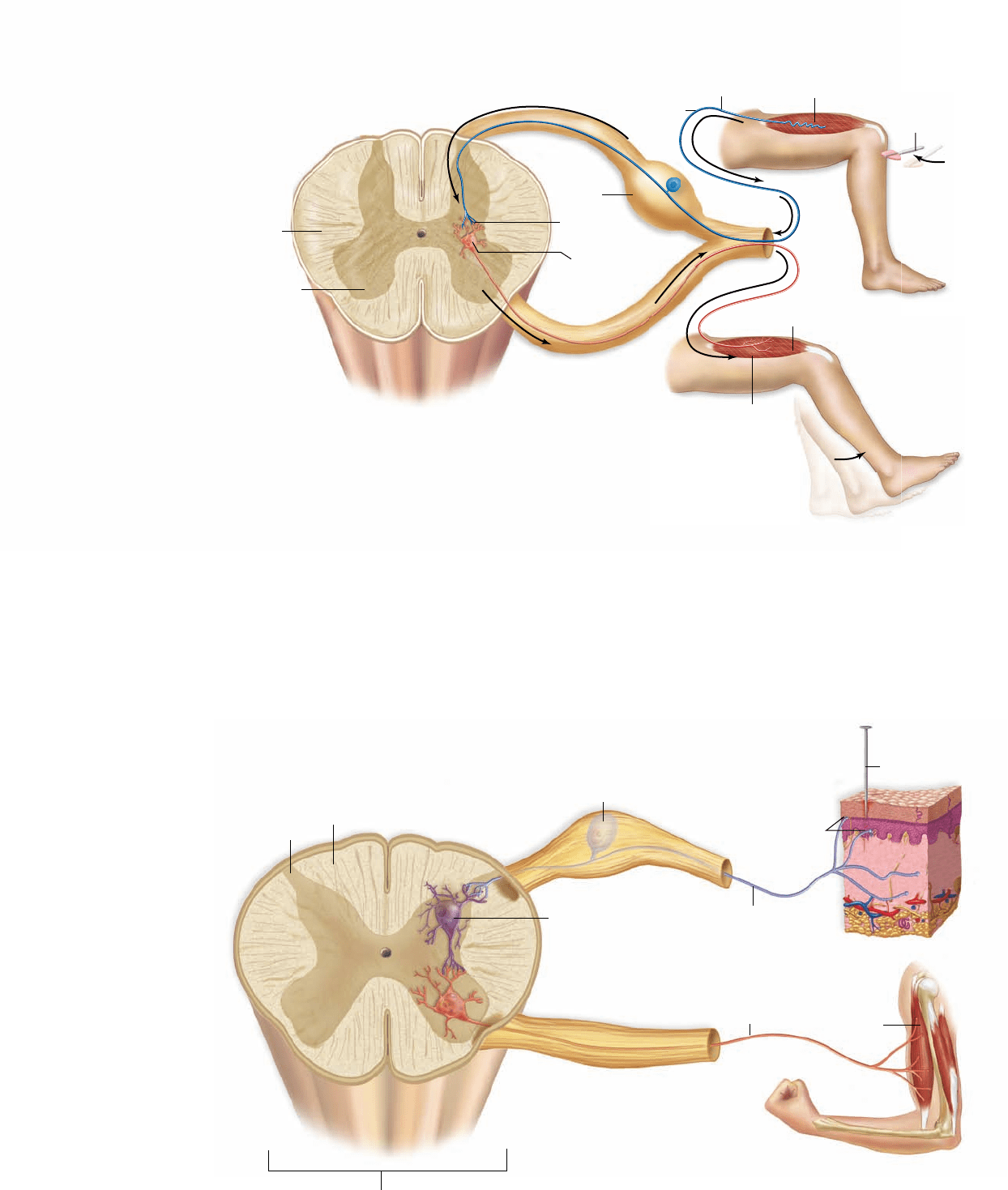
Figure 44.28
The knee-jerk re ex.
This is the simplest re ex,
involving only sensory and
motor neurons.
Figure 44.29
A cutaneous spinal
re ex. This re ex is
more complex than a
knee-jerk re ex because
it requires interneurons
as well as sensory and
motor neurons.
Interneurons connect
a sensory neuron with
a motor neuron to
cause muscle
contraction as shown.
Other interneurons
inhibit motor neurons,
allowing antagonistic
muscles to relax.
Most reflexes in verte-
brates, however, involve a single
connecting interneuron between the
sensory neuron and the motor neuron
(figure 44.29). The withdrawal of a hand from a hot stove or the
blinking of an eye in response to a puff of air involves a relay of
information from a sensory neuron through one or more inter-
neurons to a motor neuron. The motor neuron then stimulates
the appropriate muscle to contract. Notice that the sensory
neuron may also connect to other interneurons to send signals
to the brain. Although you jerked your hand away from the
stove, you will still feel pain.
neuron in the spinal cord, without higher level processing. One
of the most frequently used reflexes in your body is blinking, a
reflex that protects your eyes. If an object such as an insect or a
cloud of dust approaches your eye, the eyelid blinks before you
realize what has happened. The reflex occurs before the cere-
brum is aware the eye is in danger.
Because they pass information along only a few neurons,
reflexes are very fast. A few reflexes, such as the knee-jerk reflex
(figure 44.28) , are monosynaptic reflex arcs. In these, the sen-
sory nerve cell makes synaptic contact directly with a motor
neuron in the spinal cord whose axon travels directly back to
the muscle.
908
part
VII
Animal Form and Function
rav32223_ch44_887-914.indd 908rav32223_ch44_887-914.indd 908 11/17/09 3:32:36 PM11/17/09 3:32:36 PM
Apago PDF Enhancer
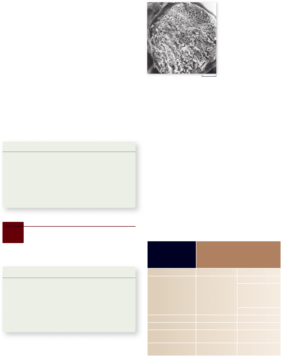
6.25 μm
Figure 44.30
Nerves
in the peripheral nervous
system. Photomicrograph
showing a cross section of a
bullfrog nerve. The nerve is a
bundle of axons bound
together by connective tissue.
Many myelinated axons are
visible, each looking
somewhat like a doughnut.
function of the PNS is to receive information from the envi-
ronment, convey it to the CNS, and to carry responses to effec-
tors such as muscle cells.
The PNS has somatic and autonomic systems
At the spinal cord, a spinal nerve separates into sensory and
motor components. The axons of sensory neurons enter the
dorsal surface of the spinal cord and form the dorsal root of
the spinal nerve, whereas motor axons leave from the ventral
surface of the spinal cord and form the ventral root of the
spinal nerve. The cell bodies of sensory neurons are grouped
together outside each level of the spinal cord in the dorsal
root ganglia. The cell bodies of somatic motor neurons, on
the other hand, are located within the spinal cord and so are
not located in ganglia.
As mentioned earlier, somatic motor neurons stimulate
skeletal muscles to contract, and autonomic motor neurons in-
nervate involuntary effectors—smooth muscles, cardiac mus-
cle, and glands. A comparison of the somatic and autonomic
nervous systems is provided in table 44.4 ; we discuss each sys-
tem in turn.
Spinal cord regeneration
In the past, scientists tried to repair severed spinal cords by
installing nerves from another part of the body to bridge the
gap and act as guides for the spinal cord to regenerate. But
most of these experiments failed. Although axons may regen-
erate through the implanted nerves, they cannot penetrate the
spinal cord tissue once they leave the implant. Also, a factor that
inhibits nerve growth is present in the spinal cord.
After discovering that fibroblast growth factor stimulates
nerve growth, neurobiologists working with rats tried “gluing”
the nerves on, from the implant to the spinal cord, with fibrin
that had been mixed with the fibroblast growth factor. Three
months later, rats with the nerve bridges began to show move-
ment in their lower bodies. Dye tests indicated that the spinal
cord nerves had regrown from both sides of the gap.
Many scientists are encouraged by the potential to use a
similar treatment in human medicine. But most spinal cord in-
juries in humans do not involve a completely severed spinal
cord; often, nerves are crushed, which results in different tissue
damage. Also, even though the rats with nerve bridges did re-
gain some ability to move, tests indicated that they were barely
able to walk or stand.
Learning Outcomes Review 44.4
The vertebrate brain has three primary regions: the hindbrain, midbrain,
and forebrain. The cerebrum, part of the forebrain, is composed of two
cerebral hemispheres in which gray matter of the cerebral cortex overlays
white matter and islands of gray matter (nuclei) called the basal ganglia.
The spinal cord relays messages to and from the brain; a refl ex occurs when
the spinal cord processes sensory information directly and initiates a
motor response.
■ What is the advantage of having reflexes?
TABLE 44.4
Comparison of the Somatic
and Autonomic Nervous
Systems
Characteristic Somatic Autonomic
E ectors Skeletal muscle Cardiac muscle
Smooth muscle
Gastrointestinal tract
Blood vessels
Airways
Exocrine glands
E ect on motor nerves Excitation Excitation or inhibition
Innervation of e ector cells Always single Typically dual
Number of sequential neurons
in path to e ector
One Two
Neurotransmitter Acetylcholine Acetylcholine,
norepinephrine
44.5
The Peripheral Nervous
System: Sensory and Motor
Neurons
Learning Outcomes
Describe the organization of the peripheral 1.
nervous system.
Explain the actions of sensory and somatic neurons.2.
Distinguish between the somatic and autonomic 3.
nervous systems.
Describe differences between the sympathetic and 4.
parasympathetic divisions of the autonomic
nervous system.
The PNS consists of nerves, the cablelike collections of axons
(figure 44.30) , and ganglia (singular, ganglion), aggregations of
neuron cell bodies located outside the CNS. To review, the
chapter
44
The Nervous System
909www.ravenbiology.com
rav32223_ch44_887-914.indd 909rav32223_ch44_887-914.indd 909 11/17/09 3:32:39 PM11/17/09 3:32:39 PM
Apago PDF Enhancer
Viscera
Autonomic
ganglion
Postganglionic
neuron
Autonomic motor reflex
Interneuron
Dorsal root
ganglion
Preganglionic
neuron
Sensory
neuron
Spinal
cord
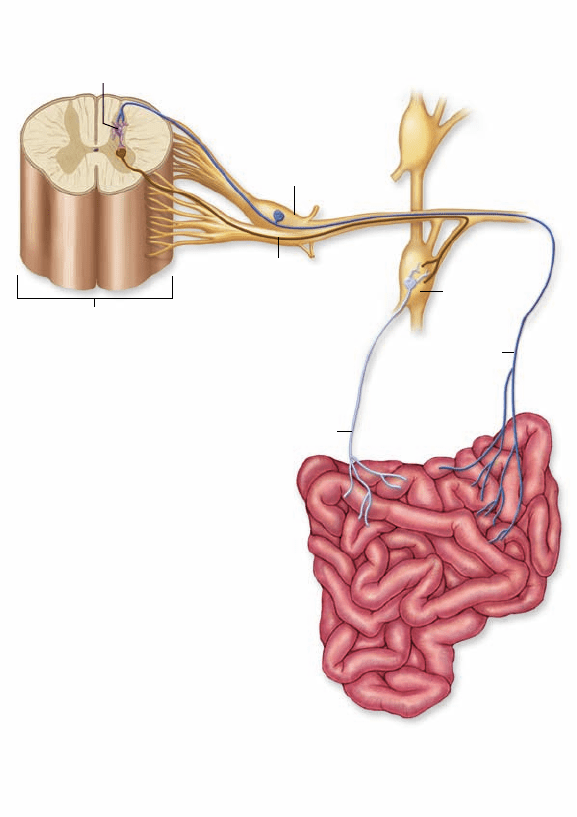
Figure 44.31
An autonomic neural path. There are two
motor neurons in the efferent pathway. The rst, or preganglionic
neuron, exits the CNS and synapses at an autonomic ganglion. The
second, or postganglionic neuron, exits the ganglion and regulates
the visceral effectors (smooth muscle, cardiac muscle, or glands).
The parasympathetic division
The actions of the sympathetic division are antagonized by the
parasympathetic division. Preganglionic parasympathetic neu-
rons originate in the brain and sacral regions of the spinal cord
(see figure 44.32, right). Because of this origin, there cannot be
a chain of parasympathetic ganglia analogous to the sympa-
thetic chain. Instead, the preganglionic axons, many of which
travel in the vagus (tenth cranial) nerve, terminate in ganglia
located near or even within the internal organs. The postgan-
glionic neurons then regulate the internal organs by releasing
ACh at their synapses. Parasympathetic nerve effects include a
slowing of the heart, increased secretions and activities of di-
gestive organs, and so on. Table 44.5 compares the actions of
the sympathetic and parasympathetic divisions.
G proteins mediate cell responses
to autonomic signals
You might wonder how release of ACh can slow the heart rate—
an inhibitory effect—when it has excitatory effects elsewhere.
The somatic nervous system
controls movements
Somatic motor neurons stimulate the skeletal muscles of the
body to contract in response to conscious commands and as
part of reflexes that do not require conscious control. Voluntary
control of skeletal muscles is achieved by activation of tracts of
axons that descend from the cerebrum to the appropriate level
of the spinal cord. Some of these descending axons stimulate
spinal cord motor neurons directly, and others activate inter-
neurons that in turn stimulate the spinal motor neurons.
When a particular muscle is stimulated to contract, how-
ever, its antagonist must be inhibited. In order to flex the arm,
for example, the flexor muscles must be stimulated while the
antagonistic extensor muscle is inhibited (see chapter 47). De-
scending motor axons produce this necessary inhibition by
causing hyperpolarizations (IPSPs) of the spinal motor neurons
that innervate the antagonistic muscles.
The autonomic nervous system controls
involuntary functions through two divisions
The autonomic nervous system is composed of the sympathetic
and parasympathetic divisions plus the medulla oblongata of the
hindbrain, which coordinates this system. Although they differ,
the sympathetic and parasympathetic divisions share several
features. In both, the efferent motor pathway involves two neu-
rons: The first has its cell body in the CNS and sends an axon
to an autonomic ganglion; it is called preganglionic neuron. These
neurons release acetylcholine at their synapses.
The second neuron has its cell body in the autonomic
ganglion and sends its axon to synapse with a smooth muscle,
cardiac muscle, or gland cell (figure 44.31) . This second neuron
is termed the postganglionic neuron. Those in the parasympa-
thetic division release ACh, and those in the sympathetic divi-
sion release norepinephrine.
The sympathetic division
In the sympathetic division, the preganglionic neurons origi-
nate in the thoracic and lumbar regions of the spinal cord
(figure 44.32, left) . Most of the axons from these neurons syn-
apse in two parallel chains of ganglia immediately outside the
spinal cord. These structures are usually called the sympathetic
chain of ganglia. The sympathetic chain contains the cell bodies
of postganglionic neurons, and it is the axons from these neu-
rons that innervate the different visceral organs.
There are some exceptions to this general pattern, how-
ever. The axons of some preganglionic sympathetic neurons
pass through the sympathetic chain without synapsing and, in-
stead, terminate within the medulla of the adrenal gland (see
chapter 46). In response to action potentials, the adrenal me-
dulla cells secrete the hormone epinephrine (adrenaline). At the
same time, norepinephrine is released at the synapses of the
postganglionic neurons. As described earlier, both of these
neuro trans mitters prepare the body for action by heightening
metabolism and blood flow.
910
part
VII
Animal Form and Function
rav32223_ch44_887-914.indd 910rav32223_ch44_887-914.indd 910 11/17/09 3:32:40 PM11/17/09 3:32:40 PM
Apago PDF Enhancer
Constrict
Secrete saliva
Dilate
Stop secretion
Dilate bronchioles
Speed up heartbeat
Increase secretion
Empty colon
Increase motility
Empty bladder
Slow down heartbeat
Constrict bronchioles
Sympathetic
ganglion
chain
Stomach
Secrete adrenaline
Decrease secretion
Decrease motility
Retain colon contents
Delay emptying
Adrenal gland
Bladder
Small intestine
Large intestine
Spinal cord
Parasympathetic Sympathetic
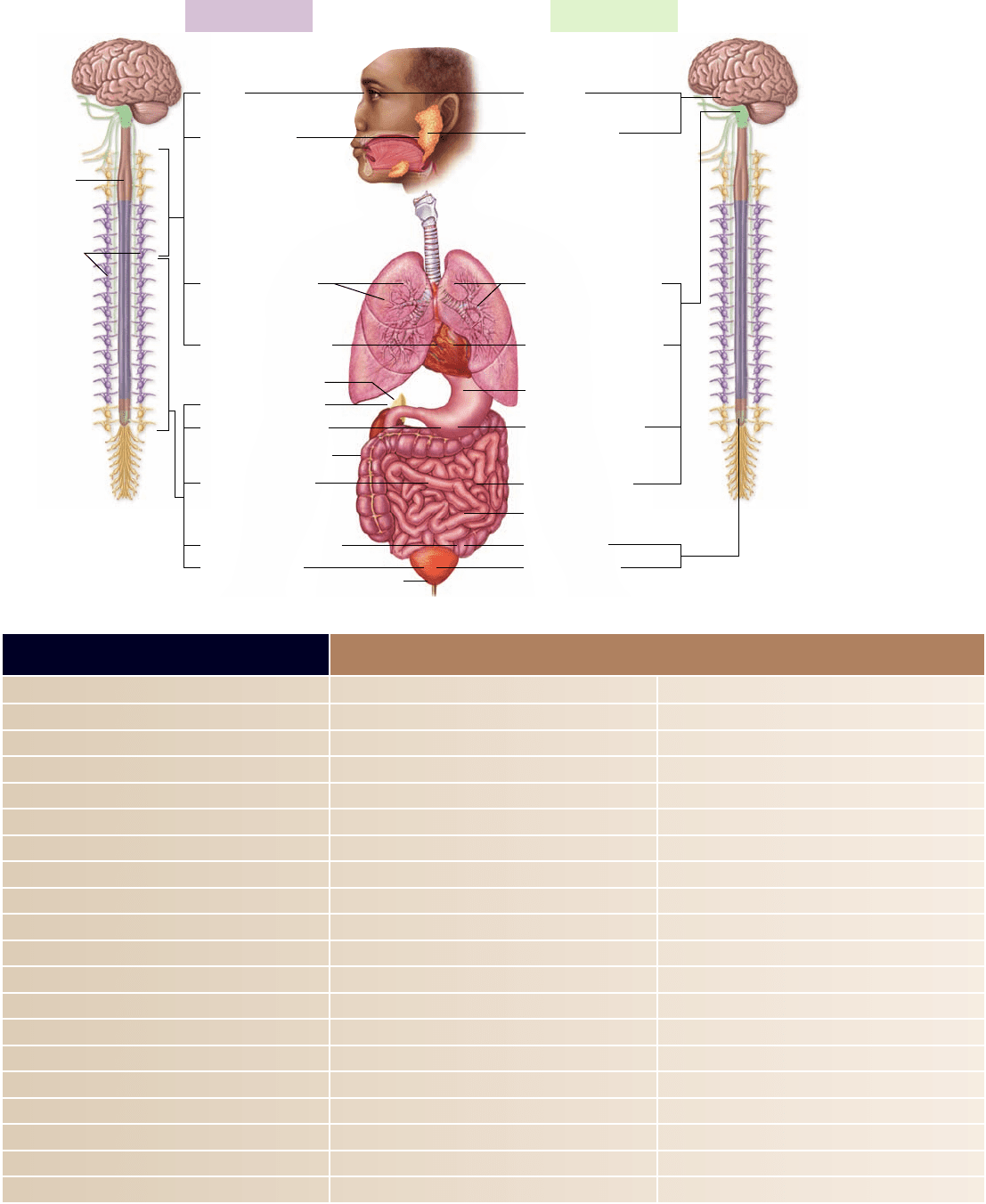
Figure 44.32
The sympathetic and
parasympathetic
divisions of the
autonomic nervous
system. The
preganglionic neurons of
the sympathetic division
exit the thoracic and
lumbar regions of the
spinal cord, and those of
the parasympathetic
division exit the brain
and sacral region of the
spinal cord. The ganglia
of the sympathetic
division are located near
the spinal cord; and
those of the
parasympathetic division
are located near the
organs they innervate.
Most of the internal
organs are innervated by
both divisions.
TABLE 44.5
Autonomic Innervation of Target Tissues
Target Tissue Sympathetic Stimulation Parasympathetic Stimulation
Pupil of eye Dilation Constriction
Glands
Salivary Vasoconstriction; slight secretion Vasodilation; copious secretion
Gastric Inhibition of secretion Stimulation of gastric activity
Liver Stimulation of glucose secretion Inhibition of glucose secretion
Sweat Sweating None
Gastrointestinal tract
Sphincters Increased tone Decreased tone
Wall Decreased tone Increased motility
Gallbladder Relaxation Contraction
Urinary bladder
Muscle Relaxation Contraction
Sphincter Contraction Relaxation
Heart muscle Increased rate and strength Decreased rate
Lungs Dilation of bronchioles Constriction of bronchioles
Blood vessels
In muscles Dilation None
In skin Constriction None
In viscera Constriction Dilation
chapter
44
The Nervous System
911www.ravenbiology.com
rav32223_ch44_887-914.indd 911rav32223_ch44_887-914.indd 911 11/18/09 4:09:51 PM11/18/09 4:09:51 PM