Raven P.H., Johnson G.B., Mason K.A. Biology (Ninth Edition)
Подождите немного. Документ загружается.

Apago PDF Enhancer
Ma Ma
S
Ar
Ar
Ar
Ar
Q
Ar
Q
Ar
Q
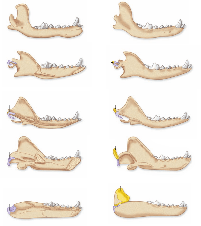
Dog
Early mammal
Cynodont
Synapsid
Morganucodon
Figure 45.7
Evolution of the
mammalian inner ear. Two of the
bones in the inner ear of modern
mammals, the stapes and malleus, are
derived from the quadrate and articular
bones, respectively, of their reptilian
ancestors. The transition from an early
ancestor of mammals, a synapsid, through
several transition forms, to a modern dog
is illustrated. Note how the bones become
smaller and change position, ultimately
disappearing from the lower jaw entirely
in modern mammals (represented by the
dog) and becoming parts of the inner ear.
During embryology in modern mammals,
these bones develop in association with
the lower jaw bone before moving inward
to the inner ear, providing further
evidence of their evolutionary origin.
The fossil record makes clear that the malleus and incus
of modern mammals is derived from the two bones in the lower
jaws of synapsid reptiles (figure 45.7) . Through evolutionary
time, these bones became progressively smaller and came to lie
closer to the stapes. Eventually, in modern mammals, they be-
came completely disconnected from the jawbone and moved
within the middle ear itself.
The middle ear is connected to the throat by the Eusta-
chian tube, also known as the auditory tube, which equalizes
the air pressure between the middle ear and the external envi-
ronment. The “ear popping” you may have experienced when
flying in an airplane or driving on a mountain is caused by pres-
sure equalization between the two sides of the eardrum.
The stapes vibrates against a flexible membrane, the oval
window, which leads into the inner ear. Because the oval win-
dow is smaller in diameter than the tympanic membrane, vibra-
tions against it produce more force per unit area, transmitted
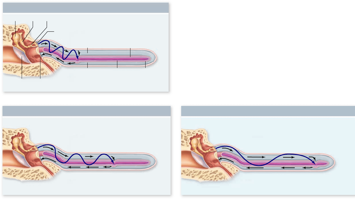
into the inner ear. The inner ear consists of the cochlea, a bony
structure containing part of the membranous labyrinth called
the cochlear duct. The cochlear duct is located in the center of
the cochlea; the area above the cochlear duct is the vestibular
canal, and the area below is the tympanic canal (figure 45.6c).
All three chambers are filled with fluid. The oval window opens
to the upper vestibular canal, so that when the stapes causes it
to vibrate, it produces pressure waves of fluid. These pressure
waves travel down to the tympanic canal, pushing another flex-
ible membrane, the round window, that transmits the pressure
back into the middle ear cavity.
Transduction occurs in the cochlea
As pressure waves are transmitted through the cochlea to the
round window, they cause the cochlear duct to vibrate. The
bottom of the cochlear duct, called the basilar membrane, is
quite flexible and vibrates in response to these pressure waves.
The surface of the basilar membrane contains sensory hair
cells. The stereocilia from the hair cells project into an over-
hanging gelatinous membrane, the tectorial membrane. This
sensory apparatus, consisting of the basilar membrane, hair
cells with associated sensory neurons, and tectorial membrane,
is known as the organ of Corti (figure 45.6d).
922
part
VII
Animal Form and Function
rav32223_ch45_915-936.indd 922rav32223_ch45_915-936.indd 922 11/17/09 3:55:09 PM11/17/09 3:55:09 PM
Apago PDF Enhancer
Vestibular
canal
Round
window
Tympanic
membrane
Malleus Incus Stapes
Oval
window
Cochlear
duct
Tympanic
canal
Apex
Base
Basilar
membrane
High Frequency (20,000 Hz)
Medium Frequency (2000 Hz) Low Frequency (500 Hz)
a.
b. c.
of other regions. When these action potentials arrive in the
brain, they are interpreted as representing a sound of a par-
ticular frequency, or pitch.
The range of terrestrial vertebrate hearing
The flexibility of the basilar membrane limits the frequency
range of human hearing to between approximately 20 and 20,000
cycles per second (hertz, Hz) in children. Our ability to hear
high-pitched sounds decays progressively throughout middle
age. Other vertebrates can detect sounds at frequencies lower
than 20 Hz and much higher than 20,000 Hz. Dogs, for example,
can detect sounds at 40,000 Hz, enabling them to hear high-
pitched dog whistles that seem silent to a human listener.
Hair cells are also innervated by efferent axons from the
brain, and impulses in those axons can make hair cells less sen-
sitive. This central control of receptor sensitivity can increase
an individual’s ability to concentrate on a particular auditory
signal (for example, a single voice) in the midst of background
noise, which is effectively “tuned out” by the efferent axons.
Some vertebrates have the ability
to navigate by sound
Because terrestrial vertebrates have two ears located on oppo-
site sides of the head, the information provided by hearing can
be used to determine the direction of a sound source with some
precision. Sound sources vary in strength, however, and sounds
are weakened and reflected to varying degrees by the presence
As the basilar membrane vibrates, the cilia of the hair cells
bend in response to the movement of the basilar membrane
relative to the tectorial membrane. The bending of these stereo-
cilia in one direction depolarizes the hair cells. Bending in the
opposite direction repolarizes or even hyperpolarizes the mem-
brane. The hair cells, in turn, stimulate the production of action
potentials in sensory neurons that project to the brain, where
they are interpreted as sound.
Frequency localization in the cochlea
The basilar membrane of the cochlea consists of elastic fibers
of varying length and stiffness, like the strings of a musical in-
strument, embedded in a gelatinous material. At the base of the
cochlea (near the oval window), the fibers of the basilar mem-
brane are short and stiff. At the far end of the cochlea (the apex),
the fibers are 5 times longer and 100 times more flexible.
Therefore, the resonant frequency of the basilar membrane is
higher at the base than at the apex; the base responds to higher
pitches, the apex to lower pitches.
When a wave of sound energy enters the cochlea from
the oval window, it initiates an up-and-down motion that
travels the length of the basilar membrane. However, this
wave imparts most of its energy to that part of the basilar
membrane with a resonant frequency near the frequency of
the sound wave, resulting in a maximum deflection of the
basilar membrane at that point (figure 45.8) . As a result, the
hair cell depolarization is greatest in that region, and the af-
ferent axons from that region are stimulated more than those
Figure 45.8
Frequency localization in the cochlea.
The cochlea is shown unwound, so that the length of the basilar
membrane can be seen. The bers within the basilar membrane
vibrate in response to different frequencies of sound, related to the
pitch of the sound. Thus, regions of the basilar membrane show
maximum vibrations in response to different sound frequencies.
a. Notice that high-frequency (pitch) sounds vibrate the basilar
membrane more toward the base whereas medium frequencies (b)
and low frequencies (c) cause vibrations more toward the apex.
chapter
45
Sensory Systems
923www.ravenbiology.com
rav32223_ch45_915-936.indd 923rav32223_ch45_915-936.indd 923 11/17/09 3:55:10 PM11/17/09 3:55:10 PM
Apago PDF Enhancer
Cochlea
Vestibular nerve
Kinocilium
Stereocilia
Sensory
neuron
Utricle (horizontal
acceleration)
Semicircular canals
Saccule (vertical
acceleration)
Gelatinous
matrix
Otoliths
Hair cells
Hair cell
Gravitational
force
Signal
Supporting
cells
Stationary Movement
a. b.
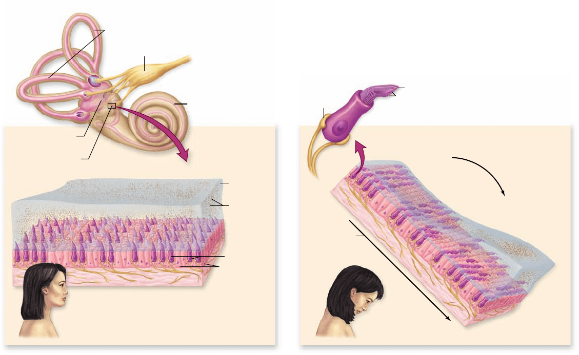
Figure 45.9
Structure and function of the utricle and saccule. a. The relative
positions of the utricle and saccule within the membranous labyrinth of the human inner
ear. Enlargement shows the gelatinous matrix containing otoliths covering hair cells.
b. When your head bends forward, gravity distorts the matrix in the direction of
movement. This causes the stereocilia in hair cells to bend, stimulating sensory neurons.
opment of sensory organs that detect body position in space
and movements such as acceleration.
Most invertebrates can orient themselves with respect to
gravity due to a sensory structure called a statocyst. Statocysts
generally consist of ciliated hair cells with the cilia embedded in
a gelatinous membrane containing crystals of calcium carbon-
ate. These stones, or statoliths, increase the mass of the gelati-
nous membrane so that it can bend the cilia when the animal’s
position changes. If the animal tilts to the right, for example,
the statolith membrane bends the cilia on the right side and
activates associated sensory neurons.
A similar structure is found in the membranous labyrinth
of the inner ear of vertebrates. This labyrinth is surrounded by
bone and perilymph, which is similar in ionic content to inter-
stitial fluid. Inside, the chambers and tubes are filled with endo-
lymph fluid, which is similar in ionic content to intracellular
fluid. Though intricate, the entire structure is very small; in a
human, it is about the size of a pea.
Structure of the labyrinth and semicircular canals
The receptors for gravity in most vertebrates consist of two
chambers of the membranous labyrinth called the utricle and
saccule (figure 45.9) . Within these structures are hair cells with
stereocilia and a kinocilium, similar to those in the lateral line
system of fish. The hairlike processes are embedded within a
gelatinous membrane, the otolith membrane, containing calci-
um carbonate crystals. Because the otolith organ is oriented dif-
ferently in the utricle and saccule, the utricle is more sensitive to
of objects in the environment. For these reasons, auditory sen-
sors do not provide a reliable measure of distance.
A few groups of mammals that live and obtain their food in
dark environments have circumvented the limitations of darkness.
A bat flying in a completely dark room easily avoids objects placed
in its path—even a wire less than a millimeter in diameter. Shrews
use a similar form of “lightless vision” beneath the ground, as do
whales and dolphins beneath the sea. All of these mammals are
able to perceive presence and distance of objects by sound.
These mammals emit sounds and then determine the time
it takes these sounds to reach an object and return to the animal.
This process is called echolocation. A bat, for example, produces
clicks that last 2 to 3 ms and are repeated several hundred times
per second. By calculating the time each click takes to hit an ob-
ject and return, bats can calculate the location, direction of move-
ment, and speed of objects in their environment. The human
inventions sonar and radar are based on the same principles
of echolocation.
The three-dimensional imaging achieved with such an
auditory sonar system is quite sophisticated. Bats can track and
intercept rapidly maneuvering aerial prey and can distinguish
one type of insect from another.
Body position and movement are detected
by systems associated with hearing systems
The evolutionary strategy of using internal calcium carbonate
crystals as a way to detect vibration has also allowed the devel-
924
part
VII
Animal Form and Function
rav32223_ch45_915-936.indd 924rav32223_ch45_915-936.indd 924 11/17/09 3:55:10 PM11/17/09 3:55:10 PM
Apago PDF Enhancer
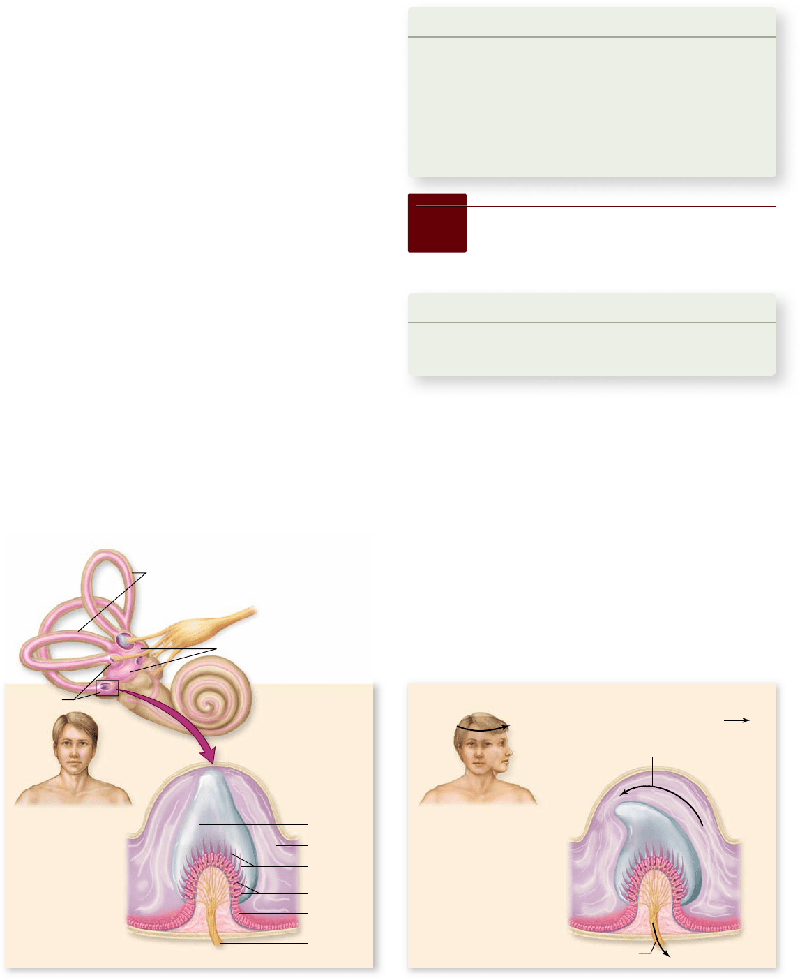
a. b.
Semicircular canals
Ampullae
Vestibule
Flow of endolymph
Cupula
Stationary Movement
Stimulation
Cilia of hair
cells
Hair cells
Supporting
cell
Vestibular
nerve
Endolymph
Direction of body movement
Vestibular nerve
horizontal acceleration (as in a moving car) and the saccule to
vertical acceleration (as in an elevator). In both cases, the accel-
eration causes the stereocilia to bend, and consequently pro-
duces action potentials in an associated sensory neuron.
The membranous labyrinth of the utricle and saccule is
continuous with three semicircular canals, oriented in differ-
ent planes so that angular acceleration in any direction can be
detected (figure 45.10) . At the ends of the canals are swollen
chambers called ampullae, into which protrude the cilia of an-
other group of hair cells. The tips of the cilia are embedded
within a sail-like wedge of gelatinous material called a cupula
(similar to the cupula of the fish lateral line system) that pro-
trudes into the endolymph fluid of each semicircular canal.
Action of the vestibular apparatus
When the head rotates, the fluid inside the semicircular canals
pushes against the cupula and causes the cilia to bend. This
bending either depolarizes or hyperpolarizes the hair cells, de-
pending on the direction in which the cilia are bent. This is
similar to the way the lateral line system works in a fish: If the
stereocilia are bent in the direction of the kinocilium, a recep-
tor potential is produced, which stimulates the production of
action potentials in associated sensory neurons.
The saccule, utricle, and semicircular canals are collec-
tively referred to as the vestibular apparatus. The saccule
and utricle provide a sense of linear acceleration, and the
semicircular canals provide a sense of angular acceleration.
The brain uses information that comes from the vestibular
apparatus about the body’s position to maintain balance
and equilibrium.
Figure 45.10
The structure of the semicircular canals. The position of the
semicircular canals in relation to the rest of the inner ear. a. Enlargement of a section of
one ampulla, showing how hair cell cilia insert into the cupula. b. Angular acceleration in
the plane of one of the semicircular canals causes bending of the cupula, thereby
stimulating the hair cells. Each inner ear contains three semicircular canals, one for each
potential axis of rotation .
Learning Outcomes Review 45.3
Sound waves cause middle-ear ossicles to vibrate; fl uid in the inner ear
is vibrated in turn, bending hair cells and causing action potentials. In
terrestrial animals, sound waves in air must transition to the fl uid in the
inner ear. Hair cells in the vestibular apparatus of terrestrial vertebrates
provide a sense of acceleration and balance.
■ Why is a lateral line system not useful to
adult amphibians?
45.4
Chemoreceptors:
Taste, Smell, and pH
Learning Outcomes
List the five taste categories.1.
Describe how taste buds and olfactory neurons function.2.
Some sensory cells, called chemoreceptors, contain membrane
proteins that can bind to particular chemicals or ligands in the
extracellular fluid. In response to this chemical interaction, the
membrane of the sensory neuron becomes depolarized and pro-
duces action potentials. Chemoreceptors are used in the senses
of taste and smell and are also important in monitoring the
chemical composition of the blood and cerebrospinal fluid.
chapter
45
Sensory Systems
925www.ravenbiology.com
rav32223_ch45_915-936.indd 925rav32223_ch45_915-936.indd 925 11/17/09 3:55:12 PM11/17/09 3:55:12 PM
Apago PDF Enhancer
Circumvallate
papilla
Taste bud
Foliate papilla
Fungiform
papilla
a. b.
Signals to brain
Different chemoreceptors
Sensory hair on foot
Pore
Proboscis
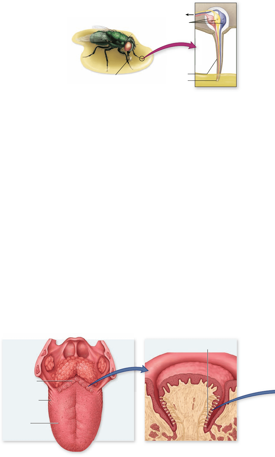
Figure 45.11
Taste. a. Human
tongues have projections called
papillae that bear taste buds. Different
sorts of taste buds are located on
different regions of the tongue.
b. Groups of taste buds are embedded
within a papilla. c. Individual taste
buds are bulb-shaped collections of
chemosensitive receptors that open
out into the mouth through a pore.
d. Photomicrograph of taste buds
in papillae.
Figure 45.12
Many insects taste with their feet. In the
blow y shown here, chemoreceptors extend into the sensory hairs
on the foot. Different chemoreceptors detect different types of food
molecules. When the y steps in a food substance, it can taste the
different food molecules and extend its proboscis for feeding.
Taste detects and analyzes potential food
The perception of taste (gustation), like the perception of color,
is a combination of physical and psychological factors. This is
commonly broken down into five categories: sweet, sour, salty,
bitter, and umami (perception of glutamate and other amino
acids that give a hearty taste to many protein-rich foods such as
meat, cheese, and broths). Taste buds—collections of chemo-
sensitive epithelial cells associated with afferent neurons—
mediate the sense of taste in vertebrates. In a fish, the taste buds
are scattered over the surface of the body. These are the most
sensitive vertebrate chemoreceptors known. They are particu-
larly sensitive to amino acids; a catfish, for example, can distin-
guish between two different amino acids at a concentration of
less than 100 parts per billion (1 g in 10,000 L of water)!
The ability to taste the surrounding water is very important to
bottom-feeding fish, enabling them to sense the presence of
food in an often murky environment.
The taste buds of all terrestrial vertebrates occur in the
epithelium of the tongue and oral cavity, within raised areas
called papillae (figure 45.11) . Taste buds are onion-shaped struc-
tures of between 50 and 100 taste cells; each cell has fingerlike
projections called microvilli that poke through the top of the
taste bud, called the taste pore (figure 45.11c). Chemicals from
food dissolve in saliva and contact the taste cells through the
taste pore.
Within a taste bud, the chemicals that produce salty and
sour tastes act directly through ion channels. The prototypical
salty taste is due to Na
+
ions, which diffuse through Na
+
channels
into cells in receptor cells in the taste bud. This Na
+
influx depo-
larizes the membrane, causing the receptor cell to release neuro-
transmitter and activate a sensory neuron that sends an impulse to
the brain. The cells that detect sour taste act in a similar fashion
except that the ion detected is H
+
. Sour tastes are associated with
increased concentration of protons that can also depolarize the
membrane when they diffuse through ion channels.
The mechanism of detection of sweet, bitter, and umami
are indirect. In this case substances that fall into these catego-
ries can bind to G protein–coupled receptors (see chapter 9)
specific for each category. The nature and distribution of these
receptors is an area of active investigation, but recent data indi-
cate that individual receptor cells in the taste bud express only
one type of receptor. This leads to cells that have receptors for
sweet, for bitter or for umami tastes. Activation of any of these
G protein–coupled receptors then stimulates a single signaling
pathway that leads the release of neurotransmitter from recep-
tor cells to activate a sensory neuron and send an impulse to the
brain. There they interact with other sensory neurons carrying
information related to smell, described next. In this model, the
different tastes are encoded to the brain based on which recep-
tor cells are activated.
Like vertebrates, many arthropods also have taste chemo-
receptors. For example, flies, because of their mode of searching
for food, have taste receptors in sensory hairs located on their feet.
The sensory hairs contain a variety of chemoreceptors that are
able to detect sugars, salts, and other tastes by the integration of
stimuli from these chemoreceptors (figure 45.12) . If they step on
potential food, their proboscis (the tubular feeding apparatus) ex-
tends to feed.
Smell can identify a vast number
of complex molecules
In terrestrial vertebrates, the sense of smell (olfaction ) in-
volves chemoreceptors located in the upper portion of the
926
part
VII
Animal Form and Function
rav32223_ch45_915-936.indd 926rav32223_ch45_915-936.indd 926 11/17/09 3:55:13 PM11/17/09 3:55:13 PM
Apago PDF Enhancer
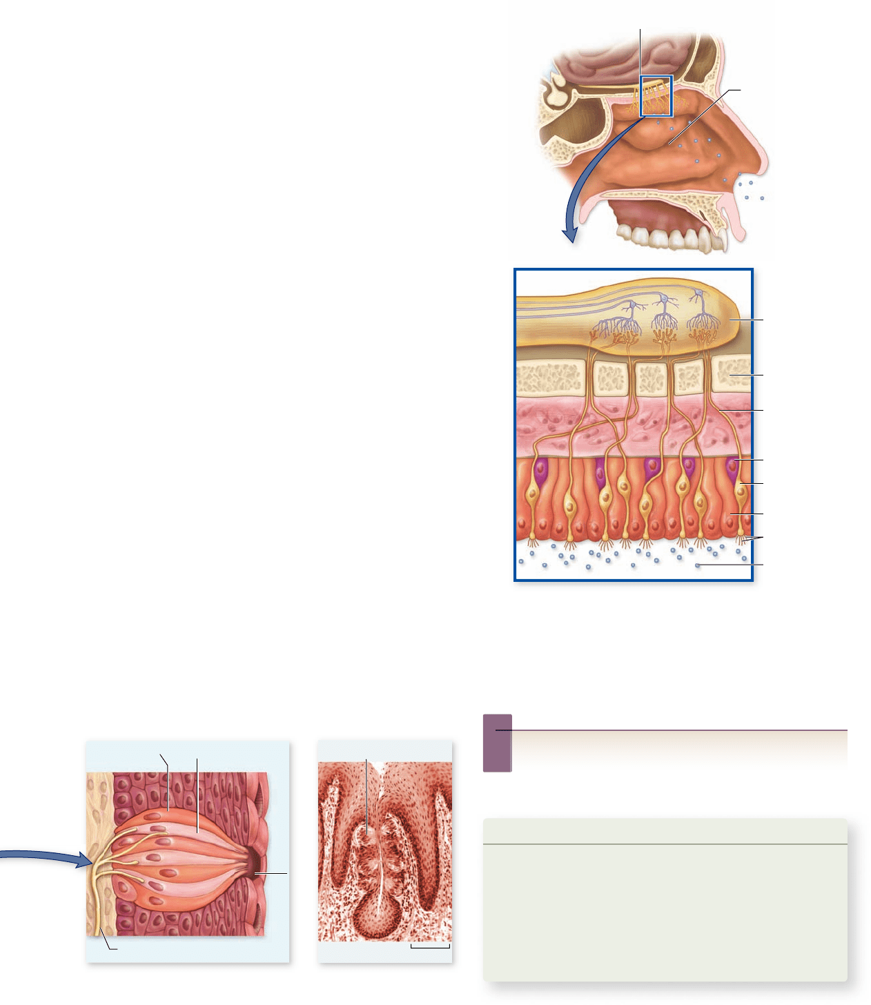
Taste bud
Support cell
Taste poreNerve fiber
Receptor cell
with microvilli
d.c.
544 µm
Olfactory bulb
Nasal passage
Olfactory
bulb
Cribiform
plate
Axon of
olfactory
nerve
Olfactory
receptor
Olfactory
hairs
Support cell
Basal cell
Odor
Learning Outcomes Review 45.4
The fi ve tastes humans perceive are sweet, sour, salty, bitter, and umami
(amino acids). Taste and smell chemoreceptors detect chemicals from
outside the body; olfactory receptors can identify thousands of diff erent
odors. Internal chemoreceptors monitor acid–base balance within the body
and help regulate breathing.
■ What are the advantages of insects’ having taste
receptors on their feet?
nasal passages (figure 45.13) . These receptors, whose den-
drites end in tassels of cilia, project into the nasal mucosa, and
their axons project directly into the cerebral cortex. A terres-
trial vertebrate uses its sense of smell in much the same way
that a fish uses its sense of taste—to sample the chemical en-
vironment around it.
Because terrestrial vertebrates are surrounded by air, their
sense of smell has become specialized to detect airborne
particles—but these particles must first dissolve in extracellular
fluid before they can activate the olfactory receptors. The sense
of smell can be extremely acute in many mammals, so much so
that a single odorant molecule may be all that is needed to ex-
cite a given receptor.
Although humans can detect only five modalities of taste,
they can discern thousands of different smells. New research
suggests that as many as a thousand different genes may code
for different receptor proteins for smell. The particular set of
olfactory neurons that respond to a given odor might serve as a
“fingerprint” the brain can use to identify the odor.
Internal chemoreceptors detect
pH and other characteristics
Sensory receptors within the body detect a variety of chemi-
cal characteristics of the blood or fluids derived from the
blood, including cerebrospinal fluid. Included among these
receptors are the peripheral chemoreceptors of the aortic
and carotid bodies, which are sensitive primarily to plasma
pH, and the central chemoreceptors in the medulla oblon-
gata of the brain, which are sensitive to the pH of cerebro-
spinal fluid. When the breathing rate is too low, the
concentration of plasma CO
2
increases, producing more car-
bonic acid and causing a fall in the blood pH. The carbon
dioxide can also enter the cerebrospinal fluid and lower the
pH, thereby stimulating the central chemoreceptors. This
stimulation indirectly affects the respiratory control center
of the brainstem, which increases the breathing rate. The
aortic bodies can also respond to a lowering of blood oxygen
concentrations, but this effect is normally not significant un-
less a person goes to a high altitude where the partial pres-
sure of oxygen is lower.
Figure 45.13
Smell.
Humans detect smells by means of
olfactory neurons (receptor cells) located in the lining of the nasal
passages. The axons of these neurons transmit impulses directly to
the brain via the olfactory nerve. Basal cells regenerate new
olfactory neurons to replace dead or damaged cells. Olfactory
neurons typically live about a month.
Inquiry question
?
In what ways do the senses of taste and smell share
similarities? How are they different?
chapter
45
Sensory Systems
927www.ravenbiology.com
rav32223_ch45_915-936.indd 927rav32223_ch45_915-936.indd 927 11/17/09 3:55:15 PM11/17/09 3:55:15 PM
Apago PDF Enhancer
Photoreceptors
Eyespot
Pigment layer
Flatworm will turn
away from light
Light
Lenses
Lens Lens
Optic
nerve
Optic
nerve
Optic
nerve
Retinular
cell
Retina Retina
Chordate Mollusk Insect
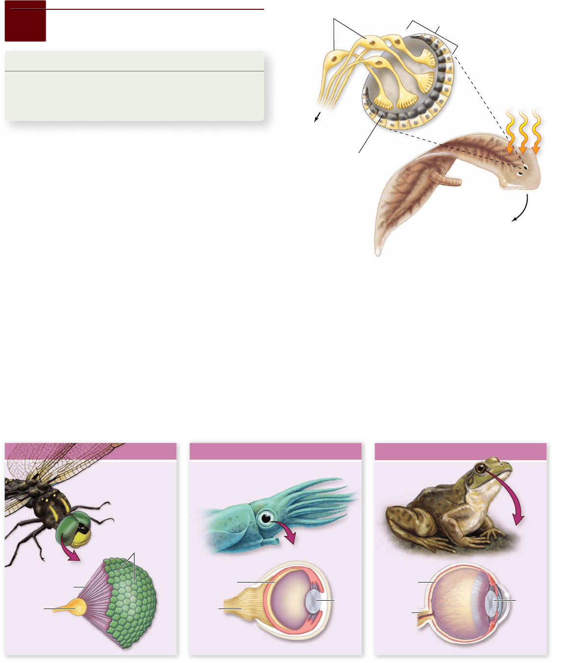
Figure 45.14
Simple eyespots in the atworm. Eyespots
can detect the direction of light because a pigmented layer on one
side of the eyespot screens out light coming from the back of the
animal. Light is thus detected more readily coming from the front
of the animal; atworms respond by turning away from the light.
Figure 45.15
Eyes in three phyla of animals. Although they are super cially similar, these eyes differ greatly in structure from one
another (see also gure 21.16 for a detailed comparison of mollusk and chordate eye structure). Each has evolved separately and, despite the
apparent structural complexity, has done so from simpler structures.
45.5
Vision
Learning Outcomes
Compare invertebrate and vertebrate eyes. 1.
Explain how a vertebrate eye focuses an image.2.
Describe how photoreceptors function.3.
The ability to perceive objects at a distance is important to
most animals. Predators locate their prey, and prey avoid their
predators, based on the three long-distance senses of hearing,
smell, and vision. Of these, vision can act most distantly; with
the naked eye, humans can see stars thousands of light years
away—and a single photon is sufficient to stimulate a cell of the
retina to send an action potential.
Vision senses light and light
changes at a distance
Vision begins with the capture of light energy by photoreceptors.
Because light travels in a straight line and arrives virtually instan-
taneously regardless of distance, visual information can be used to
determine both the direction and the distance of an object. Other
stimuli, which spread out as they travel and move more slowly,
provide much less precise information.
Invertebrate eyes
Many invertebrates have simple visual systems with photo-
receptors clustered in an eyespot. Simple eyespots can be
made sensitive to the direction of a light source by the addi-
tion of a pigment layer that shades one side of the eye. Flat-
worms have a screening pigmented layer on the inner and
back sides of both eyespots, allowing stimulation of the photo-
receptor cells only by light from the front of the animal (fig-
ure 45.14) . The flatworm will turn and swim in the direction
in which the photoreceptor cells are the least stimulated. Al-
though an eyespot can perceive the direction of light, it can-
not be used to construct a visual image.
928
part
VII
Animal Form and Function
rav32223_ch45_915-936.indd 928rav32223_ch45_915-936.indd 928 11/17/09 3:55:19 PM11/17/09 3:55:19 PM
Apago PDF Enhancer
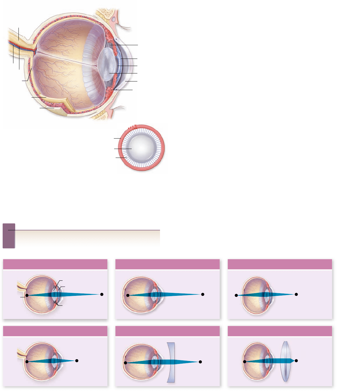
Retina
Optic
nerve
Fovea
Vein
Artery
Iris
Cornea
Lens
Lens
Suspensory
ligament
Suspensory ligament
Sclera
Ciliary muscle
Pupil
Ciliary muscle
a. b. c.
Suspensory
ligaments
Iris
Retina
Lens
Normal Distant Vision Nearsighted Farsighted
Normal Near Vision Nearsighted, Corrected Farsighted, Corrected
Figure 45.16
Structure of the human eye. The
transparent cornea and lens focus light onto the retina at the back of
the eye, which contains the photoreceptors (rods and cones). The
center of each eye’s visual eld is focused on the fovea. Focusing is
accomplished by contraction and relaxation of the ciliary muscle,
which adjusts the curvature of the lens.
Inquiry question
?
How does the human eye differ from the eye of a mollusk, and
how do these differences create a blind spot?
The members of four phyla—annelids, mollusks, arthro-
pods, and chordates—have evolved well-developed, image-
forming eyes. True image-forming eyes in these phyla, although
strikingly similar in structure, are believed to have evolved in-
dependently, an example of convergent evolution (figure 45.15) .
Interestingly, the photoreceptors in all of these image-forming
eyes use the same light-capturing molecule, suggesting that not
many alternative molecules are able to play this role.
Structure of the vertebrate eye
The human eye is typical of the vertebrate eye (figure 45.16) .
The “white of the eye” is the sclera, formed of tough connec-
tive tissue. Light enters the eye through a transparent cornea,
which begins to focus the light. Focusing occurs because light
is refracted (bent) when it travels into a medium of different
density. The colored portion of the eye is the iris; contraction
of the iris muscles in bright light decreases the size of its open-
ing, the pupil. Light passes through the pupil to the lens, a
transparent structure that completes the focusing of the light
onto the retina at the back of the eye. The lens is attached by
the suspensory ligament to the ciliary muscles.
The shape of the lens is influenced by the amount of ten-
sion in the suspensory ligament, which surrounds the lens and
attaches it to the circular ciliary muscle. When the ciliary mus-
cle contracts, it puts slack in the suspensory ligament, and the
lens becomes more rounded and bends light more strongly.
This rounding is required for close vision. In distance vision,
the ciliary muscles relax, moving away from the lens and tight-
ening the suspensory ligament. The lens thus becomes more
flattened and bends light less, keeping the image focused on the
retina. People who are nearsighted or farsighted do not prop-
erly focus the image on the retina (figure 45.17) . Interestingly,
Figure 45.17
Focusing the human eye. a. In people with normal vision, the image remains focused on the retina in both near and far
vision because of changes produced in the curvature of the lens. When a person with normal vision stands 20 feet or more from an object, the
lens is in its least convex form, and the image is focused on the retina. b. In nearsighted people, the image comes to a focus in front of the
retina, and the image thus appears blurred. c. In farsighted people, the focus of the image would be behind the retina because the distance
from the lens to the retina is too short. Corrective lenses adjust the angle of the light as it enters the eye, focusing it on the retina.
chapter
45
Sensory Systems
929www.ravenbiology.com
rav32223_ch45_915-936.indd 929rav32223_ch45_915-936.indd 929 11/17/09 3:55:21 PM11/17/09 3:55:21 PM
Apago PDF Enhancer
Outer segment Inner segment
Synaptic
terminal
Nucleus Mitochondria
Pigment
discs
Rod
Cone
Connecting cilium
100
80
60
40
20
Blue
cones
420 nm
Green
cones
530 nm
Red
cones
560 nm
Rods
500 nm
400 500 600 700
Wavelength (nm)
Light absorption (percent of maximum)
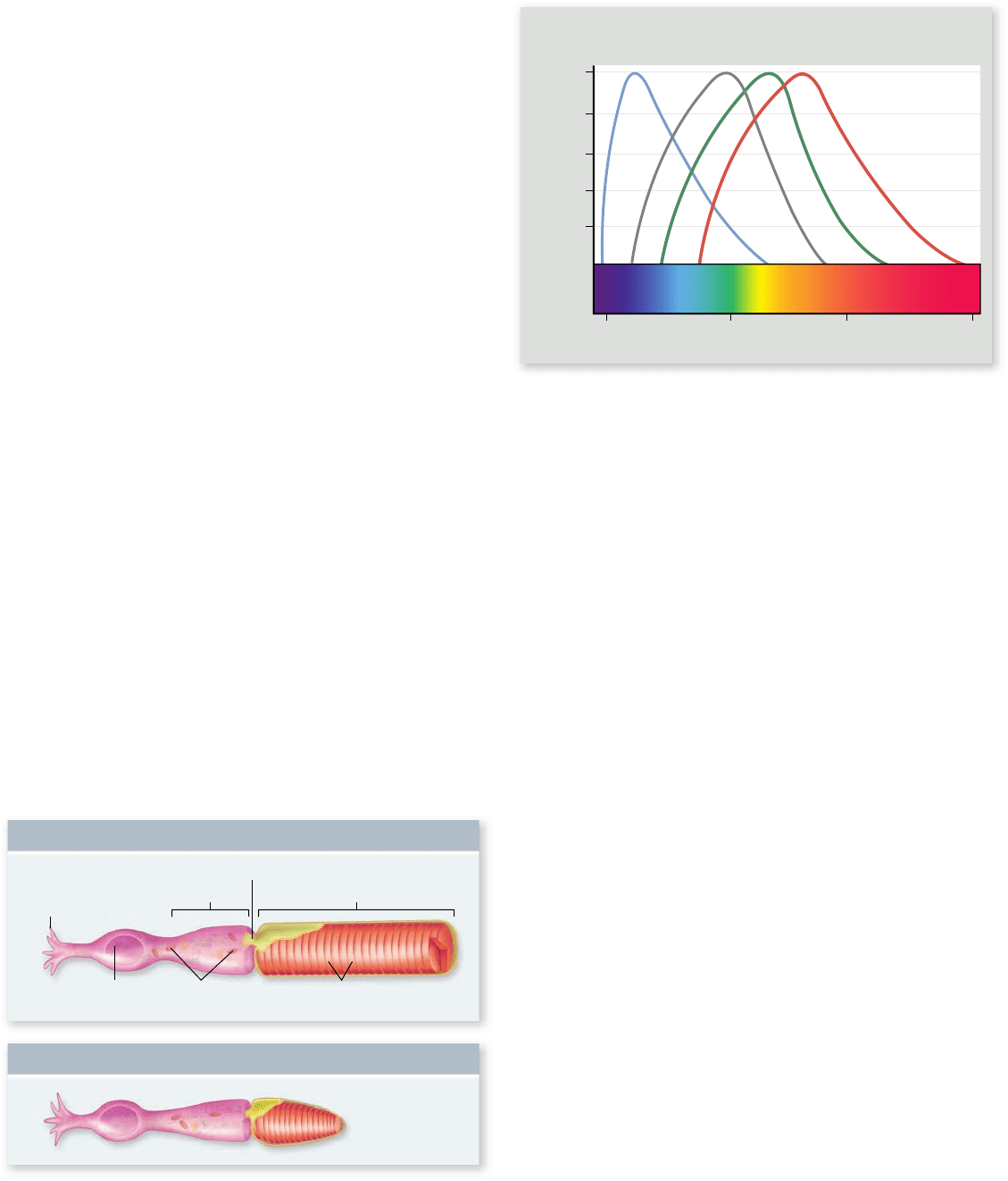
Figure 45.18
Rods and cones. The pigment-containing
outer segment in each of these cells is separated from the rest of the
cell by a partition through which there is only a narrow passage, the
connecting cilium.
Figure 45.19
Color vision. The absorption maximum of
cis-retinal in the rhodopsin of rods is 500 nm. However, the “blue
cones” have their maximum light absorption at 420 nm; the “green
cones” at 530 nm; and the “red cones” at 560 nm. The brain
perceives all other colors from the combined activities of these
three cones’ systems.
region of the electromagnetic spectrum that is best absorbed
by the pigment (figure 45.19) . The absorption maximum of
the cis-retinal in rhodopsin is 500 nanometers (nm); in con-
trast, the absorption maxima of the three kinds of cone pho-
topsins are 420 nm (blue-absorbing), 530 nm (green-absorbing),
and 560 nm (red-absorbing). These differences in the light-
absorbing properties of the photopsins are responsible for the
different color sensitivities of the three kinds of cones, which
are often referred to as simply blue, green, and red cones.
The retina, the inside surface of the eye, is made up of
three layers of cells (figure 45.20) : The layer closest to the ex-
ternal surface of the eyeball consists of the rods and cones; the
next layer contains bipolar cells; and the layer closest to the
cavity of the eye is composed of ganglion cells. Thus, light
must first pass through the ganglion cells and bipolar cells in
order to reach the photoreceptors. The rods and cones synapse
with the bipolar cells, and the bipolar cells synapse with the
ganglion cells, which transmit impulses to the brain via the op-
tic nerve. Ganglion cells are the only neurons of the retina ca-
pable of sending action potentials to the brain. The flow of
sensory information in the retina is therefore opposite to the
path of light through the retina.
Because the ganglion cells lie in the inner cavity of the
eye, the optic nerve must intrude through the retina (see
figure 45.16), creating a blind spot. You can see this blind spot
yourself by holding a finger up in front of your face. Put a col-
ored object on the finger tip, and then, with your left eye closed,
focus on a point next to, but beyond, the fingertip. Now slowly
move your finger to the right while keeping your eye focused on
the distant point. At some point, you’ll notice that you can no
longer see the colored spot on your finger. The structure of the
eye of mollusks avoids this problem by having the sensory neu-
rons attach behind, rather than in front of, the retina (see
figure 45.15).
the lens of an amphibian or a fish does not change shape; these
animals instead focus images by moving their lens in and out,
just as you would do to focus a camera.
Vertebrate photoreceptors
are rod cells and cone cells
The vertebrate retina contains two kinds of photoreceptor cells,
called rods and cones (figure 45.18) . Rods, which get their
name from the shape of their outer segment, are responsible for
black-and-white vision when the illumination is dim. In con-
trast, cones are responsible for high visual acuity (sharpness)
and color vision; cones have a cone-shaped outer segment. Hu-
mans have about 100 million rods and 3 million cones in each
retina. Most of the cones are located in the central region of the
retina known as the fovea, where the eye forms its sharpest im-
age. Rods are almost completely absent from the fovea.
Structure of rods and cones
Rods and cones have the same basic cellular structure. An in-
ner segment rich in mitochondria contains numerous vesicles
filled with neurotransmitter molecules. It is connected by a
narrow stalk to the outer segment, which is packed with hun-
dreds of flattened disks stacked on top of one another. The
light-capturing molecules, or photopigments, are located on
the membranes of these disks (see figure 45.18).
In rods, the photopigment is called rhodopsin. It consists
of the protein opsin bound to a molecule of cis-retinal, which is
produced from vitamin A. Vitamin A is derived from carotene,
a photosynthetic pigment in plants.
The photopigments of cones, called photopsins, are
structurally very similar to rhodopsin. Humans have three kinds
of cones, each of which possesses a photopsin consisting of cis-
retinal bound to a protein with a slightly different amino acid
sequence. These differences shift the absorption maximum, the
930
part
VII
Animal Form and Function
rav32223_ch45_915-936.indd 930rav32223_ch45_915-936.indd 930 11/17/09 3:55:23 PM11/17/09 3:55:23 PM
Apago PDF Enhancer
Axons to
optic nerve
Bipolar
cell
Choroid
Horizontal
cell
Amacrine
cell
Rod Cone
Ganglion
cell
Light
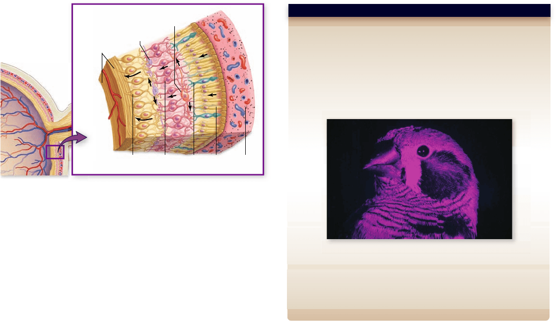
Hypothesis: Birds can see light in the ultraviolet range.
Prediction: Birds will respond to individuals differently depending on
how much ultraviolet is detected in their feathers.
Test: Zebra finch feathers reflect a moderate amount of ultraviolet light.
Female zebra finches were exposed to different males, some of which
were behind a filter that screened out UV light, whereas others were
behind a control filter that let the UV pass through.
Result and Conclusion: Females preferred to spend time near the
UV-positive males. Not only can female zebra finches see light in the UV
range, but they prefer males with UV in the feathers.
Further Experiments: What are two hypotheses about why females
prefer UV-positive males? How would you test these hypotheses?
SCIENTIFIC THINKING
Figure 45.20
Structure of the retina. Note that the rods
and cones are at the rear of the retina, not the front. Light passes
through four other types of cells (ganglion, amacrine, bipolar, and
horizontal) in the retina before it reaches the rods and cones. Once
the photoreceptors are activated, they stimulate bipolar cells, which
in turn stimulate ganglion cells. The ow of sensory information in
the retina is thus opposite to the direction of light.
nels. In the dark, many of these channels are open, allowing an
influx of Na
+
. This flow of Na
+
in the absence of light, called
the dark current, depolarizes the membrane of photoreceptor
cells. In this state, the cells produce inhibitory neurotransmitter
that hyperpolarizes the membrane of bipolar cells. In the light,
the Na
+
channels in the photoreceptor cell rapidly close, re-
ducing the dark current and causing the photoreceptor to hy-
perpolarize. In this state, they no longer produce inhibitory
neurotransmitter. In the absence of inhibition, the membrane
of the bipolar cells is depolarized, causing them to release excit-
atory neurotransmitter to the ganglion cells.
The control of the dark current depends on the ligand for
the Na
+
channels in the photoreceptor cells: the nucleotide cy-
clic guanosine monophosphate (cGMP). In the dark, the level
of cGMP is high, and the channels are open. The system is
made sensitive to light by the nature and structure of the photo-
pigments. Photopigments in the eye are actually G protein–
coupled receptor proteins that are activated by absorbing light.
When a photopigment absorbs light, cis-retinal isomerizes and
dissociates from the receptor protein, opsin, in what is known
as the bleaching reaction. As a result of this dissociation, the
opsin receptor protein changes shape, activating its associated
G protein. The activated G protein then activates its effector
protein, the enzyme phosphodiesterase, which cleaves cGMP
to GMP. The loss of cGMP causes the cGMP-gated Na
+
The retina contains two additional types of neurons called
horizontal cells and amacrine cells. Stimulation of horizontal
cells by photoreceptors at the center of a spot of light on the
retina can inhibit the response of photoreceptors peripheral to
the center. This lateral inhibition enhances contrast and sharp-
ens the image.
Most vertebrates, particularly those that are diurnal (ac-
tive during the day), have color vision, as do many insects and
some other invertebrates. Indeed, honeybees—as well as some
birds, lizards, and other vertebrates (figure 45.21) —can see
light in the near-ultraviolet range, which is invisible to the
human eye. Color vision requires the presence of more than
one photopigment in different receptor cells, but not all ani-
mals with color vision have the three-cone system character-
istic of humans and other primates. Fish, turtles, and birds, for
example, have four or five kinds of cones; the “extra” cones
enable these animals to see near-ultraviolet light and to dis-
tinguish shades of colors that we cannot detect. On the other
hand, many mammals, for example, squirrels and dogs, have
only two types of cones and thus have more limited ability to
distinguish different colors.
Sensory transduction in photoreceptors
The transduction of light energy into nerve impulses follows a
sequence that is the opposite of the usual way that sensory
stimuli are detected. In the dark, the photoreceptor cells release
an inhibitory neurotransmitter that hyperpolarizes the bipolar
neurons. This prevents the bipolar neurons from releasing ex-
citatory neurotransmitter to the ganglion cells that signal to the
brain. In the presence of light, the photoreceptor cells stop re-
leasing their inhibitory neurotransmitter, in effect, stimulating
bipolar cells. The bipolar cells in turn stimulate the ganglion
cells, which transmit action potentials to the brain.
The production of inhibitory neurotransmitter by photo-
receptor cells is due to the presence of ligand-gated Na
+
chan-
Figure 45.21
Ultraviolet vision in birds. Humans cannot
distinguish colors in the near ultraviolet range, whereas many
animals can. This photograph was taken with a special lm that
shows ultraviolet patterns on a zebra nch (Taeniopygia guttata ) that
are not detectable by humans.
chapter
45
Sensory Systems
931www.ravenbiology.com
rav32223_ch45_915-936.indd 931rav32223_ch45_915-936.indd 931 11/17/09 3:55:24 PM11/17/09 3:55:24 PM