Scheffler Immo E. Mitochondria
Подождите немного. Документ загружается.

Literally thousands of different dyes from the color photography industry
were tested. Several others will be discussed below. However, Rh123 (Figure
5.25A ) has remained a favorite for two main reasons. It appears to be the least
toxic to cells, measured by a variety of criteria: survival after weeks of expo-
surel; DNA, RNA, and protein synthesis; operation of the secretory pathway,
maintenance of the cytoskeleton; and cell motility. It may be of special interest
that it appears more toxic to cancer cells. It also appears to be unique in being
unable to stain mitochondria in the absence of ΔΨ (i.e., when azide and oli-
gomycin are used to shut down all mechanisms to produce it), while most of
the other dyes tested still give some staining of mitochondria. Differences in
lipophilicity and the presence of special lipids within the inner membrane
(cardiolipin) have been offered as explanations (253) , but the result remains
partially empirical at this time. A word of caution is in order. Studies have
clearly shown that these dyes may interact with individual complexes of the
electrontransport chain — for example, complex V (W. Allison, personal com-
muniation). Thus, inhibitory effects of these dyes in long - term studies should
not be ignored.
The concentrations of dyes, the length of exposure of the cells to the dye,
the excitation wavelength, and the emission spectrum are all parameters that
have to be considered. For Rh123 the typical conditions are: 0.1 mg/ml for 3
hours, excitation by blue light (485 nm), and a greenish fl uorescence is mea-
sured with the fi lter normally used for fl uorescein dyes (251) . For details the
original papers should be consulted.
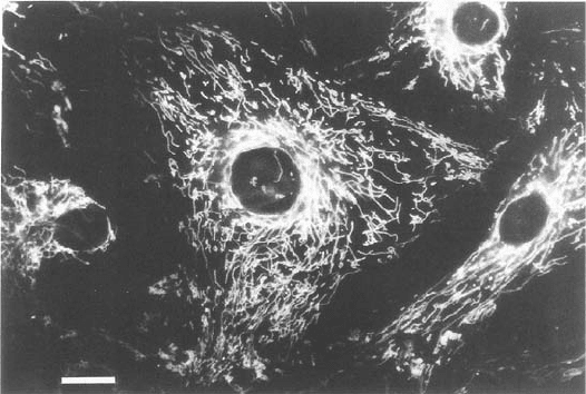
Experiments with Rh123 have shown that all mitochondria in a given cell
are homogeneous with respect to fl uorescence intensities — that is, have identi-
cal membrane potentials, regardless of shape, size, and intracellular location.
Figure 5.24 Mitochondria stained with rhodamine 123. (Photograph provided by
Dr. Lan Bo Chen.)
THE CHEMIOSMOTIC HYPOTHESIS 241

242 MITOCHONDRIAL ELECTRON TRANSFER
On the other hand, in a population of cells, signifi cant differences in Δ Ψ
between individual cells have been observed (251) (see below). Not surpris-
ingly, mitochondria in cardiac and skeletal muscle cells have the highest
membrane potentials, followed by smooth muscle, macrophages, hepatocytes,
fi broblasts, resting neuronal cells, glial cells, and keratinocytes. The lowest Δ Ψ s
were found in resting T and B lymphocytes. Differences have been observed
between resting cells and rapidly proliferating cells, and also in the course of
cell differentiation. The most dramatic change occurs when myoblasts differ-
entiate into myotubes, suggesting an activation of mitochondria which is still
under active investigation. Another signifi cant change in Rh123 fl uorescence
and hence Δ Ψ is observed at some stage in the progression of a cell toward
apoptosis (see Section 7.4 ), when the permeability transition creates large
pores for the dissipation of all ion gradients. A large number of tumor cells
have been investigated and compared with their normal counterparts (251) .
In general, carcinoma cells were found to have higher membrane potentials,
but the signifi cance is still not completely clear. It may be related to their
proliferative potential.
In recent years, several other dyes have found applications for greater sen-
sitivity and discriminations between mitochondria within a given cell and even
between regions of the inner membrane in a single mitochondrion. The dyes
can be divided into two classes: fast dyes and slow dyes. An example of the
fi rst is Rh123, a molecule that can partition into the lipid bilayer in the pres-
ence of an electric fi eld and respond almost instantly to a change in membrane
Figure 5.25 Structures of (A) rhodamine 123 and (B) the dye JC - 1.
Rhodamine
O
C
O
H
2
N
NH
2
OCH
3
5,5',6,6'-tetrachloro-1,1',3,3'-tetraethylbenzimidazolcarbocyanine iodide
JC-1
Cl
N
Cl
Cl
N
N
N
C
2
H
5
C
2
H
5
C
2
H
5
5
C
2
H
Cl
A
B
potential by a change in spectroscopic characteristics (quantum yield, absorp-
tion or emission spectrum). The slow dyes form aggregates in specifi c environ-
ments, again accompanied by dramatic changes in optical properties compared
to the monomer in solution. One of these is the cyanine dye 5,5 ′ ,6,6 ′ -
tetrachloro - 1,1 ′ ,3,3 ′ - tetraethylbenzimidazoleocarbocyanine iodide, mercifully
referred to as JC - 1 (254) (Figure 5.25B ). It was initially used as a sensitizer for
silver - halide - based photography. Theoretical treatments attempting to explain
the aggregation and spectral changes are available (see reference 255 for refer-
ences). Several aspects of the uptake and behavior of JC - 1 by isolated mito-
chondria and intact living cells were examined (254) . When coupled, energized
mitochondria were titrated with JC - 1, an absorption peak corresponding to
the monomer (527 nm — green) was observed fi rst, which was not affected by
manipulations of the membrane potential. It can be taken as a measure of
mitochondrial volume (255) . At higher concentrations, a second peak (590 nm —
red) appeared which corresponds to J - aggregate formation, and this J -
aggregate fl uorescence was proportional to the membrane potential over the
range of 30 – 180 mV. Less quantitative estimates with a fl uorescence micro-
scope are based on mitochondria appearing orange in the energized state and
green in the uncoupled state. Most surprisingly, within a certain range of
average ΔΨ , mitochondria were detected with apparently heterogeneous stain-
ing, indicative of local fl uctuations in ΔΨ within a single mitochondrion. Simi-
larly, J - aggregate formation and red fl uorescence in mitochondria of living
cells were sensitive to the electrochemical gradient; that is, the aggregates
failed to appear in the presence of either CCCP, dinitrophenol, or azide plus
oligomycin, and so on, or they could be dissociated when these uncouplers or
inhibitors were added later. Combinations of ionophores were used to show
that aggregate formation was sensitive to ΔΨ but not to Δ pH alone.
Signifi cantly, a number of different cell types examined exhibited both
intercellular and intracellular heterogeneity in staining with JC - 1, which had
not been detected previously with Rh123 or other cyanine dyes (e.g., reference
256 ). The explanation offered was that detection systems in the past (photo-
graphy, the human eye) may have been used outside of the linear response
range — that is, were saturated and hence incapable of discriminating local
differences. Therefore, even intracellular heterogeneity must now be taken
seriously, and an explanation must be sought in terms of different microenvi-
ronments for mitochondria within a cell, of the type already described by
sensitive determinations of Ca
2+
gradients. Dynamic, multichannel continuous
recordings may in the future be able to address these questions.
An even more challenging problem arises from the observation of low and
high membrane potentials (or at least an uneven distribution of J - aggregates)
within a single mitochondrion (254) . Proton diffusion is extremely rapid, and
localized proton circuits or “ respiration hot spots ” within a mitochondrion are
almost certainly transient phenomena. How can a slow dye respond to these,
and how are such states maintained at least during the interval required for
photography or other detection methods?
THE CHEMIOSMOTIC HYPOTHESIS 243
244 MITOCHONDRIAL ELECTRON TRANSFER
5.5 ATP SYNTHASE (F
1
F
0
- ATP ASE )
5.5.1 Introduction
Reference has been made repeatedly to a distinct complex (V) playing a
crucial role in oxidative phosphorylation. An enzyme had to be responsible
for the synthesis of ATP from ADP and inorganic phosphate. Somehow, this
endergonic reaction had to be coupled to redox reactions and electron trans-
port. The acceptance of Mitchell ’ s chemiosmotic hypothesis and the elucida-
tion of the biochemical and structural properties of this enzyme complex
evolved somewhat in parallel, and today we understand that a physically sepa-
rable complex can catalyze this reaction driven simply by a proton gradient
and membrane potential, which can be set up artifi cially across a vesicle mem-
brane by the choice of buffers or by the incorporation of a light - driven proton
pump from the purple membrane bacterium Halobacter halobium . ATP syn-
thase was fi rst “ seen ” by electron microscopists around 1962. Lollipop - like
structures were identifi ed by their spherical heads on the matrix side of the
inner membrane, and these were attached to the membrane by a slender stalk.
Pioneering work in Racker ’ s laboratory had succeeded in dissociating the
heads from the stalk. The heads were soluble in water, and they could be reas-
sociated with the membrane in the presence of an essential protein known as
the “ oligomycin sensitivity conferral protein ” (OSCP). OSCP had fi rst been
isolated and characterized in the laboratory of Tzagoloff (257 – 259) . Inhibition
of the ATP synthesis by oligomycin had become a diagnostic test for this
mitochondrial enzyme, and therefore the physiological signifi cance of the
required reassembly factor was apparent. Contrary to a long - held belief,
oligomycin binds to the c - ring and not to OSCP.
The complete complex was separated in the fractionation scheme devel-
oped by Hatefi (13, 14) , and the combined efforts of these early investigators
led to the now well - established separation of the complete complex V into a
soluble F
1
- ATPase, and an insoluble membrane complex referred to as F
0
. The
F
0
complex has the ability to translocate protons across the membrane from
their high potential on the outside; and when coupled to the F
1
subcomplex,
ATP synthesis can be achieved.
It was quickly apparent that this structure was signifi cantly more complex
than the fi rst ion pumps being characterized: (a) the Na
+
– K
+
pump in the
plasma membrane consisting of two subunits ( αβ ) and (b) the Ca
2+
pump in
the sarcoplasmic reticulum. The F
1
- ATPase has at least eight different peptides
(Figure 5.26 ). The relative positions of α , β , γ , δ , ε (present in the ratio 3 : 3 : 1 : 1 : 1)
were established earlier, while the positions of F6, d, and OSCP were deter-
mined only recently. The mitochondrial F
0
subcomplex (Figure 5.26 ) has a
variable number of different subunits depending on the species (11 in
mammals), of which three essential proteins (a, b, c) are homologous and
functionally related to the three proteins in the bacterial complex, and the
remainder are accessory proteins necessary for assembly, stabilization, and
control. The combination of biochemical analyses and the identifi cation of
relevant structural genes have contributed to the determination of the com-
position of the complex in various organisms (238) . All three core proteins in
F
0
are encoded by the mitochondrial genome in Saccharomyces cerevisiae and
plants, while one of these genes has been transferred to the nucleus in animals
and fungi. The establishment of the stoichiometric ratios for the F
0
subcomplex
peptides proved to be quite challenging (260) . Three distinct subunit types
(a, b, c) are present in the ratio ab
2
c
n
, where different values for n , ranging
from 10 to 14, have been reported for different organisms.
In the literature on this complex, and especially in reviews, reference is
frequently made to information from mammalian mitochondria, yeast mito-
chondria, chloroplasts, and the E. coli complex in the inner bacterial mem-
brane. The most recent and authoritative review on the composition, gene
expression and assembly with an emphasis on the yeast and E. coli systems is
by Ackerman and Tzagolof (238) . The nomenclature is not yet completely
uniform, and there is potential for some confusion. For example, the F
1
- ATPase
in all examples has fi ve subunits ( α β γ δ ε ), but the mitochondrial ε subunit has
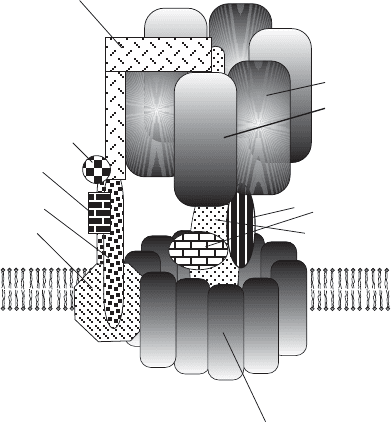
Figure 5.26 Schematic representation of complex V. The F
1
- ATPase extends into the
mitochondrial matrix and is connected to the F
0
complex in the inner membrane by a
stalk. The major peptides associated with each of the subcomplexes are discussed in
the text. The interaction of the OSCP subunit with the b
2
complex and the outside
domain of an α subunit is also emphasized.
α subunit
β subunit
ε, δ subunits
DCCD-binding
proteolipid
c-subunit
F ATPase
0
F ATPase
1
d subunit
2 b subunits
a subunit
outer stalk
inner stalk
γ subunit
OSCP
F
6
ATP SYNTHASE (F
1
F
0
-ATPase) 245

246 MITOCHONDRIAL ELECTRON TRANSFER
no counterpart (sequence, function) in either chloroplast or E. coli F
1
or F
0
,
and the δ subunits of the bacterial and chloroplast F
1
- ATPase are related to
the mitochondrial OSCP, which until recently was counted among the F
0
stalk
peptides. Thus, depending on which schematic structure is shown in various
texts, one can fi nd the OSCP subunit capping the mammalian complex, or the
δ subunit capping the bacterial complex (261) . At the same time, it is connected
to the highly elongated b subunits of the F
0
complex extending upward from
the base. The positioning of the ε subunit as part of the stalk was originally
supported by cross - linking studies. The mitochondrial δ and the bacterial ε
subunits are related. Table 5.2 is an attempt to make the cross - referencing
easier in the discussion to follow.
More than three decades after its original isolation by Racker ’ s group (see
reference 262 for an account of the early work) the studies on the bovine
F
1
- ATPase culminated in 1994 with the publication of the structure at 2.8 - Å
resolution by Walker ’ s group at Cambridge (263, 264) . This achievement was
recognized by a Nobel Prize in 1997. Over the years the structure evolved from
being the globular head of a lollipop roughly 100 Å in diameter, to an increas-
ingly refi ned description of a number of distinct features based on interpreta-
tion of data from enzyme kinetics, biochemistry, biophysics, immunochemistry,
cryo - electron microscopy, X - ray crystallography, and nanotechnology. For
TABLE 5.2 Subunit Composition of ATP Synthase
Comparison and Nomenclature of Complex V Subunits in Various Organisms
Bacteria
(E. coli )
Animal
Mitochondria
Yeast
Mitochondria
Plant
Chloroplasts
α (55.2)
a
α (55.1) α (55.3) α (56.8)
β (50.1) β (51.6) β (52.5) β (53.9)
γ (32.4) γ (30.2) γ (30.6) γ (38.1)
δ (19.3)
OSCP OSCP(20.9)
δ
ε (14.9) δ (15.1) δ (14.5) ε (14.9)
—
ε (5.7) ε (6.6)
—
— Inhibitor protein —
a a (ATPase - 6) Subunit 8 (5.87) a
b b Subunit 6 (27.9)
b, b ′
c c (ATPase - 9) Subunit 9 (7.79) c
— d d (19.66) —
— e —
— f b (P25) —
— g —
— A6L —
— F 6 —
a
The numbers in parentheses indicate the molecular mass of the peptides in kilodaltons.
example, the subunit stoichiometry and the determination of nucleotide
binding sites suggested the presence of a threefold axis of rotation, and it was
reasonable to make this axis parallel to the stalk. Now that an almost complete
crystal structure of this complex is available, the description could go into
considerable detail, but reference to schematic diagrams is still invaluable and
instructive.
5.5.2 X - Ray Structure
The single subunits γ and ε form a structure that interacts with components
of the F
0
complex in the membrane and with the α and β subunits of the F
1
on the matrix side, thus being part of the stalk seen by electronmicroscopy.
The N - and C - terminal segments of the γ subunit form extended α - helices
forming a left - handed, antiparallel, very loose coiled coil (with an extending
C - terminal), and this rod - shaped core has the α and β subunits arranged
alternately around it like the segments of an orange (Figure 5.27 ).
The δ and ε subunits appear not well - ordered and may be substoichiometric
in the original crystals (263) . In other words, the purifi cation of the F
1
- ATPase
requires a rupture of the stem structure, with the δ and ε subunits becoming
randomly distributed and disordered. Thus, their precise orientation and inter-
actions are still under investigation. It is signifi cant that the coiled - coil core of
γ with its extended C - termial helix is somewhat bent and by itself has no
threefold symmetry. Therefore the symmetrical α
3
β
3
hexamer is slipped onto
an asymmetrical camshaft, and when the camshaft rotates (see below), the
individual subunits do not make equivalent contacts at any given instant. Thus,
the nucleotide binding sites have different affi nities representing phases in the
turnover of the active site.
The α and β subunits are similarly folded: A six - stranded β barrel at the
N - terminal is connected through a nucleotide binding domain to a bundle of
7 ( α ) or 6 ( β ) helices at the C - terminal. The nucleotide binding domains are
made up of a nine - stranded β - sheet with nine associated α - helices, and they
are recognizable from similiarities with other known nucleotide binding pro-
teins such as RecA, as well as from the presence of typical loops and motifs
(263) . They are at the interfaces between the α and β subunits (i.e., between
the slices of the orange). There are three catalytic sites at which nucleotide
turnover occurs, as well as three ATP binding sites where nucleotide binding
appears to be required for the maintenance of symmetry and structure and to
prevent abortive complex formation. A proposed physiological regulatory role
for these noncatalytic sites has been questioned (265) . Some of the amino acid
side chains involved in nucleotide binding had been identifi ed from previous
studies such as affi nity labeling with 2 - azido - ATP, or site - directed mutagenesis
(see reference 266 for earlier references to the primary literature), and the
complete structure not only confi rmed previous deductions, but allowed a
detailed reconstruction of all the contacts between the phosphates, ribose, and
the adenine base.
ATP SYNTHASE (F
1
F
0
-ATPase) 247
248 MITOCHONDRIAL ELECTRON TRANSFER
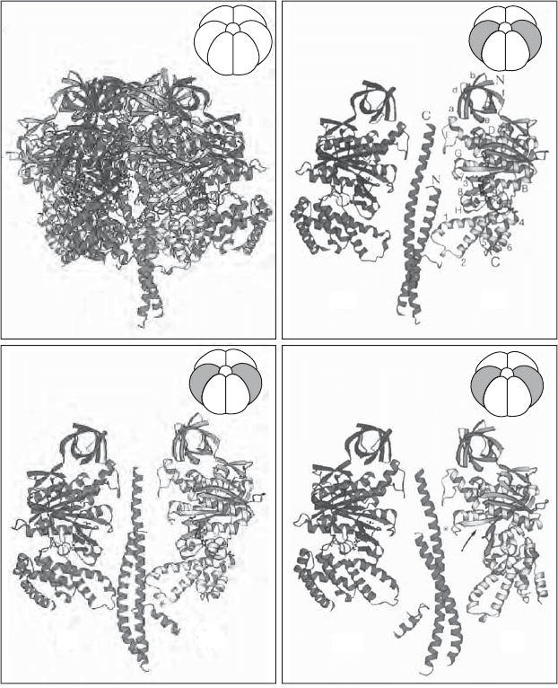
Figure 5.27 X - ray structure of the F
1
subcomplex of complex V. (A) Ribbon model
showing the α
3
β
3
hexamer and the γ subunit constituting the rotating shaft. (B – D)
Three different views of the shaft ( γ subunit) and side views of two opposite α and β
subunits. The image emphasizes the asymmetry and the different contacts made by each
of the six subunits with the central shaft. (From reference 263 with permission.) See
color plates.
AB
CD
α
DP
β
DP
β
TP
α
TP
α
E
β
E
γ
β
E
α
TP
α
TP
β
DP
α
E
β
TP
α
DP
β
E
α
E
β
DP
β
TP
α
DP
γ
β
DP
α
E
α
DP
β
TP
β
E
α
TP
γ
β
TP
α
DP
α
TP
β
E
β
DP
α
E
γ
Since the original structure of the F1 subcomplex was published, numerous
refi nements have been made. The characterization of structures binding various
inhibitors has allowed us to go from a single snapshot of the enzyme to a more
detailed view of the behavior of the enzyme during a catalytic cycle (267, 268) .
The structure of the specifi c inhibitory protein IF1 was fi rst solved as an iso-
lated protein (269) , and subsequently in a complex with the F1 - ATPase, where
it binds in the α – β interphase and also contacts the γ subunit (270) . On another
front, the structure of the peripheral stalk or stator has come into a much
better focus in recent years (271 – 273) . A partial structure at 2.8 - Å resolution
includes portions of the subunits b, d, and F6. New insights are derived from
these studies about the location of the oligomycin - sensitivity conferring protein
(OSCP). It is located near the top of the F
1
domain, interacting with the N -
terminal region of one α - chain and with the uppermost region of the b subunit
in the stator (see Figure 5.25 ).
The structure of the intact complex V has been refi ned fi rst by cryo - electron
microscopy (274) ; and as discussed below, much progress has been made
toward a high - resolution structure of the F
0
subcomplex including the c sub-
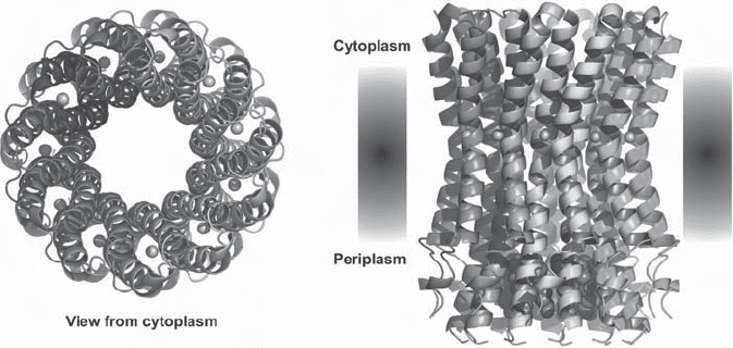
units. For practical/technical reasons the high - resolution structure of the c - ring
has been obtained so far for a related F - type Na
+
- ATPase of Ilyobacter tar-
taricus (275) (see Figure 5.28 ) and for the V - type Na
+
- ATPase of Enterococcus
hirae (276) . The structure of the c - monomer had been deduced for the E. coli
protein from MNR studies in chloroform:methanol:water (277) .
5.5.3 ATP Synthesis and Catalytic Mechanisms
Before discussing more detailed aspects of the structure, it is necessary to
become familiar with the current thinking about the mechanism by which the
ATP synthase carries out the simple reaction
ADP P ATP H O
i2
+⇒ +
Figure 5.28 Structure of the rotor ring of F - type Na
+
- ATPase from Ilyobacter tartari-
cus. (From reference 275 with permission.) (A) View perpendicular to the membrane
from the cytoplasmic side. Two of the 11 subunits are labeled. (B) Side view. Bound
Na
+
ions are shown as blue spheres, and the blue molecules inside the ring on the
periplasmic side represent detergent molecules.
AB
ATP SYNTHASE (F
1
F
0
-ATPase) 249
250 MITOCHONDRIAL ELECTRON TRANSFER
on the matrix side of the inner mitochondrial membrane. The problem is that
this reaction is endergonic and thus requires an input of energy. The chemios-
motic hypothesis identifi ed a source of this energy and introduced the general
notion that this enzyme was a proton pump running in reverse. When this
notion was verifi ed experimentally, the challenge became to understand pre-
cisely how the coupling of proton fl ow through the complex and ATP synthesis
were achieved. The fractionation of the F
1
ATPase subcomplex was a direct
indication that this complex contained the active site for ATP binding, and in
the absence of any other driving force it could catalyze the hydrolysis of ATP
(the reverse reaction). With the F
0
portion of complex V being an integral
membrane complex, it became the obvious choice for a pathway of protons
through the inner membrane. The connecting stalk/camshaft in turn must be
the structure that couples these two subcomplexes mechanically and confor-
mationally. The question is, How? The history can be found in several detailed
reviews (278, 279) that appeared prior to the publication of the crystal struc-
ture. With the information from the crystal structure the focus of the discus-
sions could obviously be sharpened considerably (263 – 265, 280 – 283) .
At this point the subunit stoichiometry of the F
1
complex and the threefold
symmetry of the arrangement of the α and β subunits come into consideration.
A “ rotating ” three - site model was fi rst proposed by Boyer in 1982 (284) , as an
extension of an “ alternating ” site model proposed earlier (285, 286) . Nucleo-
tide binding studies, chemical probes, and ligand/inhibitor binding studies had
suggested that the α and β subunits were conformationally asymmetric, and
this concept was fully substantiated by the confi rmation of the stoichiometry
α
3
β
3
γδε in the F
1
complex plus stalk. Ignoring the δ and ε peptides for the
moment, the coiled - coil of the ends of the γ peptide forms an asymmetrical
axis on which the αβ hexamer is impaled (see above); that is, each α and/or β
subunit is exposed to a different surface of this shaft. The original formulation
of the model speculation about rotations of the inner portion of the enzyme
relative to the outer portions had already been advanced (278) , and it has since
been spectacularly confi rmed that a “ central rotor of radius ∼ 1 nm, formed by
the γ subunit, turns in a stator barrel of radius ∼ 5 nm formed by the three α
and three β subunits. ” Rotary motion of the F1 - ATPase was observed directly
(Figure 5.29 ). In the presence of ATP a fl uorescent actin fi lament attached to
the γ subunit was shown to rotate for more than 100 revolutions in an anti-
clockwise direction when viewed from the membrane side (287) .
This elegant and defi nitive experiment was performed with an α
3
β
3
γ complex
from a thermophilic bacterium expressed in E. coli . The complex was attached
to a microscope slide like an upside - down mushroom, and the stalk ( γ subunit)
was coupled to fl uorescently labeled actin via a multivalent streptavidin mol-
ecule binding biotin on the actin and on the γ subunit. The model system has
demonstrated rotation of the γ subunit relative to the alternating α / β hexamer.
The system reconstituted the smallest molecular rotary motor described to
date. It can be noted here that there are now three types of rotary motors
found in nature. The fl agellar motor of bacteria is very different and will not