Townsend Courtney M.Jr., Evers B. Mark. Atlas of General Surgical Techniques: Expert Consult
Подождите немного. Документ загружается.

472 Section VI • Liver
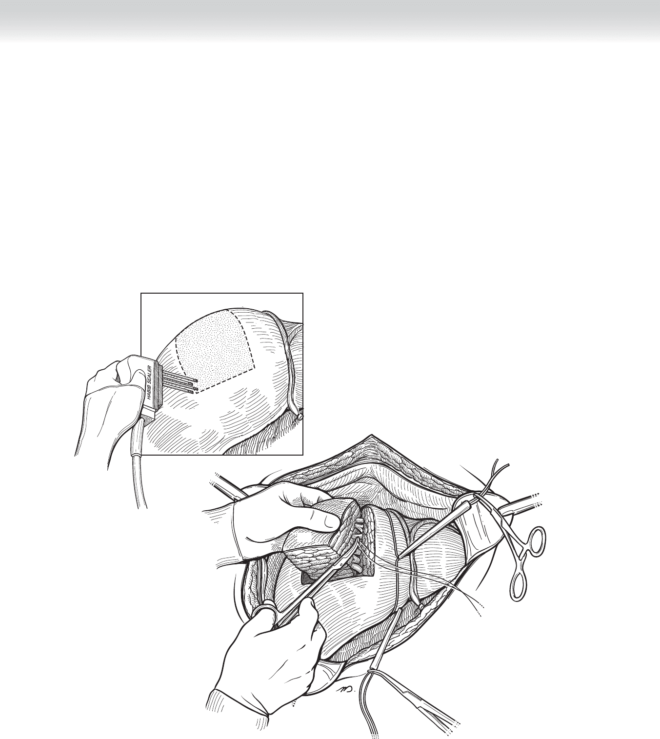
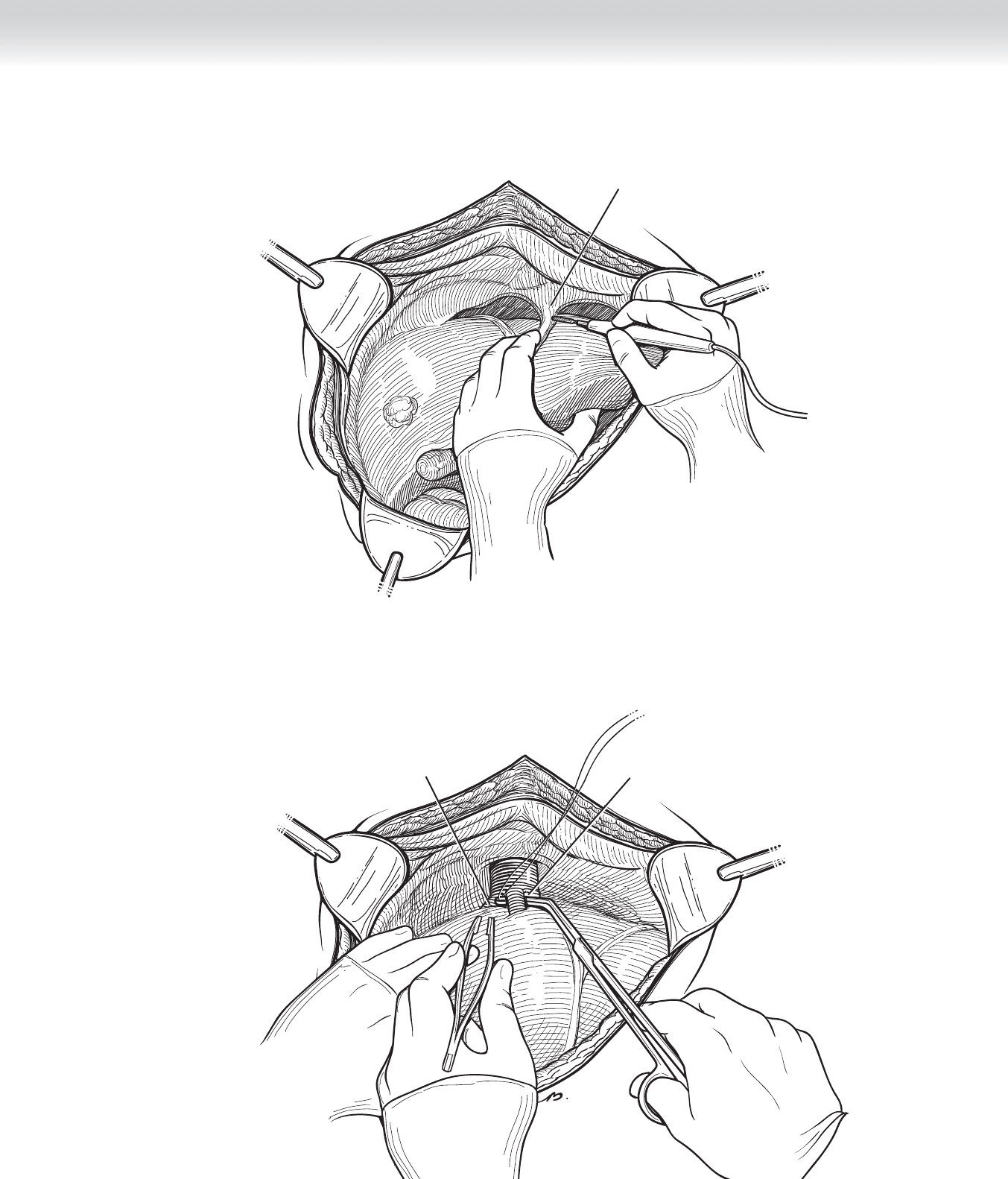
◆ Score the capsule of the liver along the line anatomically consistent with segment VIII.
Ultrasound guidance can be very helpful to defi ne anatomy and to target the lesion
(Figure 44-11).
◆ Perform dissection through the parenchyma of the liver carefully and progressively using
the handheld harmonic dissector. The recently available tissue-coagulating instrument,
based on radio-frequency energy (Habib device), may be substituted for division of
parenchyma but should not be used near major vessels. This instrument is particularly
useful in nonanatomic resections because of its effectiveness in controlling hemorrhage
during the parenchymal incision (see Figure 44-11).
V
IV
VIII
FIGURE 44 –11
CHAPTER 44 • Segmental Hepatic Resection 473
◆ Using visual inspection, and if necessary ultrasound guidance, identify major intraparenchymal
vascular tributaries and branches, and then clamp, divide, and ligate. Biliary radicals, which
must be ligated individually, are most diffi cult to identify at the time of dissection. Failure to
ligate these structures results in postoperative bile leaks (see Figure 44-11). The right and mid-
dle hepatic veins should be identifi ed and remain undisturbed during the dissection.
◆ Perform ultrasound examination intraoperatively to ensure adequate margin of resection in
the case of malignant tumor removal. A 1-cm margin is considered to be adequate, but in
major resections, margins are not typically an issue.
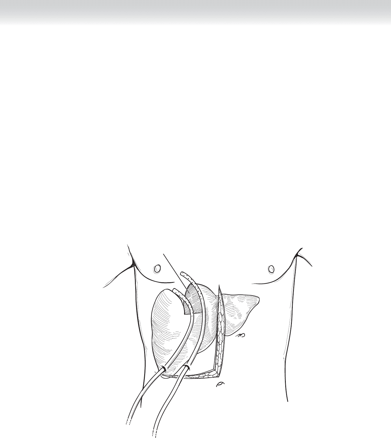
◆ Bring out two 10-mm Jackson-Pratt drains through separate stab wounds on the right side
of the abdomen and place along the divided edge of the liver (Figure 44-12). Some will
advocate taking omentum and placing it in the bed of the resected liver to act as a biologic
seal for the raw edge of the liver parenchyma.
Completed segment VIII resection
FIGURE 44 –12

474 Section VI • Liver
3. CLOSING
◆ Close the abdomen in a standard manner. We favor a horizontal mattress closure (Smead-
Jones) with heavy-gauge absorbable suture.
STEP 4: POSTOPERATIVE CARE
◆ In the fi rst 24 hours after surgery, the primary concern is hemorrhage and the related mea-
sure of coagulation status. These should be monitored by means of serial measurement of
hemoglobin and coagulation factors.
◆ In all major resections, particularly in patients with cirrhosis, one must be vigilant for any
signs of hepatic failure. A particularly ominous fi nding is the progressive rise in bilirubin
level with an enzyme pattern that supports neither obstruction nor parenchymal cell death,
such as transaminase elevations. The most ominous fi nding is a plummeting serum glucose
level, which refl ects the loss of glycogen stores in the liver and by inference the loss of via-
ble liver. Unfortunately, there is little one can do to reverse this pattern of failure. One pos-
sible cause is inadequate remaining liver after resection. This can resolve over time as the
liver regenerates, which it will do to some degree.
◆ One possibly remediable cause of this progressive demise is thrombus formation in the
portal vein. This would seem to be unlikely, because coagulation is typically inadequate in
these patients, but we have seen this phenomenon. It is possible that lysis of this clot may
restore vital fl ow.
◆ Sepsis is particularly metabolically taxing to the liver. In the compromised postoperative
liver, sepsis can be catastrophic. One should monitor and obtain cultures if necessary to
prevent infectious processes from progressing.
◆ Ascites may form, and one must be aware when this phenomenon has occurred and treat as
one would normally treat this entity with careful and judicious use of salt-containing intra-
venous fl uids and with diuresis.
◆ Remove drains if no bile is seen in the effl uent.

CHAPTER 44 • Segmental Hepatic Resection 475
STEP 5: PEARLS AND PITFALLS
◆ As with all such major operative procedures, one must exercise extreme care in patient
selection.
◆ If hemorrhage occurs at any time during the procedure, the liver can be compressed into
the spine or into the right fl ank to gain control, and another capable surgeon can be called
for assistance.
◆ Before dividing any of the major vascular structures, stop and reconfi rm that the proper
structure is being divided.
◆ If ascites forms and the drains are still in place, excessive electrolyte and fl uid loss can
occur through actively draining liters of fl uid per day. In this setting, the drains (assum-
ing they are not bile tinged) should be removed, and the skin overlying the drain tract
should be sutured.
SELECTED REFERENCES
1. Blumgart LH, Belghiti J: Liver resection for benign disease and for liver and biliary tumors. In Blumgart LH
(ed): Surgery of the Liver, Biliary Tract and Pancreas, 4th ed. Philadelphia, Saunders, 2007, pp 1341-1388.
2. Liu CL, Fan, ST, Cheung ST, et al: Anterior approach versus conventional approach right hepatic resection
for large hepatocellular carcinoma: A prospective randomized controlled study. Ann Surg 2006;244:
194-203.
3. Nanashima A, Sumida Y, Abo T, et al: Anatomic resection of segments 5, 6 and 7 of liver for hepatocellu-
lar carcinoma: Prior control of right paramedian Glisson. Hepatogastroenterology 2008;55:1077-1080.
4. Shirabe K, Shimada M, Gio T, et al: Postoperative liver failure after major hepatic resection for hepatocel-
lular carcinoma in the modern era with special reference to remnant liver volume. J Am Coll Surg
1999;188:304-309.

476
STEP 1: SURGICAL ANATOMY
◆ The liver is suspended in the right upper quadrant by avascular ligamentous attachments.
The falciform ligament is oriented vertically and suspends the liver from the anterior abdom-
inal wall at its inferior limit to the diaphragm, just anterior to the vena cava. The left and
right triangular ligaments extend in a transverse direction beginning on the lateral borders of
both the left and right liver, coursing along the diaphragm, and terminating at the vena cava,
where they join the superior extent of the falciform ligament. The triangular ligaments are
composed of both anterior and posterior leafl ets.
◆ The liver appears on gross examination to be composed of two discrete lobes. Thus there is a
traditional terminology in which the left and right lobes are defi ned by the falciform ligament.
Resection of one of these is termed a left or right lobectomy. This terminology has largely been
replaced by one based on the intraparenchymal vascular and biliary structures.
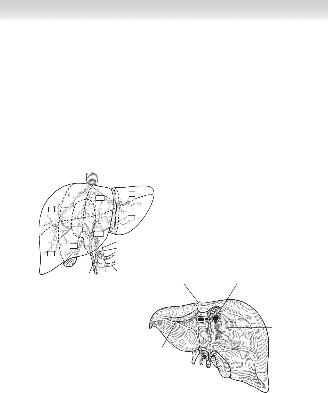
◆ Based on the intraparenchymal anatomy, the liver is divided into left and right livers, each
composed of four segments. The line of demarcation is located several centimeters to the
right of the falciform ligament and projects in a line, which transects the gallbladder bed
from anterior to posterior (Figure 45-1).
◆ Using the segmental anatomy, the liver is divided into left liver and right liver. The left liver is
served by the left portal vein, left hepatic artery, and left bile duct. It is composed of segments
I, II, III, and IV. Segments II and III represent the traditionally termed left lobe. Segment II is
attached to the left hemidiaphragm by the left triangular ligament, and segment III occupies
the inferior aspect of the left lobe. The boundary between the two extends horizontally,
approximately midway through the left lobe. Segment I, also called the caudate lobe, occupies
the posterior aspect of the liver in the midline. The segment wraps rather like a collar around
the vena cava on its left aspect. This segment is unique for its venous drainage, which is inde-
pendent of the left or middle hepatic veins and is composed of multiple tiny tributaries
between the vena cava and the segment. Segment IV, the quadrate lobe, occupies the area
between the falciform ligament medially and the gallbladder bed laterally (see Figure 45-1).
◆ The right liver is composed of segments V, VI, VII, and VIII. These four are oriented around
a horizontal line transecting the right liver at its mid-portion and similarly by a vertical line
that transects the right liver at its mid-portion. Beginning at the inferomedial segment V, the
segments follow a clockwise direction with VI inferolateral, VII superolateral, and VIII
superomedial (see Figure 45-1).
CHAPTER
45
Right Hepatectomy
William H. Nealon
CHAPTER 45 • Right Hepatectomy 477
◆ Lymphovascular and biliary structures enter the liver through the hepatoduodenal ligament
that courses between the duodenum into the base of segments IV and V, which is termed
the porta hepatis. The portal triad of microanatomy is matched by the gross anatomic ori-
entation in the hepatoduodenal ligament, composed of hepatic artery, portal vein, and bile
duct. Each structure divides into a left and right branch and then arborizes within the liver
in a pattern defi ned by the segments (see Figure 45-1).
◆ Venous drainage of the liver is primarily located at the superior aspect of the liver in short
structures between the vena cava and the liver. The left, middle, and right hepatic veins
each enter the vena cava within 2 to 4 cm of one another in a coronal orientation. One or
all of these venous elements may be intrahepatic or may have exceedingly short extrahe-
patic components. This anatomic feature raises considerably the risk of uncontrolled hem-
orrhage during dissection and resection (Figure 45-2). In addition to these three venous
structures, there are between 2 and 20 tiny tributaries between the posterior surface of the
liver and the contiguous vena cava. These must be divided to fully mobilize the right liver.
I
Common bile duct
Hepatic artery
Splenic vein
Portal vein
VII
VIII
V
VI
IV-B
IV-A
II
III
FIGURE 45 –1
Falciform ligament
Groove for vena cava
Triangular ligament
Bare area
FIGURE 45 –2

478 Section VI • Liver
STEP 2: PREOPERATIVE CONSIDERATIONS
◆ Due to the magnitude of hepatic surgery, one fi rst consideration is the medical status of the
patient and likely risk of surgery. Thus one must exclude signifi cant coronary, pulmonary,
or renal disease, or age and frailty. Of particular concern in relation to hepatic surgery is the
underlying hepatic function. Because hepatocellular carcinoma is associated with prior hep-
atitis and cirrhosis, one must determine fi rst whether cirrhosis exists and second what level
of function is apparent. Historically, this was measured by examining synthetic and excre-
tory functions and measures of portal hypertension (serum albumin level, coagulation pro-
fi le, serum bilirubin level, ascites, and mental status/serum ammonia). More recently, the
Model for End-Stage Liver Disease (MELD) score was developed as a means of segregating
candidates for liver transplant. This system incorporates prior variables, but has added and
places considerable signifi cance to renal function. Particularly when one anticipates a major
resection, one must establish that suffi cient liver will remain to support life. Unfortunately,
this estimate of “hepatic reserve” is even today an inexact science.
◆ Nutritional status, renal function, degree of ascites, and coagulation abnormalities are all
factors that may be improved by medical management before surgery. Unfortunately, we
have personal experience that such patients may thereby achieve an improved functional
grade but appear to carry a risk that exceeds the risk in patients who have had this impro-
ved functional status without a need for medical manipulation to achieve it.
STEP 3: OPERATIVE STEPS
1. INCISION
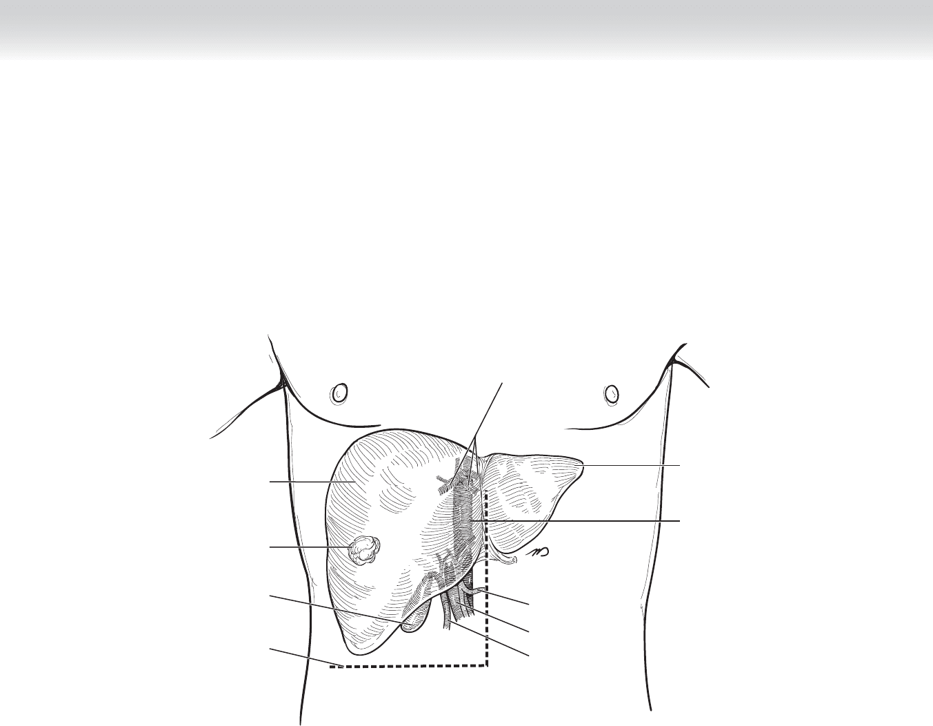
◆ Several incisions have been proposed. We favor the inverted L incision, with the option to
extend the incision vertically in the midline for added exposure, as well as extending farther
the horizontal component of the incision either laterally into the right fl ank or across the
midline (Figure 45-3).
◆ Placing self-retaining retractors maximizes the exposure of the right upper abdomen. The
incision should be extended if the operative view is inadequate.
CHAPTER 45 • Right Hepatectomy 479
Common
hepatic artery
Portal vein
Inferior vena cava
Hepatic veins
Gallbladder
Common bile duct
Incision
Lesion
Left lobe
Right lobe
FIGURE 45 –3

480 Section VI • Liver
2. DISSECTION
◆ Inferior to the liver edge, divide the falciform ligament between clamps and tie with heavy
silk suture. Cut the sutures at the caudal divided ligament. Place a hemostat on the uncut
cephalad end of the divided ligament for use in manipulating the liver during dissection.
You will discover the need for a constant balance between retracting the liver cephalad and
retracting caudad. Using this tether, you may restore some caudal exposure while the self-
retaining retractor suspends the liver toward the diaphragm.
◆ Using electrocautery, the fi lmy, avascular falciform ligament is incised beginning at its at-
tachment to the anterior abdominal wall and continuing in a cephalad and dorsal direction
until the point of convergence of this ligament meets the left and right triangular ligaments
(Figure 45-4). The hepatic veins are visualized at the superior extent of this dissection.
Carefully divide the peritoneal surface to clearly defi ne the right and middle hepatic veins.
We favor placing a vessel loop around the right and middle hepatic veins at this juncture
(Figure 45-5).
CHAPTER 45 • Right Hepatectomy 481
Diaphragm
FIGURE 45 –4
FIGURE 45 –5
Encircled middle
hepatic vein
Right hepatic vein