Westermeier R., Naven T., H?pker H.-R. Proteomics in Practice: A Guide to Successful Experimental Design
Подождите немного. Документ загружается.

1 Electrophoretic Techniques140
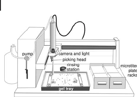
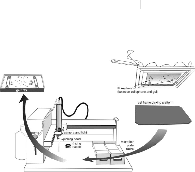
Fig. 1.53: Schematic drawing of an automatic spot picker, which
cuts the spots according to an imported picking list and transfers
the gel plugs to microtiter plates.
The picking head is coated with a hydrophobic layer to prevent gel
plugs sticking to it and protein cross-contamination. The gel plugs
are cut out by punching down to the backing plate or film, followed
by two side movements to shear the plug off the backing. Between
each spot removal the cutting head cleans itself in a rinsing station to
avoid cross contamination of proteins.
Such an instrument can also be operated in a semi-automated way
in order to cut out a few spots with eye control. In this case the cam-
era is also employed to find the spot to be removed from the gel.
1.7.1.1 Picking from Non-backed Gels
In practice it can happen that such an automated spot picker should
also be used for picking from non-backed gels. In order to prevent
shrinking or swelling of the gel it has to be stabilized during scan-
ning and spot picking. This can be done with a little accessory kit and
Cellophane
film following a procedure, which has been developed
by Burghardt Scheibe: A cellophane sheet is soaked in water and
clamped into a set of drying frames. The upper cellophane side is
dried with lint-free tissue paper and the reference markers are stuck
to the cellophane. The frame-cellophane assembly is turned upside
down to form a tray. The gel slab is soaked in water to swell as much
as possible and placed onto the cellophane. The gel is allowed to dry
on the cellophane for about 15 minutes. Then the gel is scanned, the
image is evaluated, and a picking list is created. When the gel–cello-
phane assembly is covered with a glass plate, the gel will not dry and
the assembly can be stored for several hours. A dark plastic plate is
placed into the spot picker instead of the tray as shown in Figure
1.54. The camera is recalibrated, because of the lower picking level.
GE Healthcare User Manual:
Ettan Spot Picker Nonbacked
Gel Kit. GE Healthcare Life
Sciences (2003) 11-0002-69.
1.7 Spot Handling 141
The frame with the gel is placed on the plastic plate and the spots are
picked like from a film- or glass-supported gel. In order to prevent
curling up of the gel edges, every half hour a few milliliters of water
need to be pipetted on the gel. A more detailed instruction can be
found in the GE Healthcare User Manual.
Fig. 1.54: Schematic drawing how to pick from unbacked gels
with an automatic spot picker.
1.7.2
Protein Cleavage
As the mass spectrometers become ever more sensitive, delivering
the sample of interest to the mass spectrometer in a manner which
takes advantage of the innate high sensitivity of the mass spectro-
meter will become increasingly crucial.
The most commonly used strategies for protein identification by
mass spectrometry require the protein of interest to be cleaved, pro-
teolytically or chemically, into its constituent peptides (assembly of
amino acids, see Table 1.7). In the pathway highlighted in this book
protein cleavage is performed by proteolysis after the protein has
been separated by two-dimensional gel electrophoresis. This has been
successfully applied to a wide range of proteome projects such as the
Langen H. Takcs B, Evers S,
Berndt P, Lahm HW, Wipf B,
Gray C, Fountoulakis M.
Electrophoresis 21 (2000)
411–429.
1 Electrophoretic Techniques142
analysis of the Haemophilus influenzae proteome (Langen et al. 2000).
Other methods have described the use of proteolysis without separa-
tion of the proteins by 2-D electrophoresis (see Section 2).
Tab. 1.7: 6 Twenty common amino acids detailing structure, molecular weights, symbols and
common modifications.
Residue
(3 & 1 letter symbols)
Residue structure
(R = side-chain)
Average
mass
Monoisotopic
mass
Common modifications
(and nominal molecular
weight additions)
Glycine
(Gly & G)
CH
OC NH
H
57.05 57.02146
Alanine
(Ala & A)
CH
OC
NH
CH
3
71.08 71.03711
Serine
(Ser & S)
CH
OC
NH
CH
2
OH
87.08 87.03203 Phosphorylation (80)
Glysosylation
a
Proline
(Pro & P)
CH
N
OC
97.12 97.05276
Valine
(Val & V)
CH
OC
NH
CH
2
CH
2
CH
3
99.13 99.06841
Threonine
(Thr & T)
CH
OC NH
CH(OH)CH
3
101.11 101.04768 Phosphorylation (80)
Glysosylation (O-linked
sugars)
Cysteine
(Cys & C)
CH
NH
CH
2
SH
OC
103.15 103.00919 Carbamidomethyl
b
(57)
S-propionamide
c
(71)
S-pyridylethyl
d
(105)
Leucine
(Leu & L)
CH
OC
NH
CH
2
CH(CH
3
)
2
113.16 113.08406
Isoleucine
(Ile & L)
CH
OC NH
CH(CH
3
)CH
2
CH
3
113.16 113.08406
1.7 Spot Handling 143
Residue
(3 & 1 letter symbols)
Residue structure
(R = side-chain)
Average
mass
Monoisotopic
mass
Common modifications
(and nominal molecular
weight additions)
Asparagine
(Asn & N)
CHOC NH
CH
2
C
O
NH
2
114.10 114.04293 N-linked glycosylation
e
(Sequon Asn-X-Ser/Thr)
Aspartic acid
(Asp & D)
CH
OC
NH
CH
2
C
O
OH
115.09 115.02694 Esterification, methyl
ester
f
(14)
Glutamine
(Gln & Q)
CH
OC
NH
CH
2
CH
2
C
O
NH
128.13 128.05858
Lysine
(Lys & K)
CH
OC
NH
(CH
2
)
4
NH
2
128.17 128.09496 Acetylation
g
(42)
Homo-arginine (42)
Glutamic acid
(Glu & E)
CH
OC
NH
CH
2
CH
2
C
O
OH
129.16 129.04259 Esterification, methyl
ester
f
(14)
Methionine
(Met & M)
CH
OC
NH
CH
2
CH
2
SCH
3
131.20 131.04049 Oxidation
h
(16)
Histidine
(His & H)
CH
OC
NH
CH
2
N
H
N
137.14 137.05891
Phenylalanine
(Phe & F)
CH
OC
NH
CH
2
147.18 147.06841
Arginine
(Arg & R)
CH
OC
NH
(CH
2
)
3
HN
C
NH
NH
2
156.19 156.10111
1 Electrophoretic Techniques144
Residue
(3 & 1 letter symbols)
Residue structure
(R = side-chain)
Average
mass
Monoisotopic
mass
Common modifications
(and nominal molecular
weight additions)
Tyrosine
(Tyr & Y)
CH
OC NH
O
H
CH
2
163.18 163.06333 Phosphorylation (80)
Tryptophan
(Trp & W)
CH
OC N
H
CH
2
N
H
186.21 186.07931 Oxidation (16)
Key:
a O-linked glycosylation – a range of sugars can link to Ser/Thr, including pentoses (C
5
H
10
O
5
),
hexoses (C
6
H
12
O
6
)andN-acetylhexosamines
b Modification of cysteine residue with iodacetamide
c Modification of cysteine residue with acrylamide monomer
d Modification of cysteine residue with 4-vinylpyridine
e N-linked glycosylation – a pentasaccharide core linked to the Asn residue, with a variety of
structures extending from this core
f Also esterification of the C-terminal carboxyl group of each peptide
g Also acetylat ion of the a -amino group at the n-terminus of each peptide
h Oxidation of methionine to the sulfoxide and the sulfone
1.7.2.1 Protein Cleavage – Proteolysis
Proteolysis has become the routine method of protein cleavage used
in proteomics with a range of enzymes available (see Table 1.8). Pro-
teolysis offers several practical advantages; including high specificity,
minimal side reactions and good cleavage efficiency. Important prac-
tical considerations for protein digestion include the digestion buffer
and its pH, the enzyme:substrate ratio, temperature and time.
Of all the enzymes available, the most commonly used enzyme is
trypsin. Trypsin has well defined specificity, yields tryptic peptides of
an appropriate size for efficient MS analysis and locates the basic resi-
dues at the terminus of the peptide. Though trypsin is commonly
used there may be occasions where digestion with an alternative
enzyme will be advantageous, specifically post-translational studies
such as phosphorylation analysis. The specific site of phosphorylation
may not reside on a tryptic fragment that is appropriate size for effi-
cient MS analysis.
Restricting the basic residues to
the c-terminus enables efficient
peptide fragmentation during
an MS/MS product ion experi-
ment, see Section 3.3.4.
Patterson S, Aebersold R. Elec-
trophoresis 16 (1995) 1791–
1814.

1.7 Spot Handling 145
Tab. 1.8: Enzymes commonly used in proteomics
Method of protein
cleavage
Site of cleavage Exception pH range
Trypsin C-terminus of R-X, K-X If X = P 7–9
Endoproteinase Glu-C
(V8-DE)
C-terminus of E-X, D-X If X = P 4–8
Chymotrypsin C-terminus of
F, Y, W, L, I, V, M
If X = P 7.5–8.5
Endoproteinase Lys-C C-terminus of lysine, K-X If X = P 8.5–8.8
Arg-C C-terminus of arginine, R-X If X = P 7.5–8.5
Elastase Not very specific. C-terminal
side of G, A, S, V, L, I
8–8.5
Pepsin C-terminus of F, L, E 2–4
Pronase Pronase is a mixture of endo-
and exo-proteinases. It cleaves
almost any peptide bond.
7–8,
dependent
on proteases
present
The simplest digestion would be an in-solution digestion. However,
optimal electroelution of a protein from the polyacrylamide gel
matrix into solution for digestion is difficult and highly variable from
protein to protein (Aebersold and Patterson 1995). The electrotransfer
(blotting) of the protein to a membrane (of which there were many
types) with subsequent digestion on the membrane surface (Aeber-
sold et al. 1987; Pappin et al. 1995).
A further development of the blotting technology was described by
Bienvenut et al. (1999). In this method the proteins are blotted
through a membrane of immobilised trypsin, onto a support mem-
brane where the constituent peptides are trapped and scanned
directly by MALDI peptide mass fingerprinting. This technology is
ultimately envisaged as the basis of a clinical scanner (Binz et al.
1999; Bienvenut et al. 1999; Schleuder et al. 1999).
Despite these methods the in-gel digestion methodology has
become routine for proteins separated by 2-D electrophoresis. Wilm
and co-workers (1996), and Shevchenko et al. (1996a) reported the in-
gel digestion of proteins from Coomassie Blue and silver stained gels
which was compatible with protein identification by mass spectrome-
try.
The basic theory is summarised in the following paragraphs, with
some novel developments reported. Following visualization of the
gel, the protein spots of interest can be excised from the gel manually
or automatically with commercially available gel spot pickers and be
subsequently destained. The destaining of the protein spot is particu-
Pappin DJC, Rahman D,
Hansen HF, Jeffery W, Sutton
CW. Methods in protein struc-
ture analysis (1995) 161–173.
Aebersold R, Leavitt J,
Saavedra RA, Hood LE, Kent
BS. Proc Natl Acad Sci USA 84
(1987) 6970–6974.
Bienvenut WV, Sanchez JC,
Karmime A, Rouge V, Rose K,
Binz PA, Hochstrasser DF.
Anal Chem 71(1999) 4800–
4807.
Wilm M, Shevchenko A,
Houthaeve T, Breit S, Schwei-
gerer L, Fotsis T, Mann M.
Nature 379 (1996) 466–469.
Shevchenko A, Wilm M, Vorm
O, Mann M. Anal Chem 68
(1996) 850–858.
75 mM ammonium bicarbo-
nate in 40% ethanol is an effec-
tive CBB destain.

1 Electrophoretic Techniques146
larly important. For instance, Coomassie brilliant blue binds to pro-
teins via ionic and hydrophobic interactions. The ionic interactions
between the sulfonic acid group of the CBB and the basic residues of
proteins (arginine and lysine residues) affect the trypsin digestion
efficiency, and the presence of CBB in the final sample can hamper
MS performance. This destaining step also allows the removal of
unwanted detergents such as SDS from the digestion procedure and
the MS analysis (in which detergents ionize very efficiently).
The presence of glutardialdehyde in many silver stain procedures
precluded the stained protein from subsequent analysis, as the glutar-
dialdehyde reacted with free amino groups in the protein. However,
Shevchenko et al. (1996) demonstrated that this component could be
omitted from the staining procedure and still realize high sensitive
visualization and successful MS analysis; Yan et al. (2000) also pub-
lished a silver stain method compatible with mass spectrometry.
The residual silver ions from the silver stain procedure can also be
removed using the method reported by Gharahdaghi et al. (1999).
At this stage many methods have reported the reduction and alkyla-
tion of cysteine residues contained within the protein embedded in
the gel piece (Rosenfield et al. 1992; Moritz et al. 1996; Shevchenko
et al. 1996b; Gevaert and Vandekerckhove 2000). The derivatization
comprises two steps: reduction of the disulfide bonds and alkylation
of the subsequent thiol side chain of the cysteine. This step is
included to improve the detection of cysteine containing peptides and
hence improve the potential protein coverage. However, the cysteine
thiol group can become modified as it passes through the PAGE gel
with free acrylamide monomer. Sechi and Chait (1998) noted that the
methods referred to above do not generally label the cysteine residues
post electrophoresis with the same mass addition that occurs within
the gel, hence potentially producing a heterogeneous derivatistion of
cysteine residues making protein identification complicated, as the
cysteine residues have effectively been labelled with different
reagents.
However, the 2D-electrophoresis method described in this book
includes a quantitative reduction and alkylation derivatization step of
the cysteine residues prior to the second dimension SDS-PAGE. If
the procedure is repeated post-electrophoresis and prior to digestion,
then the same reagent should be used.
Yan JX, Wait R, Berkelman T,
Harry RA, Westbrook JA,
Wheeler WH, Dunn MJ. Elec-
trophoresis 21 (2000) 3666–
3672.
Gharahdaghi F, Weinberg CR,
Meagher DA, Imai BS, Mische
SM. Electrophoresis 20 (1999)
601–605.
Additionally, the negative zinc/
imadazol stain is compatible
with mass spectrometric
analysis.
Rosenfield J, Capdevielle J,
Guillemot JC, Ferrara P. Anal
Biochem 203 (1992) 173–179.
Shevchenko A, Jensen ON,
Podtelejnikov A, Sagliocco F,
Wilm M, Vorm O, Mortensen
P, Shevchenko A, Boucherie H,
Mann M. Proc Natl Acad Sci
USA 93 (1996) 1440–1445.
Moritz RL, Eddes JS, Reid GE,
Simpson RJ. Electrophoresis 17
(1996) 907–917.
Gevaert K, Vandekerckhove J.
Electrophoresis 21 (2000)
1145–1154.
Sechi S, Chait BT. Anal Chem
70 (1998) 5150–5158.
The cysteine residues become
modified with acrylamide
monomer to form the b-propio-
namide derivative, a mass
difference of 71 Da. The modi-
fication of the cysteine with
iodoacetamide (a mass differ-
ence of 57 Da) and 4-vinylpyri-
dine, a mass difference of 105
is generally performed (see
Table 1.7).

1.7 Spot Handling 147
The in-gel digestion of the embedded protein can be performed
either manually or automatically. Staudenmann et al. (1998) reported
that the digestion of proteins above 10 pmol was routine, giving few
problems. However, once the concentration dropped below the 5
pmol level the number and yield of the peptides drops significantly,
suggesting sample loss onto vessel surfaces and digestion efficiency
as potential reasons for the disparity.
Some major considerations involved with in-gel digestion that ulti-
mately affect MS performance may include:
.
Digestion efficiency:
– Substrate-enzyme kinetics;
– Delivering the enyme to the protein whilst
minimizing autolysis.
.
Extraction efficiency of peptides from the polya-
crylamide gel matrix after digestion;
.
Sample handling;
.
Losses during the digestion procedure, for exam-
ple onto the walls of vessels;
.
Presence of detergents or staining agent;
.
Minimizing contamination with other proteins,
such as keratin.
Before addition of the enzyme to the gel plug, the gel plug (contain-
ing the embedded protein) is dehydrated using successive washes of
acetonitrile and subsequent dried under vacuum. The dehydration of
the gel plug to a completely dry dust-like material will enable efficient
uptake of the enzyme when the gel plug is rehydrated. A sufficient
volume of the enzyme in the digestion buffer (volatile buffer at the
optimum pH for enzyme activity) is applied to rehydrate the gel plug.
The enzyme has to passively diffuse through the gel matrix and digest
the protein before autolysis has a significant effect. The rehydration
step of the digestion procedure can be performed on ice (Shevchenko
et al. 1996) presumably to minimize autolysis of the trypsin whilst it
diffuses through the gel during the rehydration step. Additionally, the
modified form of the enzyme can be purchased to minimize autoly-
sis.
Upon completion of the digest reaction, between 3 and 24 hours,
the resultant digested peptides need to be extracted from the gel
matrix. To enhance peptide recovery, the inclusion of detergents such
as SDS, Tween 20, Triton X-100 or NP-40 in the extraction buffer has
been reported. However, the presence of detergent was detrimental to
MS analysis, both for matrix-assisted laser desorption ionization and
electrospray ionization (see Section 3.1). Instead, high peptide recov-
ery can be obtained from the gel matrix by passive diffusion using
Staudenmann W, Dainese-
Hatt P, Hoving S, Lehman A,
Kertesz M, James P. Electro-
phoresis 19 (1998) 901–908.
Shevchenko A, Jensen ON,
Podtelejnikov A, Sagliocco F,
Wilm M, Vorm O, Mortensen
P, Shevchenko A, Boucherie H,
Mann M. Proc Natl Acad Sci
USA 93 (1996) 1440–1445.

1 Electrophoretic Techniques148
acidic and organic solvents, namely solutions of trifluoroacetic acid
and acetonitrile.
Once extracted the peptides have to be delivered to the mass spec-
trometer in a form, which enables optimal MS performance. As
described in Section 3.1 though MALDI and ESI MS are highly
applicable to the analysis of biomolecules they are considerably differ-
ent techniques, requiring different sample preparation approaches.
MALDI involves the analysis of the sample from a remote, solid sur-
face after co-crystallization with the matrix, whilst ESI is a liquid inlet
system requiring the sample to be presented in the liquid state. Thus
ESI is effectively coupled with separation techniques such as capillary
electrophoresis and particularly high performance liquid chromato-
graphy (HPLC).
Unfortunately, the extracted peptides tend to be in a large volume
after extraction (>50 mL) and this is not ideal for MALDI or ESI analy-
sis, especially if the sample is low in abundance. Hence the sample
needs to be concentrated. However, extraction of the digested pep-
tides also extracts unwanted material from the gel which may hamper
MS ionization and reduce MS performance.. Simply concentrating
the gel extracts is insufficient, as both the contaminants and analyte
will be concentrated. Also, concentrating the sample by drying in
vacuum or by lyophilization can be problematic because of irrecover-
able sample loss on the walls of the digestion vessel; this is particu-
larly relevant for high sensitivity analysis.
An efficient approach to removing the salts, buffers and unwanted
contaminants from the digestion process is to use reversed phase
chromatography (RP-HPLC) off line prior to MALDI MS. In such a
method using a RP 300 mm 15 cm column, the peptides of interest
can generally be eluted into two or three fractions in a concentrated
volume (~4–5 mL) for subsequent MALDI MS analysis. In addition,
the eluate, if eluted with the correct solvent, is ideally suited for ESI
MS analysis.
A quicker solution for both MALDI and ESI analysis can be
achieved by binding the digested peptides to small amounts of
reversed phase resin packed into gel-loader pipette tips in a micro-
scale purification. This not only has the desired effect of desalting,
but also concentrates the sample in a suitable volume for the MALDI
analysis (Kussmann et al. 1997) Similarly, this approach can be
applied to on-target desalting, a method described by Gevaert et al.
(1998).
Stensballe and Jensen (2001) described a methodology where the
digestion and MALDI analysis was performed on the MALDI target,
minimizing sample manipulation and losses.
Alternatively a small aliquot (<0.5 mL) can be spotted directly from
the digestion mixture onto a pre-formed layer of matrix (a-cyano-4-
Kussmann M, Nordhoff E,
Rahbek-Nielsen H, Haebel S,
Rossel-Larsen M, Jakobsen L,
Gobom J, Mirgorodskaya E,
Kroll Kristensen A, Palm L,
Roepstorff P. J Mass Spectrom
32 (1997) 593–601.
Gevaert K, De Mol H,
Sklyarova T, Houthaeye T,
Vandekerckhove J. Electrophor-
esis 19 (1998) 909–917.
Stensballe A, Jensen ON.
Proteomics 1 (2001) 955–966.
Vorm O, Roepstorff P, Mann
M. Anal Chem 66 (1994)
3281–3287.

1.7 Spot Handling 149
hydroxy-cinnamic acid) and nitrocellulose. After the sample has
dried, the salts from the digestion reaction can be effectively removed
by washing with 0.1%TFA (Vorm et al. 1994; Jensen et al. 1996).
1.7.2.2 Protein Cleavage – Chemical Methods
Chemical methods of protein cleavage are complementary to proteo-
lysis, though not used as commonly. They are often used for specific
applications where no enzyme is available or proteolysis is not appro-
priate, such as cyanogen bromide cleavage of insoluble or membrane
proteins. In this instance, a chemical fragmentation in an organic sol-
vent capable of solubilizing the proteins is an attractive alternative
(Wasburn et al. 2001). Cyanogen bromide cleaves specifically at
methionine residues yielding relatively large peptides. Similarly large
peptides can be expected if cleavage at tryptophan residues is per-
formed with 2(2-nitrophenylsulfonyl-3-indolenine) (BNPS-skatole).
Acid hydrolysis of proteins with a mild formic acid solution has
been reported to be an effective method. The formic acid solution is a
good solvent for a whole range of proteins, and yields a specific clea-
vage at aspartyl residues.
See Table 1.9 for further examples of chemical methods of protein
cleavage
Tab. 1.9: Chemical reagents for protein cleavage
Chemical reagent Site of cleavage
Cyanogen bromide C-terminus of methionine
Acid hydrolysis Cleavage at aspartyl residues
Hydroxylamine Cleavage of asparagine–glycine bonds
BNPS-skatole Cleavage at tryptophan
Jensen ON, Podtelejnikov A,
Mann M. Rapid Commun
Mass Spectrom 10 (1996)
1371–1378.
Washburn MP, Wolters D,
Yates JR. Nat Biotechnol 19
(2001) 242–247.
Li A, Sowder RC, Henderson
LE, Moore SP, Garfinkel DJ
and Fisher RJ. Anal Chem 73
(2001) 5395–5402.