Westermeier R., Naven T., H?pker H.-R. Proteomics in Practice: A Guide to Successful Experimental Design
Подождите немного. Документ загружается.

3 Mass Spectrometry260
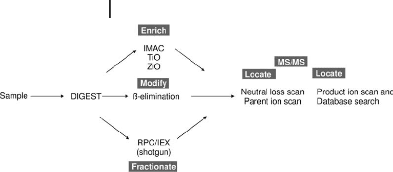
Fig. 3.29: Schematic of the methods used for phosphorylation
analysis. Unless the sample is very simple and abundant, then
enrichment of the phosphopeptide(s) is necessary prior to MS analysis.
The enrichment step is potentially very important, as unfractionated
phosphorylated peptides often suffer from signal suppression in posi-
tive ion ESI MS analysis (Mann et al. 2002).
3.4.2
Affinity Chromatography
Again there are many studies which highlight the use of affinity chro-
matography for phospopeptide isolation/enrichment, with varying
specificities. These techniques, of which common examples
described below, have been specifically applied to phosphopeptides.
3.4.2.1 Immobilized Metal Ion Affinity Chromatography
Immobilized metal ion affinity chromatography (IMAC) is a techni-
que that can be used to selectively enrich phosphopeptides in protein
digests, which resultantly increases the sensitivity of phosphopeptide
detection by MS. Traditional IMAC typically involves two compo-
nents: an immobilized chelating group and a metal species. The che-
lating group binds to and presents one face of the metal, allowing
interaction and separation of phosphorylated species. A variety of
metals have been immobolized, with Ga
3+
and Fe
3+
proving to be the
most effective. The technique has been reported on many occasions
for the enrichment of phosphorylated peptide (Nuwaysir et al. 1993;
Posewitz et al. 1999; Zhou et al. 2000; Stensballe et al. 2001).
In the technique, protein samples are reduced, alkylated and
digested, typically with trypsin. The peptide mixture is then applied
to an immobilized metal affinity chromatography column under
Mann M, Ong SE, Gronborg
M, Steen H, Jensen ON,
Pandey A.. Trends Biotechnol.
20 (2002) 261–268.
Nuwaysir LM, Stults JT. J Am
Soc Mass Spectrom 4 (1993)
662–669.
Posewitz MC, Tempst P. Anal
Chem 71 (1999) 2883–2892.
Zhou W, Merrick BA, Khaledi
MG, Tomer KB. J Am Soc Mass
Spectrom. 11 (2000) 273–282.
Stensballe A, Jensen ON.
Proteomics 1 (2001) 955–966.

3.4 Protein Characterization 261
acidic conditions (pH 2.5–3.5), unbound nonphosphopeptides are
removed from the column with an acidic wash solution and phospho-
peptides are eluted under alkaline conditions. Posewitz et al. evalu-
ated a variety of metals, including Fe
3+
,Ga
3+
,Al
3+
and Zr
3+
, for immo-
bilized metal affinity chromatography enrichment of phosphopep-
tides. Optimal performance, with respect to selectivity and recovery
was observed with iminodiacetate (IDA) resins complexed with Ga
3+
.
Selectively, enrichment and recovery of phosphopeptides using
IMAC depends heavily on the the metal ion and the chelating resin
and is biased towards multiply phosphorylated peptides (Heydon
et al. 2003). The technique is often affected by the non-selective
enrichment of acidic peptides, though this can be addressed with
methyl esterification of the peptide samples, improving the selectivity
of enrichment (Ficarro et al. 2002).
3.4.2.2 Titanium Dioxide
Several papers have been published demonstrating the ability of tita-
nium dioxide (TiO
2
) to selectively enrich for phosphopeptides from
complex peptide digests, via bidentate binding at the TiO
2
surface
(Pinkse et al. 2004; Larsen et al. 2005). When combined with multiple
protein digests and LC-MS/MS, titanium dioxide enrichment has suc-
cessfully been used for site identification at a low fmol scale.
3.4.2.3 Zirconium Dioxide
Porous zirconium dioxide (ZiO) provides high selectivity for phos-
phopeptides under an acidic binding condition (pH 2; Kweon et al.
2006).
3.4.3
Chemical Derivatization
Several methods have been reported for the enrichment of phopsho-
peptides. One common method exploits the characteristic that phos-
phoserine and phosphothreonine undergo undergo b-elimination,
losing the phosphate group which renders them susceptible to attack
by a range of nucleophilic reagents. These reagents are designed with
a tag to either enable enrichment prior to MS or to aid detection of
Increased binding of nonpho-
sphopeptides maybe observed
when loading is performed
outside the pH 2.0–3.5 range
or if overloading is allowed.
Heydon CE, Eyers PA, Lauren
D. Aveline-Wolf, Resing KA,
Maller JL and Ahn NG. Mol
Cell Proteomics 2 (2003) 1055.
Ficarro SB, McCleland ML,
Stukenberg PT, Burke DJ, Ross
MM, Shabanowitz J, Hunt DF,
White FM. Nat Biotechno. 20
(2002) 301–305.
Phosphopeptides with several
basic amino acids have been
shown to have a lower affinity
for titanium dioxide:
Pinkse MWH, Uitto PM,
Hilhorst MJ, Ooms B, Heck
AJR. Anal Chem 76 (2004)
3935–3943.
Larsen MR, Thingholm TE,
Jensen ON, Roepstorff P,
Jorgensen TJ. Mol Cell Proteo-
mics 4 (2005) 873–886.
Schlosser A, Vanselow JT,
Kramer A. Anal Chem 77
(2005) 5243–5250.
Kweon HK, Hakansson K.
Selective zirconium dioxide-
based enrichment of phos-
phorylated peptides for mass
spectrometric analysis. Anal
Chem 78 (2006) 1743–1749.
A similar b-elimination
approach could also be used to
isolate O-glycoylated Serine
and Threonine, though it
become difficult to distinguish
between the two PTMs

3 Mass Spectrometry262
the phosphopeptide in the mass spectrometer (Oda et al. 2001; Knight
et al. 2003 and Goshe MB et al,. 2001). The advantage of using a che-
mical derivatization approach is that stable isotopes can be incorpo-
rated into the mass tag, enabling relative quantification. However,
several steps are typically involved in these derivatization strategies.
Another approach, reported by Tao and colleagues, enables the
selection of methylated phosphotyrosine peptides as well as methy-
lated phosphoserine and phosphothreonine residues using an
amended, solid phase derivatization approach which reduces the
number of steps involved. With this method, the phosphate group
remains on the peptide, which supports identifying the point of mod-
ification within the peptide. Methy esterification is a key step in the
method as it protects carboxyl groups from becoming modified dur-
ing the reaction and enables stable isotopes to be incorporated for
relative quantification studies. As a result, non-phosphorylated resi-
dues are not retained on the support and selectivity for phosporylated
species is high.
Following any of these enrichment steps, the phosphopeptide sam-
ple(s) are typically analyzed by LC-MS/MS. It is advantageous if the
particular system can perform the full range of tandem mass spectro-
metry techniques: product ion scan for peptide identification and
PTM site location and neutral loss and precursor ion scanes to detect
the peptides containing the modification. As explained previously a
mass spectrometer can be configured to detect certain diagnostic
fragment ions during the fragmentation or CID process.
3.4.3.1 Precursor Ion Scan
With respect to phosphorylation analysis, a precursor ion scan can be
performed on the fragment ion 79. In this experiment, effectively the
whole sample is allowed to pass into the collsion cell to undergo frag-
mentation. Only peptides which fragment to generate a fragment ion
of 79 Da will be detected at the detector. Such a fragment is indicative
of a phosphate group and as such is diagnostic of a phosphorylated
peptide. This is a sensitive method for identifying phosphorylated
peptides and its application has been reported on a number of occa-
sions (Neubauer and Mann, 1999).
3.4.3.2 Neutral Loss Scan
With respect to phosphorylation, a neutral loss scan can be performed
to detect the loss of phosphoric acid (H
3
PO
4
) during the fragmenta-
tion process. The mass spectrometer is configured to detect this,
using knowledge of the probable charge states of the peptide (i.e. for
doubly charged phosphorylated peptides the figure should be set to
Oda Y, Nagasu T, Chait BT.
Nat Biotechnol 19 (2001)
379–382.
Knight ZA, Schilling B, Row
RH, Kenski DM, Gibson BW,
Shokat KM. Nat Biotechnol 21
(2003) 1047–1054.
Goshe MB, Conrads TP,
Panisko EA, Angell NH, Veen-
stra TD, Smith RD. Anal Chem
73 (2001) 2578–2586.
Zhou H, Watts JD, Aebersold
R. Nat Biotechnol. 19 (2001)
375–37.
No detectable interference
from other modifications such
as glycosylation was reported.
Tao WA, Wollscheid B, O'Brien
R, Eng JK, Xiao-jun L, Boden-
miller B, Watts JD, Hood L,
Aebersold R. Nat Methods 2
(2005) 591–598.
Neubauer G, Mann M. Anal
Chem 71 (1999) 235–242.

3.4 Protein Characterization 263
49; 98/2). This same fragmentation process can be utilized in a
multi-stage, MS
3
, tandem mass spectrometry experiment (Beausoleil
et al. 2004, Chang et al. 2004).
A recent publication comprehensively compared several affinity
methods for phosphoproteome wide analysis (Bodemiller et al. 2007).
The paper described the use of IMAC, titanium oxide, zirconium dox-
ide, and a solid phase chemical derivatization method for the analysis
of a cytosolic fraction of Drosophila melanogaster Kc167 cells.
They discovered that each of the methods enriched for phospho-
peptides with an acceptable level of reproducibility. However, each
method essentially enriched for different phosphopeptides, though
there was some partial overlap, which were not detectable without the
enrichment step. Using all the methods, a total of 887 unique phos-
phorylation sites were identified; ~62% of these were identified in the
IMAC samples, ~60% in the chemical derivatisated samples, ~41% in
the pTiO
2
samples and ~17% in dhbTiO
2
samples. Roughly 1/3
rd
of
the identified phosphorylation sites were identical between chemi-
cally derivatized and IMAC, 1/3
rd
beween chemically derivatized and
pTiO
2
and 1/3
rd
between IMAC and pTiO
2
. They concluded that none
one method could exhaustively represent or analyze a whole phos-
phoproteome.
3.4.4
Glycosylation
Glycosylation is an important PTM, with the composition of the gly-
cans crucial for the function of many proteins in cell signalling and
host–pathogen interactions. A number of factors make it a particu-
larly challenging PTM to analyze, including glycoprotein enrichment,
sensitivity of MS analysis, heterogeneity, occupancy, isobaric masses
for many of the sugar residues and, arguably limited chemical deriva-
tization potential (compared to phosphorylation). Glycosylation is
either N-linked or O-linked. N-linked glycosylation occurs at a very
specific sequon: Asn-Xxx-Ser/Thr, where Xxx is any residue except a
proline. In addition, N-linked glycans all have a common basic struc-
ture. On the other hand, O-linked glycosylation does not have a speci-
fic sequon, otherthan it occurs at serine or threonine residues.
Once more, identifying glycoproteins in a complex mixture can be
challenging. Glycoproteins have long been enriched using lectin affi-
nity chromatography (Gabius et al. 2002). Alternatively, digestion of
the sample in question, followed by precursor ion scanning, as
described above, can be used to locate glycosylated peptides using the
diagnostic fragment ions of hexoses and hexosamines, 163 and 204
respectively (Huddleston et al. 1993).
Beausoleil SA, Jedrychowski M,
Schwartz D, Elias JE, Villn J,
Li J, Cohn MA, Cantley LC,
Gygi SP. Proc Natl Acad Sci
USA 101 (2004) 12130–
12135.
Chang EJ, Archambault V,
McLachlin D., Krutchinsky AN,
Chait, BT. Anal Chem 76
(2004) 4472–4483.
Bodenmiller B, Mueller LN,
Mueller M, Domon B, Aeber-
sold R. Nature Methods 4
(2007) 231–237.
Gabius HJ, Andre S, Kaltner H,
Siebert HC. Biochim Biophys
Acta 1572 (2002) 165–177.
Huddleston MJ, Bean MF, Carr
SA. Anal. Chem. 65 (1993)
877–884.

3 Mass Spectrometry264
Methods combining glycoprotein enrichment with glycoprotein
digestion and tandem mass spectroimetry analysis have successfully
identified sites of glycosylation. Kaji et al. (2003) utilized lectin affi-
nity chromatography and glycoprotein digestion in
18
O-labeled water
for site-specific analysis; whilst Hagglund et al. (2004) employed lec-
tin affinity for glycoprotein enrichment and hydrophilic interaction
chromatography (HILAC) for glycopeptide enrichment prior to diges-
tion and MS/MS analysis.
The use of the enzyme PNGase F can be used to pinpoint the site
of N-linked glycosylation. This enzyme will cleave off the N-linked
glycans from every site in the protein and in the process convert the
aspartamine residue to aspartic acid, a 1 Da mass difference. A sim-
ple peptide map will then identify the peptide(s) that contained the
site of N-linked glycoyslation. Additionally, the cleaved N-linked gly-
cans can then be analyzed by MALDI-TOF to highlight the heteroge-
neity of N-linked glycosylation for that specific protein.
Methods for investigating O-linked glycosylation include b-elimina-
tion as described for phosphorylation. Wells et al. (2002) described
O-linked analysis using affinity enrichment and tandem mass spec-
trometry.
3.5
Protein Quantification Using Mass Spectrometry
Protein quantification by mass spectrometry is an exciting applica-
tion. To qualify, this is using mass spectrometry to detect the differ-
ence in protein expression between two samples. This can be per-
formed using stable isotope labeling or by a non-labeling, software
approach.
3.5.1
Stable Isotope Labeling Approaches
Stable isotope labeling is a technique that has long been used in
mass spectrometry. Mass spectrometry exploits the characteristic that
chemically identical analytes with a different stable isotope composi-
tion can be separated on a mass spectrometer, due to the mass differ-
ence associated with the different isotopes. The ratio of the different
analytes signal intensity is indicative of their relative abundance.
Many methods have been reported for just such an analysis of protein
expression. Stable isotopes have been incorporated into the protein/
peptide analyte using digestion in
18
O labeled water (Yao et al. 2001);
via metabolic labeling using heavy amino acid acids (Conrads et al.
2002); using isotope coded affinity tags (ICAT) (Gygi et al. 1999)
Kaji H, Saito H, Yamauchi Y,
Shinkawa T, Taoka M, Hira-
bayashi J, Kasai K, Takahashi
N, Isobe T. Nat Biotechnol 21
(2003) 667–672.
Hgglund P, Bunkenborg J,
Elortza F, Jensen ON, Roep-
storff P. J Prot Research 3
(2004) 556–566.
Unfortunately, if there is nore
than one N-linked site, the
specific heterogeneity at each
site cannot be assigned.
Wells L, Vosseller K, Cole RN,
Cronshaw JM, Matunis MJ,
Hart GW. Mol Cell Proteomics
1 (2002) 791–804.
Yao X, Freas A, Ramirez J,
Demirev PA & Fenselau C. Anal
Chem 73 (2001) 2836–2842.
Conrads TP, Issaq HJ & Veen-
stra TD. Biochem Biophys Res
Commun 290 (2002) 885–890
Gygi SP, Rist B, Gerber SA,
Turecek F, Gelb MH, Aerber-
sold R. Nature Biotechnol 17
(1999) 994–999.

3.5 Protein Quantification Using Mass Spectrometry 265
and more recently with isobaric mass tags (iTRAQ) which enables
both relative and absolute quantification (Ross et al. 2004). The tag-
ging of proteins as described in the latter examples have proven to be
the most common methods used, though SILAC, stable isotope label-
ing with amin acids in cell culture is a prominant method (Ong et al.
2002). Two further methods, AQUA according to Stemman et al.
(2001) and Gerber et al. (2003), and QconCAT according to Pratt et al.
(2006) have been reported for absolute quantification.
& Importantly, all the methods except iTRAQ
measure the relative abundance in MS mode,
whereas iTRAQ measures the relative abundance
in MS/MS product ion mode.
3.5.2
Isotope-coded Affinity Tags
This technique was reported by Gygi et al. (1999) describing the site-
specific labeling of cysteine residues with isotope-coded affinity tags
(ICAT; Figure 3.30). The reagent has three constituent groups; first
biotin which provides affinity for binding with an avidin column, sec-
ond a linker which incorporates stable isotopes, and third a reactive
group, iodoacetamide, which labels cysteine residues.
N
H
O
O
O N
H
NHNH
S
O
O
X
X
X
X X
X
I
O
X
X
X is either hydrogen (light chain) or deuterium (heavy chain)
Biotin
Reactive group
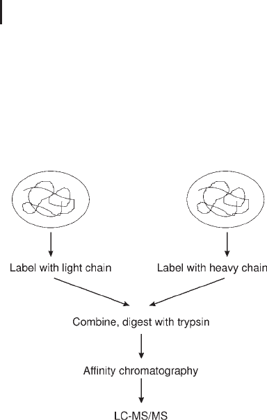
Two tags exist, one with no deuteriums in the linker (light) and one
with eight deuteriums in the linker (heavy). The strategy requires
that a two protein mixture, control and test are labeled with the light
Ross PL, Huang YLN,
Marchese JN, Williamson B,
Parker K, Hattan S, Khainovski
N, Pillai S, Dey S, Daniels S,
Purkayastha S, Juhasz P,
Martin S, Bartlet-Jones M, He
F, Jacobson A, Pappin DJ. Mol
Cell Proteomics 3 (2004)
1154–1169.
Ong S-E, Blagoev B, Kratch-
marova I, Kristensen DB, Steen
H, Pandey A. Mann M. Mol
Cell Proteomics 1 (2002) 376–
386.
Pratt JM, Simpson DM,
Doherty MK, Rivers J, Gaskell
SJ, Beynon RJ. Nat Protocols 1
(2006) 1029–1043.
Stemmann O, Zou H, Gerber
SA, Gygi SP, Kirschner MW.
Cell 107 (2001) 715–726
Gerber SA, Rush J, Stemman
O, Kirschner MW, Gygi SP.
Proc Natl Acad Sci USA 100
(2003) 6940–6945.
Gygi SP, Rist B, Gerber SA,
Turecek F, Gelb MH, Aerber-
sold R. Nat Biotechnol 17
(1999) 994–999.
3 Mass Spectrometry266
and heavy tag respectively. The two derivatized fractions are then
combined and digested before affinity isolated using an avidin col-
umn. The cysteine labeled peptides are separated by RP-HLPLC and
eluted into the mass spectrometer. The two forms of the same peptide
will differ in mass by 8 Da, and the ratio between the two determines
the relative quantification. Using data dependent analysis the product
ion MS/MS spectrum is acquired to identify the protein from the
derivatized peptide.
Fig. 3.30: ICAT reagent and workflow.
3.5.3
Stable Isotope Labeling with Amino Acids in Cell Culture
Stable isotope labeling with amino acids in cell culture (SILAC) is a
simple approach for in vivo incorporation of a label into proteins for
MS-based quantitative proteomics. This technique reported by Ong
et al. describes the metabolic labeling of two cell states, with the incor-
poration of amino acids with substituted stable isotopic nuclei (e.g.
deuterium,
13
C,
15
N); for instance normal and deuterated amino acid
leucine.
When this labeled analog is provided to cells in culture, it is incor-
porated into all newly synthesized proteins. After a number of cell
divisions, each instance of this particular amino acid will be replaced
by its isotope labeled analog. As there is negligable chemical differ-
ence between the labeled amino acid and the natural amino acid iso-
topes, cells behave normally.
In the experimental design, the samples are mixed, digested and
analyzed by LC-MS/MS. Relative abundance is derived from leucine-
Ong S-E, Blagoev B, Kratch-
marova I, Kristensen DB, Steen
H, Pandey A. Mann M. Mol
Cell Proteomics 1 (2002) 376–
386.

3.5 Protein Quantification Using Mass Spectrometry 267
containing peptides, which will appear as heavy and light peaks,
representing those leucine-containing peptides that contain deuiter-
ium and those containing hydrogen. A key limitation is that it can
only be used on cultured cells.
3.5.4
AQUA
In contrast to the strategies described above, AQUA provides abso-
lute quantification by employing synthetic peptides containing stable
isotopes. The authors describe the steps: selection of the internal pep-
tide standard selection. The peptide is selected on its amino acid
sequence and then synthesized with one of the residues being
replaced with the “heavy” version which contains the stable isotopes.
Resultantly, this peptide is chemically identical to its counterpart in
question, but distinguishable in the MS.
The AQUA peptide is analyzed by LC-MS/MS to study its fragmen-
tation pattern, which is identical to its counterpart peptide. Subse-
quent LC–MS/MS analysis using an SRM experiment produces
highly accurate measurement of both the internal standard and ana-
lyte.
3.5.5
iTRAQ
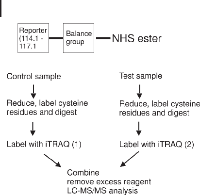
The iTRAQ reagents are designed to allow labeling of up to four sam-
ples with four reagents of the same mass (isobaric).
The reagent consists of three components: an N-hydroxysuccini-
mide (NHS) ester group which reacts with primary amines at the
amino-termini and lysine side-chains (alpha and epsilon amino
groups):
a balance group;
a reporter group.
Fragmentation of these four tags in product ion tandem mass
spectrometry, produces four reporter ions (m/z 114.1, 115.1, 116.1,
117.1 Da) which are used for quantification of the four samples. As a
result, up to four different biological samples may be labeled simulta-
neously. iTRAQ reagents label the alpha amino group of digested
peptides and epsilon amino group of the lysine side-chain. Resul-
tantly, a greater coverage of peptides is observed (also non-cysteine
containing proteins can be detected). The design of the tag and work-
flow are shown in Figure 3.31.
Stemmann O, Zou H, Gerber
SA, Gygi SP, Kirschner MW.
Cell 107 (2001) 715–726.
Gerber SA, Rush J, Stemman
O, Kirschner MW, Gygi SP.
Proc Natl Acad Sci USA 100
(2003) 6940–6945
Ross PL, Huang YLN,
Marchese JN, Williamson B,
Parker K, Hattan S, Khainovski
N, Pillai S, Dey S, Daniels S,
Purkayastha S, Juhasz P,
Martin S, Bartlet-Jones M, He
F, Jacobson A, Pappin DJ. Mol
Cell Proteomics 3 (2004)
1154–1169.
3 Mass Spectrometry268
Fig. 3.31: iTRAQ workflow for 2 samples.
3.5.6
Non-labeling Software Approaches
All of the above methods require a degree of sample manipulation to
deliver relative and absolute quantification. It can be argued that such
sample preparation can introduce error and as a result the methods
could be measuring experimental variation rather than real biological
variation. A number of software approaches have been developed
which attempt to perform relative quantification without any labeling
by comparing consecutive LC-MS profiles: Non-commercial (like
MSight) and commercial. Such a software package, DeCyder MS,
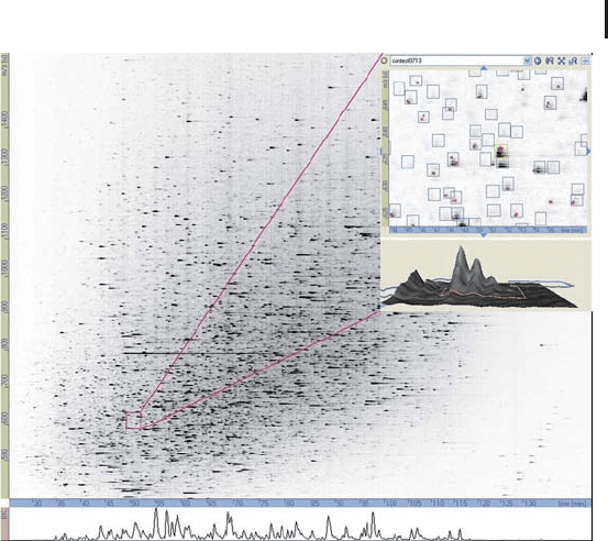
is provided by GE Healthcare. This novel software integrates visuali-
zation, detection, comparison, and statistical tools. It simplifies the
evaluation of large LC-MS and LC-MS/MS data sets for the relative
quantification of peptides, based on profile MS spectra. DeCyde MS
converts a conventional LC-MS chromatogram into a two- and three-
dimensional intensity map, as displayed in Figures 3.32 and 3.34
respectively. Identification results from the MS/MS spectra can be
imported into the software and used for simple sorting of peptides
belonging to the same protein.
This software package is based
on similar algorithms like the
Decyder 2D software for the
evaluation of DIGE gels, which
are described in Section
1.6.2.2.
3.5 Protein Quantification Using Mass Spectrometry 269
Fig. 3.32: Two-dimensional intensity map of an LC-MS run generated
by DeCyder MS. The inset illustrates a zoomed section where the
charge states of detected peptides can be seen as well as the MS/MS
events (red stars). Courtesy of A. Parbel GE Healthcare.
DeCyder MS performs two main analysis procedures:
.
Peptide detection. The PepDetect module pro-
vides accurate m/z signal detection, background
subtraction, isotope and charge-state deconvolu-
tion, and reporting of the eluted peptides from
the column. Relative quantification is obtained
by summing up the relevant peak volumes for
each peptide in all charge states.
.
Run-to-run matching. The PepMatch module
aligns the individual charge states from the
same peptide in different runs. According to the
experimental setup ratio values and statistical
significance for each of the peptides is calcu-
lated. Various normalization and filtering techni-
ques can be applied to report easily the requested
results.
A workflow is highlighted in Figure 3.33, and an example of differ-
ence in protein expression is shown in Figure 3.34. This intensity
map approach also acts as a valuable visualization tool enabling quali-
tative analysis of an LC-MS run as shown in Figure 3.35.