Yao N. Focused Ion Beam Systems: Basics and Applications
Подождите немного. Документ загружается.

12
Ion beam implantation of surface layers
daniel recht and nan yao
Princeton University
12.1 Introduction
Ion implantation is a method for the direct, controlled introduction of
impurities into solids. In ion implantation, a beam of dopant ions is aimed at
a target material (the substrate) so that the ions are incident with sufficient
energy to become permanently embedded. Because ion implantation is an
essentially nonequilibrium process, it allows for the creation of concentration
profiles that would be impossible to achieve using equilibrium techniques
such as diffusion. The advent of focused ion beam (FIB) systems spawned a
host of new applications for ion implantation. The ability to create high-
resolution (feature sizes of order 10 nm [1,2]) doping configurations without
the use of a mask allows not only for rapid prototyping, but also for unique
devices whose fabrication would not otherwise be feasible. FIB implantation
has been used to make a wide variety of experimental devices including low-
dimensional transistors, single photon detectors, subwavelength optics, and
quantum computers.
This chapter covers the basics of ion implantation in general and of FIB
implantation in particular. Next it considers the challenge of measuring
the ion dose introduced into the substrate. It then discusses the major
parameters relevant to the FIB implantation process and presents a sample of
their complex interrelationships. Finally, it describes the aforementioned
applications of FIB implantation to the fabrication of novel devices.
12.2 Basics
In the years since its development in the 1950s [3], ion implantation has
become the preferred method for the introduction of impurities into solid
Focused Ion Beam Systems: Basics and Applications, ed. N. Yao.
Published by Cambridge University Press. ª Cambridge University Press 2007.
318
substrates. Because it introduces ions mechanically instead of by thermally
activated transport processes, ion implantation is capable of producing
concentration profiles that are unachievable via diffusion. Specifically,
standard ion implantation can create impurity concentrations far above
the solid solubility and concentration gradients along the implantation
direction much steeper than could be produced by any equilibrium process.
The ability to produce supersaturated solid solutions and sharp junctions was
a substantial boon to the semiconductor industry. However, standard ion
implantation systems have proven unsuitable for many nanoscale applica-
tions because their broad beams prevent the creation of sufficiently small
lateral features. Although masking of the beam using a layer of photoresist
or other material atop the target can compensate for this to some degree, it
is not ideal for many applications. Focused ion beam (FIB) implantation
systems represent a significant advance in implantation technology because
their narrow beams confer the ability to produce sharp gradients in directions
perpendicular to the implantation vector as well as parallel to it.
FIB implantation can be performed on most FIB systems. The standard
‘‘point’’ ion sources such as the liquid-metal and gas field ion sources are
adequate for implantation, as are typical column designs, focusing optics,
etc. [4 ]. In general, the only major differences between FIB implantation and
other FIB processes come in the handling of the target material. Since the
purpose of ion implantation is to inject a precise amount of dopant into a
substrate, it is vital that one be able to measure and control that amount
(called the dose) with high precision and accuracy. As will be described in the
next section, there are many ways to accomplish this.
12.3 Dosimetry
Precise and accurate dosimetry, i.e., measurement of the amount of impurity
ions implanted into the substrate, is important in all types of ion implantation.
Particular challenges in dosimetry are as varied as the applications of ion
implantation. Focused ion beam systems, in particular, require great lateral
(horizontal) resolution to match the feature sizes that they can produce. In
practice, two sorts of dosimetry are required for successful implantation. First,
in what is aptly named dose control, the number of ions introduced into the
target must be monitored in real time during the implantation process. Second,
after implantation is complete, the three-dimensional distribution of ions in the
material must be ascertained. This process will here be referred to as
characterization.
Ion beam implantation of surface layers 319
Dose control technology used in FIB implantation is quite similar to that
applied to standard implantation. Current technology is such that dose
control is much more of a solved problem than characterization. Thus, most
commercial FIB implanters include dose controllers that rely on the same
basic principles. After passing through the column, the ion beam is ideally
free of neutral particles and uniform in composition. Standard dose con-
trollers thus begin with a measurement of the instantaneous electric current
flowing in the sample. This current is almost entirely due to the flux of ions
into the substrate. The current measurements are summed by an integrator
and converted to a dose value under the assumption that all the incident ions
carry the same charge. This assumption holds well when the beamline
(the space through which the ion beam travels) is maintained at a good
vacuum pressure. Otherwise, the ions will collide with residual gas molecules
in the beamline and either lose or gain electrons, affecting their response to
electric and magnetic fields. A net neutralization or charge exchange of ions
in the beamline will cause the actual implanted dose to differ from the dose
calculated by integration of the current. However, beamline vacuums of
5 · 10
7
torr or less can easily be achieved today, while techniques exist to
compensate for dose error at up to 5 · 10
4
torr [5]. Thus, the uniform current
assumption is valid for all purposes. Alternatively, the integrator can be
excluded to obtain the instantaneous dose rate. These measurements can then
be sent into a feedback loop to regulate the dose rate and thus the total dose.
Furthermore, if the size and position of the beam are known, these mea-
surements can also be used to produce a map of the dose applied to each
beam-sized pixel of the substrate. This is the basis behind patterned
implantation using FIBs. In practice, it is often inadvisable to measure the
current directly on the substrate. Thus many modern implanters monitor the
ion beam using Faraday cups independent of the substrate [6,7].
A complete characterization of the dose in an implanted sample consists of a
three-dimensional map of the concentration of impurity ions throughout it. As
described above, after implantation is complete the dose controller is capable
of outputting a planar map of the total dose applied to each beam-sized pixel
of the substrate. In addition to completely ignoring the vertical impurity dis-
tribution, this does not take into account the random horizontal scattering of
ions. Evidently, the information available from the dose controller is insuffi-
cient to determine if an implantation step has met a set of specifications to
within appropriate tolerances. Consequently, post-processing for implanted
substrates must include a characterization step to determine the actual spatial
ion distribution resulting from implantation.
Focused ion beam systems320
The primary characterization method used to determine the horizontal
distribution of ions in conventional implantation is based on the effect of
lattice damage on the photomodulation via thermal waves of the reflectance
of the substrate. The most common commercial devices using this effect are
known as Therma-Probe dosimeters. Practically speaking, thermal wave
characterization of ion implanted samples operates as follows. Light from a
laser, called the pump beam, is focused onto a spot on the substrate, and its
intensity is modulated at a high frequency (usually tens of MHz). A second
laser beam called the probe, operating at lower power, is focused to an
adjacent spot. A photodetector is used to measure the reflected light from the
probe beam. Thermal and plasma waves caused by the periodic heating of
the modulated pump beam cause the optical parameters of the substrate
to also vary periodically [8]. This leads to small but regular oscillations in
the reflected power, which increases in magnitude as more damage is intro-
duced into the crystal structure of the substrate [9]. Consequently there is
a readily observable (nonlinear) correlation between dose and oscillation
amplitude [9]. With appropriate calibration measurements of the reflected
probe light can thus be converted to dose. Advances in thermal wave tech-
nology since its original application to ion implantation in 1985 have enabled
it to make precise measurements over a wide range of doses yielding 2D maps
of the type described above much more accurate than those provided by the
dose controller [10,11].
Because it relies on light, this technique is not yet fully suitable for use with
FIB implantation. The resolution of its measurements is limited by the
wavelength of the probe laser. Given the operating wavelength of most lasers
today, thermal wave characterization cannot resolve pixels much finer than
0.5 mm in size, whereas FIB implantation was developed to produce features
nearly two orders of magnitude smaller. However, a 157 nm wavelength
fluorine laser was developed in 2002 [13], Thales Laser is developing 13.5 nm
Extreme UV light (EUV) [14], and currently the shortest wavelength reached
is 5.9 nm, an achievement matching FIB resolution that was made by the UK
X-ray laser consortium. The latter is extremely bulky, but a laser that emits
X-rays in the 14–20 nm wavelength range is sufficiently small to fit on a
tabletop at the Lawrence Livermore National Laboratory [15]. For now,
however, an alternative technique is still necessary to determine the hor-
izontal concentration profiles of FIB implanted materials. The remainder of
this section is devoted to examining three possible candidates: secondary ion
mass spectrometry (SIMS), Rutherford backscattering spectrometry (RBS),
and positron annihilation spectroscopy (PAS).
Ion beam implantation of surface layers 321
In SIMS an FIB is used to sputter atoms from the surface of the sample.
Some of these atoms will become charged through what is known as sec-
ondary ionization. These secondary ions can be analyzed using a standard
mass spectrometer to determine the composition of the sample. Because this
technique relies on sputtering, it is possible to use a time series of data from
the spectrometer to create a depth profile of concentration as the incident
ions slowly eat through the substrate. In general, SIMS can produce 3D
concentration maps with 50 nm lateral resolution, which is sufficient for use
with FIB implantation. The major drawback to using SIMS is that its reli-
ance on sputtering makes it a destructive characterization process. A sample
analyzed with SIMS becomes worthless. Thus SIMS is useful only when
naturally occurring sample-to-sample variation can be neglected [12].
RBS is a largely nondestructive technique that is quite similar to SIMS. In
RBS an FIB is used to produce a stream of light ions such as He
þ
focused
onto the substrate. A detector is placed to catch ions that backscatter at an
angle of nearly 180
. The energy of these ions depends on the depth of their
penetration and the mass of the atom or ion off which they scattered. Thus
the energy spectrum can be combined with calibration data and the atomic
masses of the implanted and substrate elements to derive a 3D concentration
map largely equivalent to that produced by SIMS. The main issue with RBS
is that it cannot distinguish between heavy elements with similar masses
despite being most useful for detecting heavy ions in a heavy substrate (since
there is little risk of the ‘‘measuring’’ ions causing lattice damage). A second
nondestructive technique called particle induced X-ray emission (PIXE)
analysis needs to be used in conjunction with RBS for accurate mass iden-
tification and distinction of such medium to heavy elements [17]. A further
complication is that most of the incident ions in RBS do not backscatter but
are instead implanted. These combine to limit the usefulness of RBS for the
characterization of FIB implanted samples. This is because the small features
created by FIB are especially susceptible to the small amounts of damage
introduced by RBS of light substrates and implanted species. In addition, the
electrical properties of these features are more likely to be degraded by the
presence of additional impurities [16,18,19].
Traditionally PAS has been used to characterize the defect distribution
in implanted materials, but it is currently being adapted to provide infor-
mation about dose as well as correlations between the two maps. The basic
premise behind the most applicable type of PAS is that the gamma rays
produced when a positron and an electron annihilate each other experience
Doppler broadening, the extent of which is determined by the momentum of
the electron before annihilation. Since free, vacancy-trapped, and surface
Focused ion beam systems322
electrons all have different momenta, the observed gamma ray spectrum can
be used to determine the relative concentration of each. This can in turn yield
a 2D map of defects in the material with resolution comparable to that of an
electron microscope. The energy of the incident positrons can be tuned to
modify the depth to which they penetrate, thus extending this map into the
third dimension. Limitations arise from the fact that the relatively new
technique has not been perfected yet, and the concentration profiles it pro-
duces have a vertical resolution that degrades linearly with depth due to
scattering of the positron beam after impinging on the sample. However, it
was recently shown that for depths of 50–500 nm beam based PAS has a
sensitivity of 10 nm [ 21], a good resolution for FIB characterization. Thus,
PAS is well suited for surface or near-surface analysis [ 20].
In summary it is apparent that although satisfactory methods for dose
control exist (as they must for FIB implantation to be possible at all), there is
room for improvement in post-implantation characterization. Thermal wave
analysis, the standard post-processing dosimetry technology in conventional
implantation, will not be well suited to measuring FIB implanted samples
until short-wavelength lasers or EUV are successfully employed. While sev-
eral techniques exist that are capable of providing dose information with the
spatial resolution necessary for use with FIBs, none is yet applicable under all
circumstances. Thus, it is necessary to choose a dosimetry method based on
the specifics of the FIB implantation step being performed. Selecting an
appropriate characterization procedure allows implantation to be tuned (via
trial and error) to a precision far greater than is achievable using the dose
controller alone. As will be discussed in the next section, dose is just one of
several parameters that must be tightly controlled in order to fabricate
devices using FIB implantation.
12.4 Important parameters
As is described in Chapter 2, the interactions of ions in matter, though
mediated by a host of complex relationships, have been modeled with rea-
sonable accuracy. Although these same relationships govern FIB implanta-
tion, it is often useful to conceptualize the distribution of normally incident
implanted ions using the first-order approximation that the concentration
profile as a function of depth is given by a Gaussian distribution with mean R
(the range) and standard deviation 1R
P
(the longitudinal straggle) and that
the profile as a function of lateral position is a Gaussian surface of revolution
centered at the middle of the ion beam with standard deviation 1R
L
(the
lateral straggle). Along with the distribution of defects formed during
Ion beam implantation of surface layers 323
implantation, which is discussed fully in Chapter 2, these are the main
variables of FIB implantation that cannot be directly controlled [22].
The objective of a scientist or engineer using FIB implantation to produce
a precise concentration profile of a certain impurity species in a particular
substrate is to achieve the desired value of R at all points on the target while
minimizing 1R
P
and 1R
L
. This minimization is required since 1R
P
and 1R
L
determine the maximum achievable pattern resolution. The free variables
over which this three-fold optimization is performed are the implantation
energy, implantation angle, dose rate, scan rate (speed at which the beam
travels over the target surface), and temperature.
As is well known, R, 1R
P
, and 1R
L
are all increasing functions of the
implantation energy, but their precise dependences on it are quite complex [23].
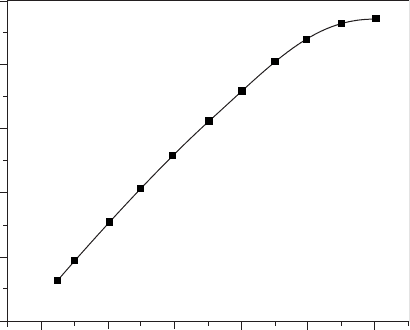
The best understood of the three is R. In fact, to a first approximation, the
implantation depth R increases linearly with ion energy [25], as seen in
the graph for diamond film (Figure 12.1). The scattering that occurs as the ions
enter the substrate limits R in diamond film to less than 500 nm, and leads to
a lateral profile that broadens with depth, not unlike that shown in Figure 12.5
[26]. Most often, given a target material and a species to be implanted,
experimenters select an implantation energy to provide the desired value
of R and then attempt to adjust the other parameters to improve resolution.
Jager et al. modeled the combined effects of implantation energy and ion mass
on the pattern resolution and found, as can be seen in Figure 12.2, that, all
500
400
300
200
100
0
0
20
40
60 80 100
Ion ener
gy
(keV)
Implantation depth (angstrom)
Figure 12.1 Simulation results of ion implantation depth versus ion energy
in diamond film [25].
Focused ion beam systems324
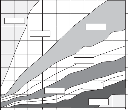
other things being equal, heavy ions can achieve a given resolution at higher
energies than light ones [24]. Nakagawa and his colleagues have published
several interesting papers on the effect of implantation energy on the ratio of
1R
L
to 1R
P
[26,27]. They have found through modeling and comparison with
data in the literature that the behavior of this ratio with respect to energy in
crystalline substrates depends on the implantation angle and the ratio of the
masses of the implanted and target species. With an implantation angle of 7
,at
which the arrangement of target atoms appears to be random, this ratio
increases with energy while, with normal incidence, the ratio remains relatively
constant or decreases.
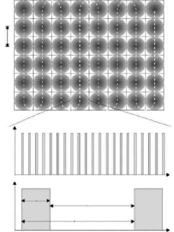
Because focused ion beams have finite resolution, the rastering systems
used for FIB implantation must divide the target into an array of discrete
pixels, which are implanted one by one. Typically, the ion beam scans
meander-like (Figure 12.3) across the sample spending a time, t
d
, implanting
each pixel. This time is called the dwell time and it is inversely related (by the
beam diameter) to the scan rate. It is immediately apparent that the dose is
equal to the product of the dose rate, dwell time, and number of scans. In
other words, for a given dose rate, it is possible to implant the same dose in
a large number of fast scans or a small number of slow ones. Hausmann et al.
5
15
25
35
45
55
65 75
85
95
Ener
gy
(keV)
4
12
20.2
28.1
39.9 Mass (amu)
47.8
55.8
65.4
79
10–20 nm
20–30 nm
40–50 nm
50–70 nm
70–80 nm
>90 nm
0–10 nm
Figure 12.2 Results of Jager’s model of the effect on ion mass and
implantation energy on the achievable pattern resolution. This graph defines
resolution as the lateral range in which 50% of ions are implanted, this
corresponds to roughly 0.67 1R
L
on each side [17].
Ion beam implantation of surface layers 325
found in several studies on the formation of cobalt disilicide layers in silicon
via FIB implantation of cobalt that dwell time has a significant effect on
the results of implantation [28,29]. Specifically, they found that, for dwell
times greater than some critical value, the CoSi
2
layers that formed after
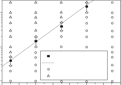
implantation were no longer smooth but instead quite irregular. Further-
more, they demonstrated that the critical dwell time increases exponentially
with temperature (Figure 12.4), suggesting that some sort of dynamic
annealing process is behind the effect. Dynamic annealing refers to the healing
of implant damage even as the implantation process is still occurring, when
the heat applied to the substrate makes the point defects more mobile [31]. In a
similar vein, Tamura et al. observed improved post-anneal electrical activa-
tion of boron ions implanted into silicon at low (of order 10
2
cm/s instead of
order 1) scan speeds [30]. They attributed this to the increased amorphization
of the silicon substrate due to the longer dwell time. So while scan rate has not
been shown to have a clear effect on the resolution of FIB implantation, it is
d
Current
Current
t
d
t
p
t
r
t
t
Figure 12.3 Schematic picture of the serial nature of FIB implantation. The
upper image gives a top-down view of the implantation area. It is divided
into pixels with diameter d. The beam is scanned meander-like over this
area. The lower image gives a current vs. time graph as seen by a single pixel.
Implantation can be done with a short dwell time, t
d
, and thus short pauses
between implantations as the beam targets the other pixels, or with a long
dwell time and thus long pauses [20].
Focused ion beam systems326
nonetheless an important variable in determining the end result of an
implantation step.
Although heating of the substrate may be an inadvertent effect of the ion
beam, for FIB lithography with implantation, the substrate with implanted
pattern actually needs to be thermally treated by a rapid thermal annealing
step (700
C for 60 s) to prevent ions from spreading continuously in the
implanted area (Figure 12.5). This would cause defined lines on the integrated
circuit, microoptical, or other device to broaden. Avoiding such loss of lateral
resolution is especially crucial for fabrication of Bragg gratings in DFB
(distributed feedback) laser diodes, in which the thermal annealing selectively
intermixes the quantum wells in the implanted areas. The intermixing
increases the band gap of the energy by about 40 meV. The modulation of the
band-edge absorption between implanted and nonimplanted areas forms an
absorption grating where spacing must be precise and uniform [33].
The dose rate affects FIB implantation in a way similar to the scan rate,
though not necessarily through a similar physical mechanism. Dose rate is
defined as the number of ions per unit area per unit time incident on the
target. As described in the previous section, it is usually obtained by dividing
the observed current density by the charge per ion. The narrow beams used in
FIB implantation allow for typical dose rates several orders of magnitude
larger (order 10
17
instead of 10
14
ions/cm
2
s) than those of unfocused ion
beams. In their study, Tamura’s group also noted that the effect of increasing
the dose rate was quite similar to that of decreasing the scan rate. Both
Critical dwell-time
Fit of critical dwell-time
Continuous layers
Disrupted layers
350
360 370
380
390
400
410
420
Tem
p
erature
(
˚C
)
100
10
1
Dwell-time (s)
Figure 12.4 Plot showing the quality of CoSi
2
layers as a function of dwell
time and temperature for FIB implantations of Co
2
þ
performed at 70 keV
with a current of 0.52 nA and a dose of 1.0 · 10
17
cm
2
[20].
Ion beam implantation of surface layers 327