Yao N. Focused Ion Beam Systems: Basics and Applications
Подождите немного. Документ загружается.

Scanning electron microscopy (SEM) and transmission electron microscopy
(TEM) have both been widely used in conjunction with serial sectioning to
produce three-dimensional tomographic reconstructions [18–20]. In these
studies, SEM was used to look at metal precipitates while TEM was used for
looking at cellular structures and organelle. In the case of SEM the sample
needs to be removed from the microscope and polished between images. This
allows slices to be taken at 200–300 nm depth increments. Another way SEM
has been used is by looking at backscattered electrons as a function of
primary electron energy [21]. Backscattered electrons come from increasing
depths as the primary beam energy is increased. Noise for a particular depth
image can be reduced by energy filtering the backscattered electrons. In
this case the method is nondestructive. In the TEM method serial sections of
1–2 mm in thickness were taken from the structure of interest and then ana-
lyzed slice by slice in the microscope [20].
Another tec hniqu e that can be used for serial section t omog raphic
reconstructi ons with high spatial resolut ion is scann ing force microsc opy
(SFM). Proof of concept for the SFM was done on a block copolymer
sample. Sectioning of this sample was accomplished using plasma etch
processes. An advantage of this tech ni que i s t hat changes in to pogr aphy due
to preferential etching can be followed with the SFM so that planarization
is not needed. Lateral resolution for this experiment was found to be as high
as 10 nm [22].
A number of ways have been found to use primary ion beams for recon-
structing three-dimensional volumes from both shape and elemental infor-
mation obtained by serial sectioning. A nondestructive method using particle
induced X-ray emission (PIXE) and scanning transmission ion microscopy
(STIM) has been developed using focused ion beams (FIB) [ 23,24]. An ion
beam, usually a proton beam, is rastered over the surface of a sample and the
emitted X-ray intensity is recorded as a function of position. Since X-ray
attenuation is a nonlinear process, local X-ray attenuation factors are cal-
culated from STIM density reconstructions to account for differences in
X-ray production cross sections [25]. Through accurate modeling of the
experiment and advanced algorithms for concatenating experimental output,
accurate three-dimensional representations can be obtained with spatial
resolutions of around 0.5 mm[26]. This technique has been used to study both
biological samples [25] as well as metallurgical samples [27].
Secondary ion mass spectrometry (SIMS) can be used in a mode that
is equivalent to serial sectioning in order to measure three-dimensional
elemental information from a solid sample [28]. A primary ion beam is used
to sputter material from a surface. Ions produced in this way are extracted
Focused ion beam systems298
into a mass filter and their yield as a function of mass is recorded. When
an ion microscope is used as a detector, a specified mass is collected and
imaged on a detector that can measure ion intensity as a function of position
[29]. Since the primary beam is sputtering the surface away, images can be
collected at given time intervals corresponding to depths in the sample [30].
Slices can then be reconstructed into a three-dimensional volume with a
lateral resolution of 2 mm and a vertical resolution of 20 nm [31–34]. One
drawback of this method is that sputtering efficiency varies dramatically with
both composition and crystallographic orientation [28] making topo-
graphical changes as the sample is sputtered an increasing problem with
increased depth. This can be compensated for through use of atomic force
microscopy data or by milling the surface perpendicular to the imaging plane,
but this involves removing the sample from the ion microscope and can lead
to contamination problems [35].
These techniques leave an important part of dimensional space inacces-
sible, corresponding to length scales of tens of nanometers to tens of
micrometers. As the field of nanotechnology advances, these length scales
become increasingly critical. Further, the ability to engineer on the nanoscale
(i.e., on length scales of the order of tens to hundreds of atomic dimensions)
implies control (and therefore measurement) of properties in three dimen-
sions. It is to this important regime that focused ion beam tomography is
ideally suited.
The essence of focused ion beam tomography is the serial acquisition of
images at different depths in a structure during sputtering along the surface
normal. These images may be created by secondary electrons, by secondary
ions, or by mass-filtered secondary ions to create chemical maps. The reso-
lution of these images is determined by a combination of the primary ion
beam diameter at the sample surface, secondary electron and ion escape
depth, and implant straggle of primary ions in the sample. For a state-of-the-
art primary ion beam diameter of 10 nm, and a beam energy of 30 keV, this
translates into a practical spatial resolution of the order of 20–30 nm for most
inorganic materials, both parallel and perpendicular to the sample surface
normal, as will be described later in this chapter. Sets of images recorded at
different depths from the original sample surface are then aligned and con-
catenated in a computer using appropriate interpolation algorithms between
slices. This forms a three-dimensional representation of the sample which can
contain more than 10
7
individual volume elements (voxels), each of which are
separately addressable. Three-dimensional structural and chemical relation-
ships between components in the structure may thus readily be investigated.
Three-dimensional visualization of nanostructured materials 299
A major issue in the generation of such three-dimensional reconstructions
is signal to noise. The secondary electron yield (number of secondary electrons
emitted per incident ion) is relatively high, generally greater than one for
conducting or semi-conducting materials. Thus, even at 10 nm resolution
there are high numbers of secondary electrons to detect. In contrast,
secondary ion yields are many orders of magnitude lower, and this affects the
signal to noise ratio in secondary ion mass spectroscopy (SIMS) elemental
maps. Collection efficiencies and transmission through quadrupole mass
filters typically are also relatively low. This means that chemical mapping
using mass-filtered secondary ion species may often be signal limited. Com-
pounding this issue is the fact that there is only one opportunity to detect a
sputtered ion. This is in contrast to techniques such as electron energy loss
spectroscopy in the transmission electron microscope, where low interaction
cross sections between the primary electron and the appropriate chemically
sensitive inelastic scattering event in the sample may be overcome by
acquisition of spectra from literally billions of primary electrons incident on
the same volume of sample. This ‘‘one shot’’ aspect of FIB-SIMS recon-
structions means that efficient strategies for secondary ion detection are
crucial. Despite these limitations, it is our experience that extremely useful
chemical reconstructions may be obtained for inorganic materials at local
atomic concentrations of the order of a few percent or greater, and at spatial
resolutions of the order of tens of nanometers.
Focused ion beam microscopy (FIB) is clearly well suited for tomographic
reconstructions using serial sectioning methods. Several groups have
attempted to use the FIB for three-dimensional analyses but were limited by
the capability of computers to handle the amount of data generated [36].
With the advent of fast microcomputers, images could be acquired at
increasing depths and elemental distributions compared as a function of
depth. This methodology has the same inherent topography problem
encountered in the ion microscope but has better lateral resolution. Since the
FIB is an ion mill and material can be removed with nanometer precision
in-situ, serial sectioning can be performed. This is done by first milling a flat
surface parallel to the incident ion beam, and then rotating the sample to
image the newly formed surface [37–41]. This process is repeated to obtain
images at various depths in the sample. These images can then be compared
to investigate changes in composition. The microstructure of a multi-layer
device as well as embedded particles have been examined in this manner
[37,38]. In the case of the layered device a trench was milled in the surface of
the sample at the position of the device of interest. The sample was then
rotated 70
so that the newly cleaned surface could be imaged and ion
Focused ion beam systems300
mapped. Distributions of various elements were then compared. Since the
surface being imaged is not normal to the primary ion beam, the image is a
shortened projection of the actual surface. This can easily be compensated for
in various commercial software packages.
FIB tomography has also been used to examine fracture and bonding
surfaces [42–44]. In one case images were taken between serial milling and the
morphology compared slice to slice. Since the new surfaces were easily
recognized due to their smoothness, elemental mapping was not needed and
image reconstruction was adequate. For the bond pad investigation ele-
mental maps were desired for comparison to the images. To minimize the
ambiguity due to surface morphology the sample was imbedded in molding
resin prior to analysis. This minimized surface damage at deeper layers and
allowed images to be more easily compared to elemental maps.
Another advancement in viewing three-dimensional volumes is in the area
of computer aided tomographic reconstructions. Volume reconstructions for
medical applications have fueled a good deal of research into interpolation
routines as well as algorithms for noise reduction [45–49]. Many of these
algorithms can be used for rendering volumes of FIB generated data [50].
Volumes have been reconstructed from both secondary electron images and
ion maps so that the distribution of elements in semiconductor via structures
could be compared. A more advanced algorithm using shape based inter-
polation was used to look at metal precipitates with irregular features. These
reconstructions can be combined with animation routines so that the whole
of a complex structure can be easily studied and visualized. While limits to
resolution come from both beam interactions and redeposited material
during milling [51], a lateral resolution of 10 nm can be obtained.
11.2 Quantitative three-dimensional tomographic reconstructions
using focused ion beams
Fundamentally, FIB tomography is a nanoscale application of classic serial
sectioning that is capable of yielding quantitative three-dimensional insight
into problems in materials science. An implicit assumption in FIB tomo-
graphy is that the physical properties of a feature can be accurately recon-
structed in three dimensions from discrete two-dimensional slices. Ideally,
one might like to disassemble a feature of interest atom by atom using a
continuous sputter process, sort atoms by species and position, and then
display physical and chemical information graphically for analysis. Unfor-
tunately, continuous sputter processes yield poor spatial resolution for most
materials because differential sputtering and redeposition seriously degrade
Three-dimensional visualization of nanostructured materials 301
resolution and chemical sensitivity. Problems due to differential sputtering
and redeposition can be overcome by discretely sectioning a three-dimensional
feature of interest as a function of depth and then interpolating geometric
and chemical information between sections. In this case, a section or slice
is taken at discrete depths specifically chosen to avoid ambiguities and
distortions due to continuous sputter processes.
For each slice, images are collected using either secondary electron or ion
images. Chemical information is also collected for each slice using SIMS,
energy dispersive X-ray analysis (EDX), Auger electron spectroscopy (AES),
or any other spectroscopic technique capable of producing two-dimensional
elemental maps. These slices are then concatenated and a sampled volume is
reconstructed using interpolative algorithms. As such, focused ion beam
tomography can be thought of as a two-step process, data collection and
volume reconstruction. In the following sections, we explain an algorithm for
focused ion beam tomography that uses secondary electron images and SIMS
elemental maps. The reader should keep in mind that these are only two of
many possible signals that can be used to generate tomographic recon-
structions. The volume reconstruction methods discussed below are useful for
a variety of imaging and spectroscopic signals.
11.2.1 Data collection
Data collection is the most crucial step in focused ion beam tomography
because it determines the ultimate accuracy and resolution of three-dimen-
sional reconstructions. A typical data collection algorithm used for focused
ion beam tomography is as follows.
First, an area of interest is found in a sample and fiducial marks are cut
into the periphery. These marks will subsequently be used to align slice data
during volume reconstruction and are re-cut at several depths to avoid slice
misalignment.
Next, secondary electron images and/or SIMS elemental maps are collected
as a function of depth into a feature of interest. Each image and elemental
map is divided into pixels, where pixel size is chosen to be approximately
equal to the probe size. A probe of appropriate size is then scanned over the
image area and held at each pixel for a user-selected dwell time, typically
ranging from 20–40 ms for images and up to 4 ms for SIMS elemental maps.
At each pixel, secondary electron and SIMS signals are collected and stored.
The feature of interest is then sputtered down to a specified depth with the
beam parallel to the data collection surface, and another set of images and
Focused ion beam systems302
elemental maps is collected. To a first approximation, slice thickness is
determined by the amount of material removed by the beam when collecting
secondary electron images and SIMS elemental maps. Secondary electron
images and SIMS elemental maps are repeatedly collected as a function of
depth until a volume of interest has been sampled.
There are several aspects of this procedure that warrant further explana-
tion. First, it is very important that a feature of interest be sectioned with the
beam parallel to the surface for data collection because this minimizes
deviations from a planar section due to differential sputtering. If one chooses
to remove material between slices using normal incidence sputtering, surface
undulations due to differential sputtering will increase in magnitude and
severely degrade depth resolution.
Second, it is important that fiducial marks used for alignment remain sharp
and undistorted in each slice. Typically, at least three square fiducial marks
are cut into the periphery of each slice so that both rotational and transla-
tional drift can be corrected.
Third, care must be taken to ensure that the depth of each slice is measured
accurately. The most straightforward method for measuring the depth of
slices is to measure sputtered depth, edge-on using secondary electron
images. In particular, the depth from the original surface is measured with
the beam parallel to the sputtered surface. Inherent in this method is the
assumption that the image plane is flat and not inclined along the beam.
A second method for determining slice depth is to measure the final sputtered
depth using scanning tunneling microscopy or atomic force microscopy. The
total depth is then divided by the number of slices to estimate the average
spacing between slices.
Finally, it is important to choose a probe size that does not over or under
sample image and elemental map signals in a particular slice. Since focused
ion beam imaging and SIMS are inherently ion processes, one has to take
account of the normal and lateral extents of signal generation. A typical FIB
image or elemental map is divided into pixels with an image plane area
determined by the digital-to-analog characteristics of beam scan coils. Since
only those ions within 0.5 nm of the surface have enough energy to escape
[52], a more important consideration is the lateral range of ions once they
have impacted a sample surface. Quick estimates of lateral range for most
materials can be made using a typical spreadsheet and approximations to
Linhard, Scharff, and Schiott (LSS) theory [53]. Using this information, an
appropriate probe size can be chosen to ensure that signals measured do in
fact arise from within voxel bounds.
Three-dimensional visualization of nanostructured materials 303

The ultimate resolution of FIB tomographic reconstructions is directly
determined by instrumental factors and physical aspects of sputter events.
Since we are discussing a sputter based technique, both lateral and depth
resolution must be considered.
Lateral resolution is controlled by two factors, the smallest ion probe
achievable in a given FIB and the lateral range of probe ions in a feature of
interest. The smallest achievable probe is determined by the FIB column
contrast transfer function and Coulombic interactions of ions within the
beam [54]. Using modern FIB instrumentation, it is possible to achieve ion
probes 10 nm in diameter. High current ion probes smaller than 10 nm in
diameter are susceptible to perturbations due to Coulombic interactions [54].
If dual-beam FIB systems are used to collect data, higher resolution data are
achievable with modern secondary electron microscopy columns. Ultimate
resolution of these columns is, however, still limited by the contrast transfer
function of the particular column used but can be as high as 1 to 2 nm.
Since ion probes are used to sputter surfaces, the effect of ion–sample
interaction has to be taken into account when considering the ultimate
resolution of image and SIMS elemental maps. Because secondary electron
images and SIMS elemental maps are incoherent, lateral resolution is directly
proportional to effective probe size. As was mentioned earlier, the effective
probe size is increased by the lateral range of probe ions in features of interest.
To estimate the increase in probe size due to lateral straggle, one can use a
variety of techniques. The simplest method is to use LSS theory. Table 11.1
shows estimates for 30 keV Ga ions in Al, Ti, and Si.
The effective probe size can then be estimated by adding the lateral range
to probe ions in sample materials to the smallest achievable incident probe.
Using the lateral ranges in Table 11.1 and a 10 nm incident probe, the ultimate
lateral resolution of images and elemental maps from these materials is
approximately 24 nm for Al and Ti and less for Si. As was mentioned earlier,
lateral range is strongly dependent upon sample material and should be
calculated on a case-by-case basis.
Depth resolution in FIB tomography is affected by differential sputtering
and damage profiles produced as ions traverse sample materials. To minimize
Table 11.1. Estimated lateral ranges for 30 keV Ga ions.
Material Lateral range (nm)
Al 7
Ti 7
Si 3
Focused ion beam systems304
differential sputtering, it is important to section a feature of interest with the
ion beam parallel to the surface for data collection. This minimizes differ-
ential sputtering during sectioning.
Damage profiles along the beam direction during data collection directly
affect the minimum distance between slices for a feature of interest. Knock-on
displacement events can distort sample features, so the minimum depth
between slices is limited by depth of maximum damage for a given sample
material. For 30 keV Ga ions, this depth ranges from a few nanometers to
tens of nanometers depending upon sample material.
11.2.2 Volume reconstruction
Once data from a feature of interest have been collected, they are processed
to reconstruct three-dimensional image and chemical information. There are
several methods that can be used to reconstruct volumes from these data, but
we will focus on linear intensity interpolation and shape based interpolative
methods.
11.2.3 Linear intensity interpolation
In linear interpolative schemes, collected data are concatenated as a function
of depth to produce a three-dimensional set of discretely sampled data. Voxels
between slices are then interpolated using linear interpolation algorithms.
In linear interpolative schemes, secondary electron intensities or SIMS inten-
sities are measured at each voxel in a slice and then voxels are interpolated
from slice to slice.
Linear interpolative schemes are useful for objects that can be generated by
extrusion operations such as columnar grains and vias used in multilevel
interconnect structures for integrated circuits. For example, Figure 11.1
shows a 3 · 4 array of vias from a structure in an integrated circuit. This
volume reconstruction was done using secondary electron image data from
eight slices through these vias, parallel to the top surface of the reconstructed
volume.
Using the same interpolative methods, three-dimensional chemical maps
can be reconstructed using linear interpolative schemes. Shown in Figure 11.2
is a set of Al vias reconstructed using linear interpolation. These vias are cut
into SiO
2
interlayer dielectric, lined with Ti, and then filled with Al.
It is clear from Figure 11.2 that linear intensity interpolation is capable
of accurately reconstructing via structures. However, as was mentioned
Three-dimensional visualization of nanostructured materials 305
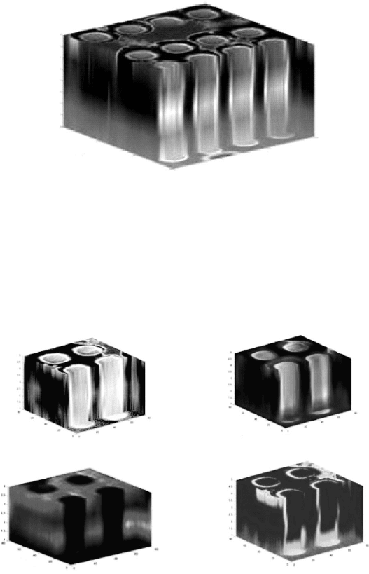
Secondary electron image reconstruction
(a)
3.0 µm
3.0 µm
0.91 µm
3.0 µm
3.0 µm
0.91 µm
3.0 µm
3.0 µm
0.9 µm
3.0 µm
3.0 µm
1.0 µm
(b)
(d)(c)
Al SIMS reconstruction
O SIMS reconstruction
Ti SIMS reconstruction
Figure 11.2 Image and SIMS chemical maps reconstructed from secondary
electron images and SIMS elemental maps. (a) Secondary electron image
reconstruction calculated using linear interpolation from five secondary
electron images. (b) Al elemental reconstruction calculated using linear
interpolation from five SIMS Al elemental maps. In this figure, light gray
indicates the highest Al signal, while dark gray and black indicate low Al
intensity. (c) SIMS elemental reconstruction using negative O ion elemental
maps. In this reconstruction, gray represents highest O concentration, while
black represents background. (d) Ti SIMS reconstruction using five Ti SIMS
elemental maps. In this figure, light gray represents the highest Ti signal and
dark gray represent background level signals [50].
5
4.5
4
3.5
3
2.5
2
1.5
1
1000
1000
100
10
0
5.6 mm
5.6 mm
100
0.9 µm
10
0
Figure 11.1 A secondary electron image volume reconstruction of a 3 · 4
array of vias in an integrated circuit macro. This reconstruction was done
using eight slices that were cut parallel to the top surface of this volume. In
this reconstruction, contrast is observed from differences in secondary
electron yield due to different materials and crystallographic orientations.
Focused ion beam systems306
previously, this type of interpolation does not accurately reconstruct features
that cannot be generated by extrusion operations. If for instance, these vias
contained voids, void edges would be blurred during the interpolation pro-
cess. This would make it nearly impossible to make quantitative estimates of
void volume, surface area, connectivity, or morphology.
11.2.4 Shape based interpolation
Shape based interpolation is a scheme developed for medical tomographic
imaging that uses interfaces present in three-dimensional data sets to
reconstruct complex features. Shape based interpolation is more accurate
than intensity based reconstruction techniques because the shape of the
object, rather than image intensity, is being interpolated, thus inaccuracies and
edge blurring often observed in intensity interpolation are avoided [55,56].
Since FIB images and elemental maps consist primarily of spatially varying
intensities, shape based interpolation can be readily adapted to three-
dimensional tomographic reconstructions of geometric and chemical data
from FIB microscopy data. Shape based volume reconstructions can be used
to not only establish three-dimensional chemical and geometric relationships,
but also to obtain quantitative information such as the sharpness of inter-
faces, surface area, volume, volume fraction, and connectivity of features of
interest.
Shape based interpolation is a method in which the shortest distance of a
voxel to the edges of a feature within a slice is calculated using either
Euclidean or city-block distances [55] as is shown in Figure 11.3. If a voxel is
inside the edges of a feature, this distance is entered into a voxel as a positive
distance; if a voxel is outside the edges of a feature, its closest distance to
feature edges is entered into a voxel as a negative distance. Voxels that fall on
the edges of a feature, by definition, have zero distances. The determination
of whether a voxel is inside or outside of a feature is done using standard
inside-out tests [57].
Once each experimentally collected slice has been processed and edge
distances recorded, distances for slices between recorded data slices are
interpolated using bi-linear interpolation [55]. After interpolation has been
completed, voxels that have negative edge distances are turned off (set to
zero) and the resulting volume is represented by a volume of voxels that have
zero or positive distances to feature edges.
The accuracy of tomographic reconstructions calculated using shape
based interpolation is directly dependent upon the accuracy with which the
Three-dimensional visualization of nanostructured materials 307