Givan A.L. Flow Cytometry. First Principles
Подождите немного. Документ загружается.

THE STRATEGY OF GATING
In ¯ow cytometry the term gating is applied to the selection of cells
(according to their ¯uorescence and/or scatter characteristics) that
will be carried forward for further analysis. The process of gating
corresponds to the decisions made by the microscopist about what
particles in a ®eld to include in a count of any particular type of cell.
Gating is generally acknowledged to be one of the most powerful, but
also one of the most problematic, aspects of ¯ow cytometry (right up
there with spectral compensation). It is problematic primarily because
¯ow cytometrists like to think of their technique as objective and do
not like to admit that much of ¯ow cytometric analysis rests on the
foundation of a few subjective initial decisions. The ideal strategy for
gating should therefore move us toward two goals: First, gating needs
to become as objective as possible, and second, ¯ow cytometrists
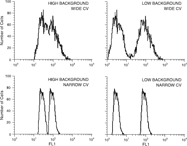
Fig. 6.7. The ability to distinguish stained cells from unstained cells depends on both
the breadth of the distributions of light intensities of the two populations as well as
their relative average intensities.
Leukocytes, Surface Proteins, and the Strategy of Gating 99
need to recognize explicitly those aspects of gating that continue to
require subjective decisions.
To continue with the example drawn from leukocyte techniques,
gating has been employed e¨ectively in the use of FSC and SSC
characteristics for the selection of lymphocytes from within a mixed
population of cells from peripheral blood. This use of gating is de-
rived from two causes: First, a frequent question asked by immunol-
ogists (whether of a microscope or of a ¯ow cytometer) concerns the
distribution of lymphocytes into subpopulations. For example, ``what
percentage of lymphocytes are B lymphocytes?'' or ``what percentage
of lymphocytes are CD8-positive lymphocytes?'' Second, as discussed
at the beginning of this chapter, lymphocytes possess physical charac-
teristics that generally allow them to be distinguished from other types
of leukocytes in both ¯ow and microscopic analysis. Therefore, if one
wants to know what percentage of the lymphocytes are CD8 positive,
one simply de®nes the FSC and SSC characteristics of lymphocytes
(by ¯ow) or the nuclear and cytoplasmic patterns of lymphocytes (by
microscope) and then analyzes the particles within this gate (i.e., with
the de®ned characteristics) to see which of these stain with an anti-
CD8 monoclonal antibody more intensely than an unstained control
(Fig. 6.8). The ability to de®ne a lymphocyte gate takes a bit of
practice, but it is usually an easy decision for a skilled ¯ow cyto-
metrist (and also for a skilled cytologist using a microscope) when
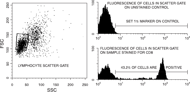
Fig. 6.8. A common type of ¯ow analysis. The cells within a lymphocyte gate are
analyzed, ®rst in an unstained control sample and next in a stained sample, to
determine how many cells within that gate are positively stained.
Flow Cytometry100
blood is relatively normal. It can, however, be a very di½cult deci-
sion, by either technique, when blood is abnormal (e.g., blood from
immunosuppressed patients, who have relatively few and possibly ac-
tivated lymphocytes).
Figure 6.9 shows the light scatter pro®les from cell preparations
from a normal donor and from patients with leukemia and receiving
chemotherapy, just to show diverse examples of light scatter pro®les.
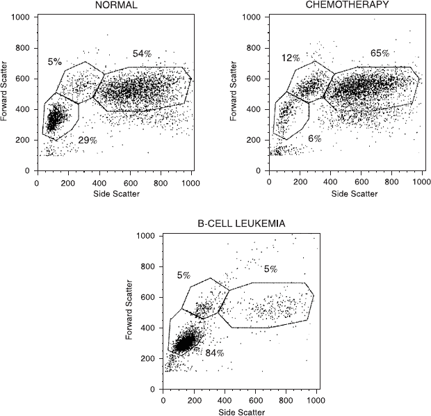
Fig. 6.9. Peripheral blood cells from a normal volunteer, a patient receiving chemo-
therapy, and a patient with B-cell leukemia. Normal cells appear as a tight cluster of
lymphocytes with a di¨use group of monocytes on their shoulder possessing brighter
FSC and SSC and a large cluster of cells with high SSC (neutrophils). The patient
receiving chemotherapy has clusters in similar positions, but with far fewer lympho-
cytes that merge at their top end into the monocytes. The leukemic patient's cells are
almost exclusively lymphocytes, which have slightly lower FSC than normal cells.
Data ®les were provided by Marc Langweiler and Sharon Rich.
Leukocytes, Surface Proteins, and the Strategy of Gating 101

Figure 6.10 indicates the change in light scatter that occurs when
lymphocytes become activated or stimulated to divide; both FSC and
SSC intensities increase, and the lymphocytes ``move out of the lym-
phocyte gate.'' When few lymphocytes are present or those that are
present have enlarged after immunological challenge, decisions about
what is or is not a lymphocyte become di½cult for both microscopists
and cytometrists. The most important message here is that this type
of decision, whether it be microscopic classi®cation or a cytometrist's
gating, is essentially a subjective decision. It is based on training,
skill, and practice. Its subjective nature can lead to di¨erent results
from di¨erent operators, particularly when operators are inexperi-
enced or when abnormal specimens are being analyzed, because the
percentage of cells within a gate that are stained (with any given
stain) will vary depending on the range of cells included within that
gate (see Table 6.2). If we want to analyze lymphocytes, then the gate
should ideally include all the lymphocytes from the sample and
should include only lymphocytes.
Recognition of the need for some objectivity in gating decisions
(both because operators may not be experienced and because not
all samples are ``normal'') has led to attempts both to automate the
de®ning of the gate and to describe the goodness of the gate, once
de®ned. Progress toward automation with lymphocytes brings us
to the technique of back-gating, which has proved informative as a
strategy for ¯ow cytometric analysis in general. Historically, gating
has been performed on the SSC and FSC characteristics of cells (by
Fig. 6.10. The changes in patterns of scattered light that occur when lymphocytes
become activated.
Flow Cytometry102

analogy with a microscopist's use of physical criteria to classify cells),
and this gate has then been used to inquire about the ¯uorescence
properties of the gated cells. Back-gating is really just a di¨erent sort
of gatingÐit reverses the usual protocol by using a stained sample,
placing a gate around particles with certain ¯uorescence character-
istics, and then asking what the physical characteristics (i.e., FSC and
SSC) of these particles are. Once the scatter characteristics of a
population have been ascertained in this way, the subsequent placing
of the scatter gate can be done on the basis of rational and de®ned
criteria.
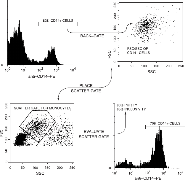
For example, if one stains leukocytes with a PE-conjugated
monoclonal antibody speci®c for the CD14 determinant (a monocyte
marker) and puts a gate around the cells that are brightly ¯uorescent,
it can be determined where these bright cells fall in a plot of FSC
versus SSC (Fig. 6.11). As it turns out, most of the CD14-positive
cells have moderate SSC and moderate FSC and lie in a cluster on
the FSC/SSC dot plot. It also turns out, however, that there are a
few CD14-positive cells that lie outside this region. We are now in
a position to count the total number of CD14-positive cells in the
sample; to draw what we think is an appropriate gate within the
FSC/SSC plot; and ®nally to ask two questions that will tell us how
good a gate we have drawn: (1) How many of the CD14-positive cells
have we excluded by the drawing of that gate? (2) What percentage
of the cells within the gate are not monocytes? Thus the technique of
back-gating has allowed us to make an ``educated guess'' in drawing
TABLE 6.2. The E¨ect of Size of the FSC/SSC Gate on Determination of the
Characteristics of Cells Within that Gate: Peripheral Blood Mononuclear Cells
from a Heart/ Lung Transplant Patient
Small gate (691 cells) Large gate (1074 cells)
Lymphocytes 646 cells 928 cells
94% of gated cells 86% of gated cells
CD3 cells (T cells) 81% of gated cells 70% of gated cells
IL-2r
(activated) T cells 12% of T cells 16% of T cells
CD20 cells (B cells) 9% of gated cells 8% of gated cells
CD4 cells (helper T cells) 56% of gated cells 51% of gated cells
CD8 cells (cytotoxic T cells) 24% of gated cells 20% of gated cells
CD16 (NK cells) 8% of gated cells 11% of gated cells
Leukocytes, Surface Proteins, and the Strategy of Gating 103
a monocyte gate in the FSC/SSC plot; and it has then allowed us to
evaluate that gate in terms of both purity and inclusivity. A perfect
monocyte gate would contain only monocytes (100% purity) and
would include all the monocytes (100% inclusivity). As we shall see,
most gates are a compromise between these two goals. In any case,
with back-gating to help us de®ne a monocyte (FSC/SSC) gate, that
scatter gate can be used in subsequent samples when staining the
monocytes to analyze their phenotype.
By extension from this simple example, the technique of back-
gating has been applied quite elegantly to leukocytes by the use of
Fig. 6.11. Back-gating from CD14 ¯uorescence to determine the scatter character-
istics from monocytes. Such back-gating facilitates the placing and then evaluation
of a monocyte scatter gate.
Flow Cytometry104
a combination of two stains and two-color analysis. When a PE-
conjugated monoclonal antibody speci®c for monocytes (as described
above) is mixed with a ¯uorescein isothiocyanate (FITC)-conjugated
antibody speci®c for a determinant found on all white cells (CD45 is
a so-called leukocyte common antigen), this mixture can be used to
stain a white cell preparation. After dividing the FITC/PE plot into
four quadrants, it can be imagined that any erythrocytes or debris in
the preparation will appear in the lower left quadrant (double nega-
tives); any white cells will appear in the upper or lower right quad-
rants (FITC positive); and any monocytes will appear in the upper
right quadrant (double positive). One might imagine that lympho-
cytes and granulocytes would appear in the lower right quadrant, as
they are leukocytes but not monocytes (and should be FITC positive,
PE negative).
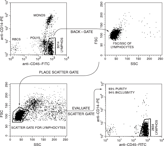
The results from such a staining protocol are given in Figure 6.12.
It turns out that they are even more useful than we might have
imagined. It happens that granulocytes express a lower density of the
common leukocyte antigen on their surface than do lymphocytes, and
these two types of cells can be distinguished from each other as well
as from the PE-positive monocytes in this staining mixture. Thus this
protocol allows us to do the two things we have set as goals for our
lymphocyte gating strategy. We can back-gate from the lymphocyte
cluster in the FITC/PE plot to help us ®nd the FSC/SSC region in
which to draw our gate, and we can use the staining pro®le to eval-
uate that gate in terms of purity and inclusivity. In the case of the
example shown in Figure 6.12, back-gating from the FITC-bright/
PE-negative cluster leads us to a region for the FSC/SSC gating of
lymphocytes. If we draw a reasonable scatter gate based on the scatter
signals of the lymphocytes, we ®nd that we have included most of the
lymphocytes and that almost everything in that gate is a lymphocyte.
A smaller gate might have higher purity but miss some lymphocytes; a
larger gate might include 100% of the lymphocytes but some mono-
cytes, neutrophils, and red blood cells as well. This kind of staining
and back-gating protocol has led the way toward a fully automated
gating procedure in which the gating and evaluation of that gate are
done by computer. A computational gating algorithm can be written
to adjust the scatter gate to aim for some desired level of purity and/
or inclusivity.
However, any automated procedure will have some degree of
Leukocytes, Surface Proteins, and the Strategy of Gating 105
di½culty with abnormal samples, and laboratories will tend to have
their own (subjective) methods for compromising when the sample is
such that a gate with both high purity and high inclusivity is not
possible. By way of illustration of this situation, we can look at a
sample of blood cells from an immunosuppressed patient (Fig. 6.13).
Back-gating from the FITC-bright/PE-negative cluster leads us to
placing a gate in a region of the FSC/SSC plot. We can see, however,
that there appear to be lots of lymphocytes that have abnormally
high FSC and SSC; these could be activated lymphocytes. If we draw
a gate large enough to include these lymphocytes in our subsequent
Fig. 6.12. Back-gating from CD14/CD45 ¯uorescence to determine the scatter
characteristics of lymphocytes. Such back-gating facilitates the placing and then
evaluation of a lymphocyte scatter gate within a peripheral blood mononuclear cell
preparation.
Flow Cytometry106
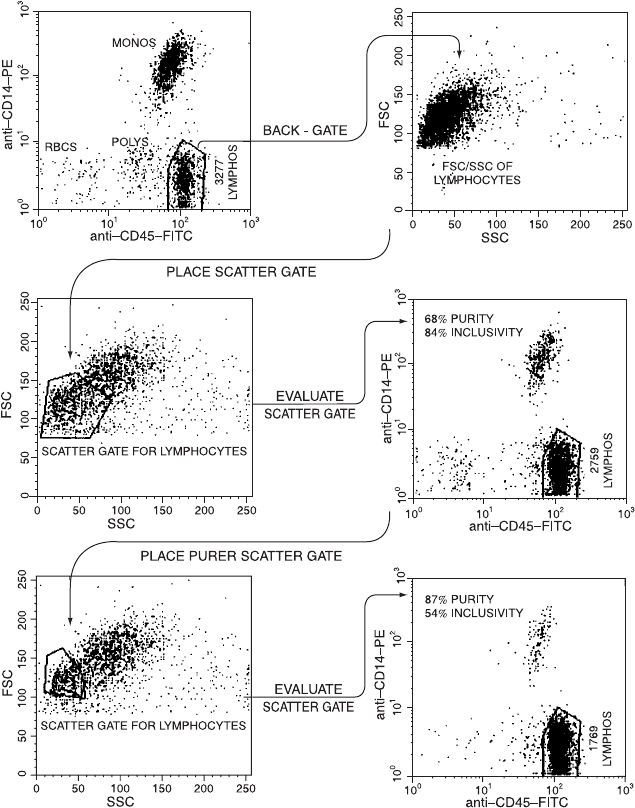
Fig. 6.13. Di½culties in placing a pure and inclusive lymphocyte scatter gate on a
peripheral blood mononuclear cell preparation from a transplant patient with few
and blasted lymphocytes.
Leukocytes, Surface Proteins, and the Strategy of Gating 107
analysis (high inclusivity), we ®nd that the gate also includes many
monocytes (low purity).
Unfortunately there is really no entirely satisfactory way out of
this dilemmaÐthe simple fact is that activated lymphocytes look, by
¯ow cytometric scatter measurements, rather similar to monocytes
(see Fig. 6.10). Any automated software will have trouble handling
the type of situation when it is impossible to draw an FSC versus SSC
gate with both high purity and high inclusivity. Practice will di¨er
from lab to lab, with some operators tending to aim for high purity
and accepting low inclusivity while others aim in the opposite direc-
tion. In either case, if one is expressing results as the percentage
of lymphocytes that stain with a given marker, these results should
be corrected for contamination by other particles (e.g., monocytes)
within the lymphocyte gate. In fact, the situation is even more com-
plicated than that. Because one may be staining lymphocytes for
markers that appear on, for example, only activated cells (e.g., the
interleukin-2 receptor), the inclusion or exclusion of the larger, more
activated cells in the lymphocyte gate may have a profound e¨ect on
the result obtained even after this correction. Thus, the upshot is that
we can aim for objectivity, but our decisions are often, by necessity, a
subjective compromise between con¯icting goals.
GATING ON FLUORESCENCE
As we have seen, one of the problems in gating lymphocytes accord-
ing to their FSC and SSC characteristics is that it can be di½cult,
according to these scatter parameters, to distinguish lymphocytes
from red blood cells, from platelets, from monocytes, and from de-
bris. Immunologically activated patients may have lymphocytes that
overlap monocytes; samples from some donors often have large
numbers of red blood cells that are not easily removed by lysis or by
centrifugation. Gating these preparations according to FSC and SSC
may result in ¯uorescence analyses being reported as the stained per-
cent, not of lymphocytes, but of an unknown mixture of cells.
For example, the more red blood cells that overlap lymphocytes in
the scatter gate, the lower will be the percentage of those gated cells
that are scored as CD4-positive (even though the percentage of lym-
phocytes that are CD4-positive may be identical in all cases). Now
Flow Cytometry108