Givan A.L. Flow Cytometry. First Principles
Подождите немного. Документ загружается.

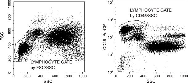
that many benchtop ¯ow cytometers have ``parameters to spare,''
current clinical protocols for lymphocyte analysis suggest that anti-
bodies against CD45 be added to all tubes as a third color to be used
for gating. By gating on CD45 positivity along with SSC, the gated
cells (Fig. 6.14) will not include high numbers of erythrocytes, debris,
or platelets (all CD45 negative). In this way gating can be re®ned if
you are using a ¯ow cytometer with extra ¯uorescence parameters.
The distinction between lymphocytes and monocytes is not, however,
helped by this procedure (and the CD45/SSC gate in Fig. 6.14 in-
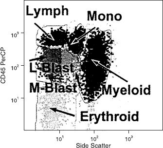
dicates ambiguity here). Figure 6.15 illustrates CD45/SSC gating in a
bone marrow sample, where this type of analysis has proved partic-
ularly useful in identifying clusters that are related to various mature
and immature hematological lineages.
In fact, this CD45/SSC gating marks the general trend away from
using scatter parameters toward a quite di¨erent strategy for ¯ow
analysis. The basic problem, as seen above, is that lymphocytes (or
any other taxonomic group) are not a homogeneous collection of
cells with perfectly delineated physical characteristics; they are mainly
homogeneous, but they usually contain at least some cells at the
fringes with marginal characteristics. Any gating based on FSC/SSC
forces us either to exclude these fringe cells or to include many ex-
traneous cells. With the availability of multicolor instrumentation
Fig. 6.14. The use of CD45 to exclude erythrocytes, platelets, and debris from a
lymphocyte gate. The distinction between monocytes and lymphocytes can be am-
biguous with either the FSC/SSC or the CD45/SSC gate. Data ®le provided by Marc
Langweiler.
Leukocytes, Surface Proteins, and the Strategy of Gating 109
and multicolor stains, it has become possible to avoid making any of
these di½cult gating decisions and to include all cells in the analysis
by using stain itself either to gate in or to gate out the cells of interest.
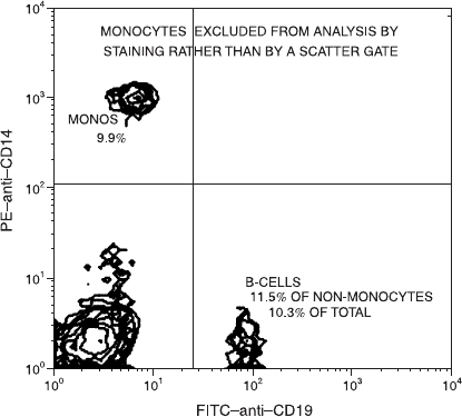
For example, if one is studying the prevalence of a particular
subpopulation among lymphocytes, one could stain every peripheral
blood mononuclear cell sample with a PE stain for monocytes in
addition to an FITC conjugate of the marker of interest. Then, in the
analysis stage, one could simply gate out (exclude) from analysis any
PE-positive particles and analyze all the PE-negative particles for the
percentage that are FITC-positive. Figure 6.16 shows an example
of this protocol. By the use of three-color analysis, there are even
greater possibilities. This type of analysis avoids the necessity of prior
decisions about the FSC and SSC characteristics of the cells of choice
(e.g., lymphocytes). It becomes particularly important when the cells
of interest are less homogeneous in physical characteristics than lym-
phocytes. For example, by gating on a stain that is speci®c for cyto-
keratin (a protein found on tumor cells of epithelial origin), tumor
cells within a mixed population from a breast tumor biopsy specimen
can be selected for further analysis (e.g., DNA content) without any
prejudgement about the FSC or SSC of a poorly de®ned and hetero-
geneous population of abnormal cells.
Fig. 6.15. Bone marrow from a normal donor showing CD45/SSC clusters.
CD45 expression varies as cells mature. Lymph lymphocytes; Mono mono-
cytes; L-Blast lymphoblasts; M-Blast myeloblasts; Myeloid neutrophils; and
Erythroid cells of the erythroid lineage. From Loken and Wells (2000).
Flow Cytometry110
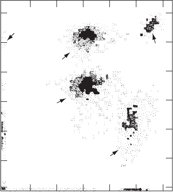
A logical extension of this kind of technique can be seen in the
methodology proposed by PK Horan in 1986: No decisions based on
the scatter characteristics of cells have been made. Cells are stained
simply with a cocktail of conjugated monoclonal antibodies at ap-
propriate (sometimes not saturating) concentrations, and all the cells
in the mixture are classi®ed according to their staining characteristics
(Fig. 6.17). Staining cells from leukemic patients currently follows a
similar strategyÐwhere abnormal and normal cells are de®ned by the
way they cluster in a two-dimensional dot plot. In other words, cells
are de®ned by their relative intensities more than by their negativity
or positivity for a given antibody.
At the beginning of this section, a strategy was described for plac-
ing a scatter (FSC/SSC) gate and then evaluating it in terms of purity
and inclusivity. We were then forced to admit that gating is often an
uneasy and subjective compromise between these con¯icting criteria.
We ®nd therefore that we must conclude that using FSC and SSC
characteristics to gate cells may not be a good thing after all. The
availability of multicolor analysis has led to a trend toward using
staining characteristics to de®ne the cells of interest without regard to
Fig. 6.16. Rather than a scatter gate, a PE-anti-CD14 stain can be used to exclude
monocytes from a count of B and non-B lymphocytes. Data courtesy of Jane
Calvert.
Leukocytes, Surface Proteins, and the Strategy of Gating 111
their possibly variable physical (light scatter) properties. By using one
or more colors in the analysis either to select or to exclude (gate in or
gate out) particular groups of cells, we can avoid any prejudgement
on the physical characteristics of those cells. This overall strategy
makes sense when our system allows for multicolor analysis (with
parameters to spare) and when antibodies are available to de®ne the
cells of interest. It is, nevertheless, still true that antibodies to de®ne
subsets of cells are not perfect: Cells of di¨erent phenotypes react
with similar antibodies (sometimes, thankfully, at di¨erent intensities),
and several antibodies are often required to fully de®ne the taxonomy
of a cell.
As a summary comment on gating, we need simply to remember
that in ¯ow cytometry our questions are usually formulated in terms
of ``what percentage of a certain population of cells is positive for a
certain set of characteristics?'' The choice of a gate de®nes that ``cer-
tain population'' of cells. The choice of that gate will therefore a¨ect
the answer to the question (the percentage positive). Whether gating
10
10
20
30
40
50
60
20 30
log green fluorescence intensity
log red fluorescence intensity
40 50
60
NK cells
T
s
cells
T
h
cells
B cells
M
Fig. 6.17. The two-color ¯uorescence pro®le of peripheral blood mononuclear cells
stained simultaneously with six di¨erent monoclonal antibodies to delineate ®ve dif-
ferent populations of cells. From Horan et al. (1986).
Flow Cytometry112
is applied by means of scatter characteristics, by means of staining
characteristics, or not at all, the procedure still needs to be described
and quanti®ed if the results are to be meaningful and reproducible. It
is only when we have stated exactly which ``certain population of
cells'' we are analyzing that we have ful®lled our goals of objectivity
and/or explicit subjectivity in ¯ow analysis.
FURTHER READING
Chapters 3 and 5 in Ormerod, Chapters 3.2 and 3.3 in Diamond and
DeMaggio, Chapters 10 and 11 in Darzynkiewicz, Chapter 6.2 in Current
Protocols in Cytometry, and Chapters 17 and 34 in Melamed et al. are all
good discussions of general lymphocyte staining methodology for ¯ow
analysis.
Chapter 1.3 in Current Protocols in Cytometry and Chapter 14 in Darzyn-
kiewicz (both by Robert Ho¨man) are excellent discussions of sensitivity
and calibration.
Volume 33, number 2 (1998) of Cytometry is a special issue devoted to
``Quantitative Fluorescence Cytometry: An Emerging Consensus.''
Leukocytes, Surface Proteins, and the Strategy of Gating 113

7
Cells from Within:
Intracellular Proteins
Although many applications of ¯ow cytometry involve the staining of
cells for proteins expressed on the outer membrane, cells also have
many proteins that are not displayed on their surface. With appro-
priate procedures, ¯ow cytometry can provide a means to analyze
these intracellular proteins. The outer cell membrane is impermeable
to large molcules like antibodies; however, if we intentionally ®x cells
to stabilize proteins and then disrupt the outer membrane, the cells
can be stained with ¯uorochrome-conjugated monoclonal antibodies
against intracellular proteins. After time to allow the antibodies to
pass through the now-permeabilized membrane, the cells are washed
to remove loosely bound antibodies and then are run through the
¯ow cytometer to measure their ¯uorescence intensity.
This intensity should, under good conditions, be related to the
amount of the intracellular protein present. However, in describing
our ability to stain cells for surface proteins, we mentioned that it is
best to stain viable cells. Dead cells have leaky outer membranes;
they often show high nonspeci®c staining because antibodies get
through the disrupted membrane and become trapped in the intra-
cellular spaces. Therein lies a con¯ict in our ability to stain cells for
intracellular proteins. Because antibodies of all types are easily
trapped in the cytoplasm, there is greater potential for nonspeci®c
staining of permeabilized cells than intact cells. The very procedure
that we carry out to give access of the staining antibody to its target
(intracellular) antigen actually increases the access of all antibodies
to nonspeci®c targets. To lower this nonspeci®c background, antibody
titers are critical and washing steps are important. Unfortunately,
115
Flow Cytometry: First Principles, Second Edition. Alice Longobardi Givan
Copyright
2001 by Wiley-Liss, Inc.
ISBNs 0-471-38224-8 (Paper); 0-471-22394-8 (Electronic)
even with low antibody concentrations and careful washing, back-
ground ¯uorescence from isotype-control antibodies is often consid-
erably higher on permeabilized than on intact cells.
There is, in addition, a second problem. The procedures used for
®xing and permeabilizing cellsÐto give the staining antibodies access
to intracellular proteinsÐcan modify or solubilize some antigens,
thus destroying the stainability of the very proteins that are being
assayed. To make matters worse, the protocol that works best for one
antigen may entirely destroy a di¨erent antigen. This should not be
surprising after consideration that ``intracellular'' includes proteins of
many types and in many di¨erent environments. Some intracellular
proteins are soluble, some are bound to organelle membranes, and
some are in the nucleus. Therefore, methods for staining cells for
intracellular proteins cannot be as standard or as dependable as the
methods for staining surface proteins. They have to be individually
optimized for the cells and the proteins in question.
METHODS FOR PERMEABILIZING CELLS
While not attempting to describe possible methods in detail, I feel it is
important here to point out the issues involved in intracellular stain-
ing because they highlight some general issues that a¨ect all of ¯ow
cytometric analysis. Methods for permeabilizing and ®xing cells are
various and must be optimized for the particular intracellular antigens
being detected because some antigens are more robust than others in
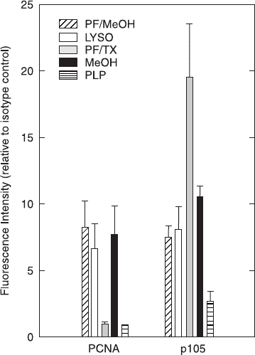
the face of di¨erent agents. Figure 7.1 gives an example comparing
®xation/permeabilization e¨ects on two di¨erent intracellular anti-
gens: Five di¨erent ®xation/permeabilization protocols have been
used, and their e¨ects on staining PCNA (proliferating cell nuclear
antigen) and p105 (a mitosis-associated protein) have been compared.
The good news is that you can stain for intracellular antigens. The
bad news is that it may be di½cult to stain cells optimally for
two di¨erent antigens at the same time (and the relative intensity of
staining for two di¨erent antigens may tell you little about the actual
relative proportions of these proteins in the cell).
The general protocol for intracellular staining involves, ®rst,
staining the cells for any surface (outer membrane) antigens, as de-
scribed in the previous chapter. Then the surface proteins with their
Flow Cytometry116
bound antibodies, as well as the intracellular proteins, are ®xed gently
to stabilize them. The purpose of the ®xation is to cross-link the
proteins well enough that they are not removed or washed out of the
cells after the cells are permeabilized, but not so well that the intra-
cellular antibody binding sites are masked or destroyed. Although
ethanol and methanol can be used for ®xation (by themselves or
following another ®xative), the most common ®xative used prior to
intracellular staining is formaldehyde. Formaldehyde is generally
used at lower concentration and/or for a shorter period of time than
for routine ®xation of surface-stained cells (where ®xation overnight
in 1% formaldehyde is the [optional] last step of the procedure before
¯ow cytometric analysis). Formaldehyde (at 0.5±1.0%) for 10 min is
a good suggested concentration and time for cell ®xation, but lower
or higher concentrations, for shorter or longer periods of time, might
be required.
Fig. 7.1. The e¨ects of di¨erent ®xation protocols on the relative amounts detected
of two di¨erent intracellular proteins. Modi®ed from A McNally and KD Bauer as
published in Bauer and Jacobberger (1994).
Intracellular Proteins 117
This formaldehyde ®xation does permeabilize the cytoplasmic
membrane a bit (formaldehyde-®xed cells are permeable to small
molecules), but proteins are often cross-linked too tightly for staining
of intracellular proteins with antibodies. Therefore the ®xation step
is followed by a permeabilization step. Permeabilizing agents are
usually detergents, such as Triton X-100, digitonin, NP40, or saponin,
at concentrations of about 0.1%. Combined ®xation/permeabilization
reagents are also available as proprietary commercial reagents. With
luck, the detergent will open up the cell enough so that the now-®xed
proteins are accessible to the antibodies used for staining.
What are the criteria by which we can determine whether a ®xation/
permeabilization procedure has been optimized for an antigen in
question? This optimization is, in essence, no di¨erent from opti-
mization of a protocol for surface staining of cells. It is ®rst necessary
to maximize the ¯uorescence intensity of cells that are known to pos-
sess the intracellular antigen (the positive control); ®xation time and
concentration need to be altered in combination with di¨erent deter-
gent concentrations to increase the positive staining. It is then neces-
sary to decrease the background staining (using cells stained with
isotype-control antibodies) as much as possible; this is done by trying
increasing detergent concentrations and washing the cells thoroughly
in bu¨er that contains the detergent. In other words, the goal is to
increase the signal-to-noise ratio. Because antibody concentration,
®xative agent, ®xative concentration, ®xation time, choice of per-
meabilization agent, and concentration of that permeabilizing agent
are all variables in this protocol (and the optimal characteristics of
each may be di¨erent for di¨erent antigens), staining for intracellular
antigens requires some persistence on the part of the investigator. The
following examples (in this chapter and in the following chapter on
DNA) will demonstrate, however, that it is certainly possible.
EXAMPLES OF INTRACELLULAR STAINING
From the point of view of a ¯ow cytometer, surface, cytoplasmic, and
nuclear proteins are similar. The ¯ow cytometer cannot ascertain the
location of the source of ¯uorescence. In addition, the nuclear mem-
brane has large enough pores that it provides little or no obstacle to
staining once the outer, cytoplasmic membrane has been breached.
Flow Cytometry118
Cells have been stained successfully for nuclear proteins related to
proliferation (for example, PCNA, Ki-67, and various cyclins, which
will be discussed in the chapter on DNA) and to tumor suppression
(for example, p53, c-myc, and the retinoblastoma gene product).
They have also been stained for proteins bound to interior membrane
surfaces (e.g., Bcl-2, multidrug resistance protein [MDR], and P-gly-
coprotein), and many strictly cytosolic proteins have been analyzed
(like tubulin, hemoglobin, surface proteins that exist intracellularly at
various stages of di¨erentiation, and many cytokines).
As an example of one of the more complex biological situations,
we can use the staining of cytokines as an illustration. Cytokines are
a diverse class of proteins that, in response to cell stimulation, are
synthesized and then secreted by leukocytes. For example, when T
lymphocytes are stimulated, either nonspeci®cally or by immuno-
logical triggers, they begin to synthesize interferon-g in their endo-
plasmic reticulum, send the proteins to the Golgi apparatus, and then
secrete the molecules into the environment for stimulation of neigh-
boring cells. To stain for intracellular interferon-g, the usual technique
is to stimulate cells with a biological trigger and then to incubate them
with an inhibitor (brefeldin A or monensin) for several hours. These
inhibitors block the normal secretion of proteins from the Golgi
apparatus and thus allow the cytokine concentration to build up in
the cell to levels that are detectable. After the incubation period, the
cells are stained for any surface antigens of interest, ®xed brie¯y in
formaldehyde, permeabilized with saponin, and, ®nally, stained with
a monoclonal antibody against interferon-g.
Figure 7.2 shows an example of the way in which cells can be
stained for a phenotypic surface marker (CD8) as well as the intra-
cellular cytokine, interferon-g. The ¯ow data indicate that interferon-
g is associated, after PMA-ionomycin stimulation, primarily with
CD8-negative cells. More of the CD8-negative than the CD8-positive
cells have intracellular interferon-g, and those that have that cytokine
have more of it per cell. The tricks in the procedure for staining in-
tracellular cytokines are as much biological as chemical (because the
stain is for the end result of a functional process). In addition to a
knowledge of how to ®x and permeabilize a cell and how to avoid
nonspeci®c staining, we require knowledge of how to trigger the
cytokine production, knowledge of the time course of cytokine syn-
thesis after stimulation, and knowledge of how long cells can survive
Intracellular Proteins 119