Givan A.L. Flow Cytometry. First Principles
Подождите немного. Документ загружается.


5
Seeing the Light: Lasers,
Fluorochromes, and Filters
Because ¯ow cytometry involves the illumination of particles by a
light source and the subsequent analysis of the light emitted by par-
ticles after this illumination, an understanding of some of the princi-
ples behind light production, light absorption, and light emission is
important for the e¨ective design and interpretation of experimental
protocols. The principles of photochemistry apply both to the gener-
ation of light by a light source and to the absorption and emission of
light by a ¯uorochrome. It is best to get these concepts straight before
we begin to describe the staining of cells.
GENERAL THEORY
We need to begin with a brief review of atomic structure. Atoms
consist of relatively compact nuclei containing protons and neutrons.
At some distance from these dense nuclei each atom has electrons
moving in a cloud around the central nucleus. The electrons move
in shells or orbitals or probability waves (di¨erent words derived
from more or less classic or quantum mechanical terms of reference)
around the nucleus, and the number of electrons circulating in these
orbitals depends on the element in question. Four things are particu-
larly important for ¯ow cytometrists to understand about these elec-
trons: First, atoms have precisely de®ned orbitals in which electrons
may reside. Second, an electron can reside in any one of the de®ned
orbitals but cannot reside in a region that falls between de®ned orbi-
tals. Third, the energy of an electron is related to the orbital in
59
Flow Cytometry: First Principles, Second Edition. Alice Longobardi Givan
Copyright
2001 by Wiley-Liss, Inc.
ISBNs 0-471-38224-8 (Paper); 0-471-22394-8 (Electronic)
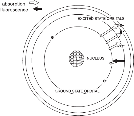
which it resides at any given moment. Fourth, an electron can absorb
energy and be pushed to an excited (higher energy) orbital, but it
will quickly give back that energy as it rapidly returns to its stable,
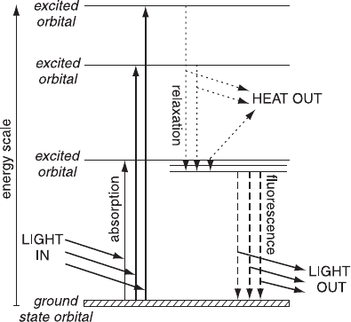
ground-state con®guration (Fig. 5.1). The energy given back as an
electron returns from an excited orbital to its ground-state orbital can
be in the form of light.
With my apologies to all physical chemists for simplifying elec-
tronic structure to four facts, we can now add to our knowledge one
more fact about light itself. Light is a form of energy made up of
photons, and the color of the light is related to the amount of energy
in the photons of that light. For example, when red light is emitted by
an object, that object is releasing photons of relatively low energy;
blue light, in contrast, is made up of photons of higher energy.
Wavelength (expressed in nanometers, abbreviated to nm) is a term
often used to describe the color of light. The wavelength is inversely
related to the amount of energy in the photons of that light: Blue
light has wavelengths in the range of 400±500 nm, and its photons
have relatively large amounts of energy; red light has wavelengths
Fig. 5.1. An ``old-fashioned'' but conceptually easy diagram of an atom with elec-
trons circling the nucleus. Electrons can absorb energy to raise them to an excited
state orbital. When they fall back to their ground state, they may emit light, which
we call ¯uorescence.
Flow Cytometry60
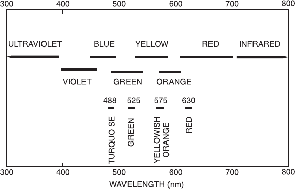
of about 600±650 nm and photons with less energy than those in
blue light. The other colors of the visible spectrum fall between these
values (Fig. 5.2). Infrared light has a longer wavelength than red light
(and less energetic photons); ultraviolet light has a shorter wavelength
than blue light (and more energy). With this information, we can
start to apply our knowledge of light and atomic structure to ¯ow
cytometry.
LASERS
The illumination of particles as they ¯ow past a light source is re-
sponsible for generation of the signals upon which ¯ow cytometric
analysis is based. The illumination of particles can be provided by an
arc lamp or by a laser. In either case, electrons within the light source
are raised to high-energy orbitals by the use of electricity, and energy
is given o¨ in the form of photons of light when the electrons fall
back to their lower energy orbitals. The color of that light is deter-
mined by the energy di¨erences between the excited and lower orbi-
Fig. 5.2. Light, in the region to which our eyes respond, can be described by a
``color'' or, more precisely, by a wavelength. Photons with longer wavelengths have
less energy.
Lasers, Fluorochromes, and Filters 61
tals of the atoms in the lamp or laser. Lasers and arc lamps each have
certain advantages. Because lasers are the light source of choice in
most current ¯ow systems, it is important to understand the de®nite
restrictions on experimental ¯exibility that are imposed by the way in
which a laser works.
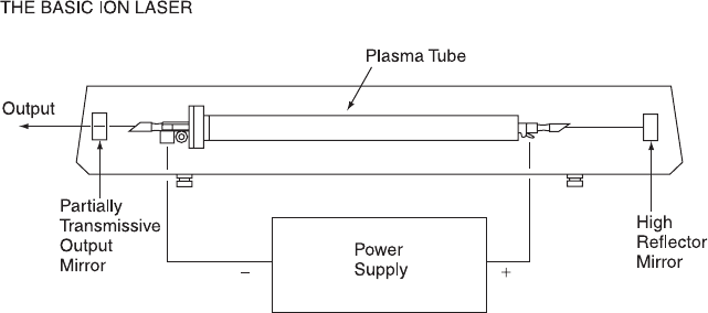
Gas lasers consist of tubes (called plasma tubes) ®lled with gas; a
cathode lies at one end and an anode at the other (Fig. 5.3). A volt-
age is applied across the plasma tube in order to raise the electrons in
the atoms of gas to excited orbitals. As the electrons fall back to
lower energy ground states, they give o¨ energy in the form of pho-
tons of light; the color of the emitted light is determined by the type
of gas used and is a function of the di¨erences between the energy
levels of its atomic orbitals. Re¯ecting mirrors are placed outside the
tube at either end. If the re¯ecting mirrors are aligned precisely with
the plasma tube, then the photons given o¨ by the gas will be re-
¯ected back and forth through the tube between the two re¯ecting
mirrors. The applied voltage maintains the electrons in the gas in
excited orbitals. The re¯ected photons, as they oscillate back and
forth within the tube, interact with these excited gas ions to stimulate
them to release more photons of identical energy (in a way predicted
by Einstein). An ampli®cation system thereby results: The photons
oscillating back and forth between the two end mirrors cause more
and more light to join the beam. By allowing a small percentage of
Fig. 5.3. The structure of a basic gas ion laser. Reproduced with permission of
Spectra Physics.
Flow Cytometry62
this oscillating beam to leave the system at the front mirror, we have
generated what is known as a coherent light source.
A laser light source is coherent in three respects. It is coherent as
regards its direction in that a beam is generated that diverges little
and remains compact and bright for great distances (as in laser light
shows and ``star wars''). It is coherent with respect to polarization
plane (of possible relevance for some specialized aspects of cytometry).
Finally, it is coherent with respect to color because the electrons in a
gas atom or ion are restricted in the orbitals that they use under
plasma tube conditions and are therefore restricted in the amount of
energy that is emitted when they fall from a higher energy to a lower
energy state.
Coherence with respect to direction is the reason lasers are useful
in ¯ow cytometry: They provide a very bright, narrow beam of light
allowing particles ¯owing in a stream to be illuminated strongly for a
very short period of time so that measurable signals from one particle
can be generated and then separated by darkness from signals gen-
erated by the following particle (refer back to Figs. 3.1 and 3.5). The
disadvantage of this spatial coherence is that cells in a stream must be
well aligned in the center of that stream if they are going to be uni-
formly illuminated.
Although spectral purity has certain advantages, coherence with
respect to color is actually one of the major limitations of a laser
system. Whereas an ordinary light bulb will put out light of a wide
range of colors (a white light bulb puts out a mixture of the whole
range of colors in the visible spectrum), the color of the output from
a laser is restricted by the restricted range of electron orbitals that
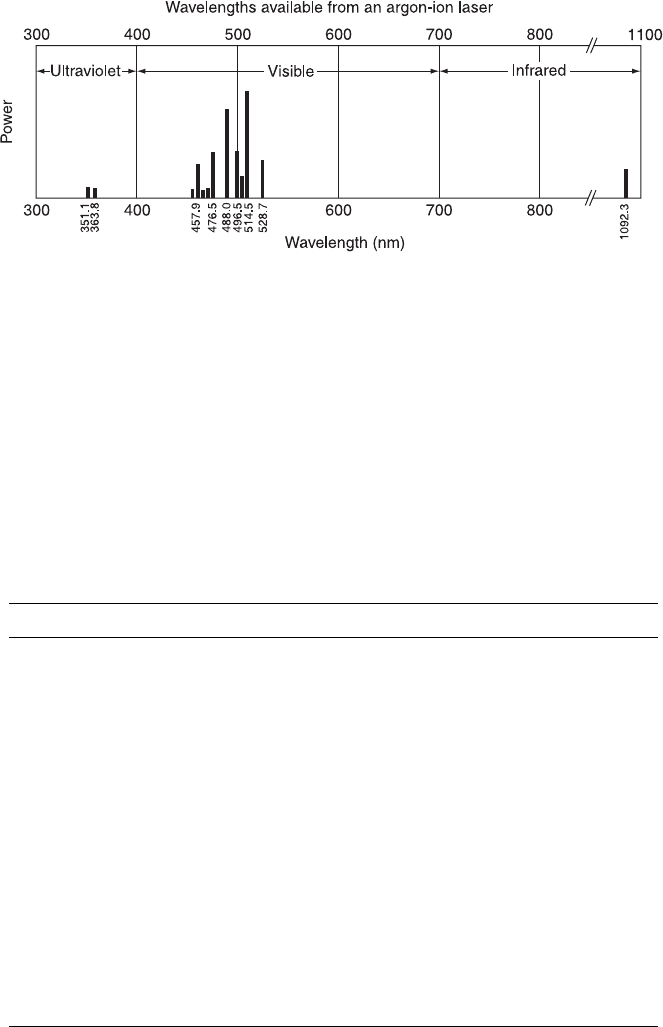
will support lasing in any particular gas. Argon ion lasers are the
most common lasers used in ¯ow cytometry today; they emit useful
amounts of light at 488 nm (turquoise) and at 514 nm (green), as well
as small amounts of ultraviolet light. By using a prism or a wave-
length-selective mirror, the operator or manufacturer can choose one
or the other of these colors, but there is relatively little light available
in other regions of the spectrum (Fig. 5.4). Helium-neon lasers or red
diode (solid-state) lasers are also used (often in conjunction with an
argon ion laser in a dual laser benchtop ¯ow system); a helium-neon
laser puts out red light at 633 nm and a red diode laser at 635 nm.
Research instruments may have three lasers, with one or two argon
lasers for 488 nm and ultraviolet excitation plus a third HeNe laser
Lasers, Fluorochromes, and Filters 63
or dye laser for red excitation. As a general principle, ¯ow cytometers
all have argon lasers emitting light at 488 nm; more advanced in-
struments will add additional lasers with additional wavelengths as
required (Table 5.1).
A laser is therefore very good at providing bright, narrow, stable
Fig. 5.4. The wavelengths of light emitted by an argon ion laser. The most powerful
(and therefore most useful) wavelengths are 488 and 514 nm. High-power argon ion
lasers can provide some useful light in the ultraviolet range.
TABLE 5.1. Laser Options with Examples of Some Possible Fluorochromes
Laser type Wavelengths (nm) Possible ¯uorochromes
Argon ion 488 Fluorescein, R-phycoerythrin, PerCP,
PE-tandems, EGFP, EYFP, propidium
iodide, Alexa 488, acridine orange
514 Rhodamine, propidium iodide, EYFP,
R-phycoerythrin
Ultraviolet
(351/364)
Hoechst dyes, DAPI, Indo-1, AMCA,
Cascade Blue, EBFP
Helium-neon
(HeNe)
Usually 633 Allophycocyanin, Cy5, TO-PRO-3
Krypton ion 568 Cy3, Texas Red, Alexa 568
647 Allophycocyanin, Cy5, TOPRO-3
Red diode 635 Allophycocyanin, Cy5, TOPRO-3
Helium-cadmium
(HeCd)
325 Indo-1, propidium iodide
442 Mithramycin, chromomycin A3, ECFP
Flow Cytometry64
light beams of well-de®ned color and intensity. It is in¯exible, how-
ever, in terms of the color of that light. In addition, large lasers are
demanding in their requirement for routine, skilled maintenance;
careful alignment between plasma tube, re¯ecting mirrors, output
beam, and stream is essential. Furthermore, lasers are expensive;
plasma tubes have limited lives and cost considerably more than an
arc lamp when they need replacing. (It is worth noting that a well-
aligned plasma tube with clean mirrors will last longer than a poorly
aligned and dirty one because it will draw less current to produce the
same amount of light; loving care of a laser can make a considerable
di¨erence to the overall running costs of a ¯ow cytometer.) Many
¯ow systems use small, air-cooled lasers. Air cooling is feasible when
the laser is of low power and does not generate much heat (gas lasers
are all relatively ine½cient and generate a good deal of waste heat).
However, cytometers with a stream-in-air analysis point have space
and refractive index±mismatch surfaces between the cells and the
light collecting lenses; as a result, detection of the light signals gen-
erated by the cells is ine½cient. For this reason, sorting cytometers
may require large, high-power lasers that put out very bright beams
of light but also generate a great deal of waste heat. These sorting
cytometers usually need a large supply of cold water to keep their
lasers cool and also need skilled cleaning and alignment. The main
fact to be kept in mind about lasers, as we move to a discussion of
¯uorochromes, is that, because lasers make use of electrons excited to
a limited range of orbitals, they are restricted in the color of the light
they emit.
FLUOROCHROMES
In a laser, electrons are excited by means of energy from an applied
voltage. We then take advantage of the energy given o¨ as light when
the electrons fall back to lower energy states. With a ¯uorescent
molecule (a ``¯uorochrome'' or ``¯uorophore''), light itself (called the
excitation light) is used to excite the electrons initially. We then ana-
lyze the emitted light (of a di¨erent color) that is given o¨ as the
electrons of the ¯uorochrome return to their ground-state orbitals.
On the basis of our knowledge about electron orbitals together with
our knowledge about laser output, it should now be apparent that,
when a ¯uorochrome is illuminated, the electrons in that ¯uorochrome
Lasers, Fluorochromes, and Filters 65
can absorb energy if, and only if, the excitation light contains pho-
tons with just the right amount of energy to push an electron from
one de®ned ¯uorochrome orbital to a higher (more energetic) de®ned
orbital. Thus the di¨erence between orbital energy levels of a ¯uoro-
chrome will strictly determine what color of light that particular
¯uorochrome can absorb. When electrons fall back from their excited
(more energetic) levels to their ground-state or less energetic orbitals,
light is emitted of a color that depends on the di¨erence in energy
levels between the two ¯uorochrome orbitals in question. Because
energy is not created and is, in fact, always lost to some extent as
heat, the color of the light emitted when an electron falls back from
an excited state to its ground state is always of a somewhat lower
energy (that is, of a longer wavelength) than the energy absorbed in
raising the electron to the higher orbital in the ®rst place (Fig. 5.5).
The light given o¨ when an electron falls back from an excited state
to its ground state orbital is called ¯uorescence. The time required
for ¯uorescence to take place is approximately 10 ns after the initial
activation of the ¯uorochrome by the excitation beam.
If we plot the colors of light that can be absorbed and the colors
that are then emitted by various compounds (called absorption and
Fig. 5.5. The absorption of light by an electron and the subsequent emission of the
energy from that light as both light and heat. Because some of the absorbed energy is
lost as heat, the emitted light always has less energy (and is of longer wavelength)
than the absorbed light.
Flow Cytometry66
emission spectra), we can see how these principles work in practice
(Fig. 5.6). The wavelengths of light absorbed by a compound will
depend exactly on the electronic orbitals of its constituent atoms; the
light then given o¨ as ¯uorescence is always of a longer wavelength
than the absorbed light. The di¨erence between the peak wavelength
for absorption and the peak wavelength for ¯uorescence emission is
known as the Stokes shift. Some compounds (for example, PerCP)
have a larger Stokes shift than others (for example, ¯uorescein).
We are now in a position to understand why the use of a laser to
provide the illuminating beam in a ¯ow system restricts the choice of
¯uorochromes that can be used for staining cells. If we are using an
argon ion laser with an output of light at 488 nm, we can consider as
suitable stains those and only those ¯uorochromes that absorb light
at 488 nm. Rhodamine, a stain used extensively by microscopists,
absorbs light poorly at 488 nm and is therefore not useful in con-
junction with a 488 nm laser. Stains like DAPI and Hoechst can be
used with a high-energy argon ion laser tuned to its ultraviolet line,
but cannot be used if the laser is tuned to 488 nm. Appropriate stains
for 488 nm excitation include DiOC
n
(3) for looking at membrane
potential and propidium iodide and acridine orange for looking at
nucleic acid content. With the 488 nm light from an argon laser, the
situation is also ideal for staining cells with ¯uorescein (another
standby of microscopists). In fact, this traditional allegiance to ¯uo-
rescein (sometimes abbreviated as FITC; ¯uorescein isothiocyanate is
the chemically active form of the dye that will conjugate to proteins)
is the principal reason that argon ion lasers were initially selected for
the ®rst laser-based ¯ow cytometers. Fluorescein absorbs light in the
range of 460±510 nm and then ¯uoresces in the range of 510±560 nm,
with a peak at about 530 nm (green); it can also be readily conjugated
to antibodies, thereby providing speci®c ¯uorescent probes for cell
antigens (Table 5.2).
Because multiple photodetectors are available, a ¯ow cytometer
has the ability to measure two or more ¯uorescence signals simulta-
neously from the same cell. To use several ¯uorochromes at the same
time, cytometrists with only one laser required a group of stains, all
of which absorb 488 nm light but which have di¨erent Stokes shifts
so that they emit ¯uorescent light at di¨erent wavelengths and thereby
can be distinguished from each other by the color of their ¯uores-
cence. Propidium iodide and ¯uorescein are a pair of ¯uorochromes
that ful®ll these criteria (having di¨erent Stokes shifts) and can be
Lasers, Fluorochromes, and Filters 67
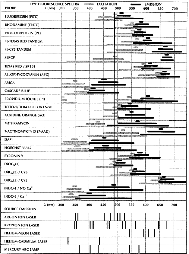
Fig. 5.6. The absorption (gray bands) and emission (black bands) spectra of various
¯uorochromes. The wavelength widths of the bands for each ¯uorochrome indicate
the range of wavelengths that will be absorbed and emitted. Laser (excitation) wave-
lengths are indicated at the bottom of the chart. From Shapiro (1995).
Flow Cytometry68