Hogg S. Essential microbiology
Подождите немного. Документ загружается.

JWBK011-03 JWBK011-Hogg August 12, 2005 15:52 Char Count= 0
68 CELL STRUCTURE AND ORGANISATION
apparatus is to package newly synthesised substances such as proteins and assist in
their transport away from the cell. The substances are contained in vesicles that are
released from the main part of the complex, and fuse with the cytoplasmic membrane.
The Golgi apparatus is poorly defined in certain fungi and protozoans.
Lysosomes
Another function of the Golgi apparatus is to package certain hydrolytic (digestive)
enzymes into membrane-bound packets called lysosomes. The enzymes are needed to
digest nutrient molecules that enter the cell by endocytosis (Figure 3.14), and would
break down the fabric of the cell itself if they were not contained within the lyso-
somes. Peroxisomes are similar to lysosomes, but smaller, and also contain degradative
enzymes. They contain the enzyme catalase, which breaks down the potentially toxic
hydrogen peroxide generated by other breakdown reactions within the peroxisome.
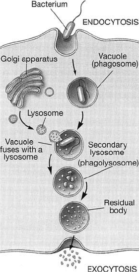
Figure 3.14 Endocytosis. Membrane-bound vacuoles surround a food particle and inter-
nalise it in the form of a phagosome. This fuses with a lysosome, which releases digestive
enzymes, resulting in the breakdown of the contents. The process of endocytosis is unique to
eucaryotic cells. From Black, JG: Microbiology: Principles and Explorations, 4th edn, John
Wiley & Sons Inc., 1999. Reproduced by permission of the publishers
JWBK011-03 JWBK011-Hogg August 12, 2005 15:52 Char Count= 0
THE EUCARYOTIC CELL 69
Outer membrane
Inner membrane
Intermembrane space
Crista
Matrix
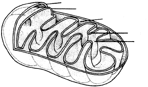
Figure 3.15 Mitochondrial structure. The inner membrane, the location of the electron
transport chain in aerobic respiration, is formed by the invagination of the more permeable
outer membrane. Mitochondria have similar dimensions to many bacteria (approx. 1–3µm),
but may vary in shape due to the plasticity of their membranes
Mitochondria
Whereas in procaryotes the enzymes involved in adenosine triphosphate generation (see
Chapter 6) are associated with the plasma membrane, in eucaryotes they are found
in specialised organelles called mitochondria. These are generally rod-shaped and may
be present in large numbers. They are enclosed by a double membrane, the inner sur-
face of which is folded into finger-like projections called cristae. Respiratory enzymes
are located on the increased surface area this provides, while other metabolic reactions
take place in the semi-fluid matrix (Figure 3.15) (see also Chapter 6).
The mitochondrial cristae of algae, fungi and protozoans each have their own char-
acteristic shapes. Until very recently, a few primitive protozoans, such as Giardia, ap-
peared to lack mitochondria completely, and were thought to represent an intermediate
stage in the evolution of the eucaryotic condition. Recent research, however, has shown
them to possess highly reduced remnants of mitochondria, which have been given the
name mitosomes. It seems that such organisms did, after all, once possess mitochondria,
but have subsequently lost much of their function – an example of so-called reductive
evolution.
Chloroplasts
Chloroplasts are specialised organelles involved in the process of photosynthesis, the
conversion of light into cellular energy. As such, they are characteristic of green plants
and algae. Like mitochondria, chloroplasts are surrounded by a double membrane,
and serve as the location for energy-generating reactions. Inside the chloroplast are
JWBK011-03 JWBK011-Hogg August 12, 2005 15:52 Char Count= 0
70 CELL STRUCTURE AND ORGANISATION
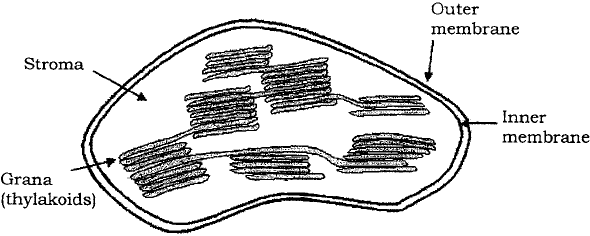
Figure 3.16 Chloroplast structure. Adenosine triphosphate generation from photosyn-
thesis occurs on the thylakoid membranes. In green algae these take the form of discrete
structures called grana. The enzyme ribulose bisphosphate carboxylase, responsible for fix-
ing carbon dioxide via the Calvin cycle (see Chapter 6) is located in the stroma. The outer
membrane of chloroplasts is relatively permeable, allowing the diffusion of the products
of photosynthesis into the surrounding cytoplasm. Reproduced by permission of Dr Lance
Gibson, Iowa State University
flattened membranous sacs known as thylakoids, which contain the photosynthetic
pigment chlorophyll. Thylakoids are arranged in stacks called grana (Figure 3.16).
Mitochondria and chloroplasts both contain 70S ribosomes (similar to those found in
procaryotes), a limited amount of circular DNA and the means to replicate themselves.
This is seen as key evidence for the endosymbiotic theory of eucaryotic evolution, which
envisages that specialised organelles within eucaryotic cells arose from the ingestion of
small procaryotes, which over a long period of time lost their independent existence.
Vacuoles
Vacuoles are membrane-covered spaces within cells, and derive from the Golgi appara-
tus. They act as stores for various nutrients, and also for waste products. Some types of
vacuole are important in regulating the water content of the cell.
Plasma membrane
Many eucaryotes do not have cell walls, so the plasma membrane represents the out-
ermost layer of the cell. The sterols mentioned earlier are important in helping these
cells to resist the effects of osmotic pressure. The only procaryotes to contain sterols
are the mycoplasma, which are unusual in not possessing the typical bacterial cell wall.
Although the eucaryotic plasma membrane does not have the role in cellular respira-
tion associated with its procaryotic counterpart, it does have additional functions. The
process of endocytosis (and its reverse, exocytosis), by which particles or large soluble
molecules are enveloped and brought into the cell, is carried out at the plasma mem-
brane. Also, carbohydrate residues in the membrane act as receptors for cell-to-cell
recognition, and may be involved in cell adhesion.
JWBK011-03 JWBK011-Hogg August 12, 2005 15:52 Char Count= 0
THE EUCARYOTIC CELL 71
CH
2
OH
CH
2
OH
OH OH
O
(a)
(b)
O
O
O
CH
2
OH
OH
O
O
CH
2
OH
NHCOCH
3
CH
2
OH
CH
2
OH
OH OH
O
O
O
O
CH
2
OHNHCOCH
3
NHCOCH
3
NHCOCH
3
OH OH
O
O
O
O
CH
2
OH
OH
O
O
CH
2
OH
CH
2
OH
OH OH
O
O
O
O
NHCOCH
3
OO
OO
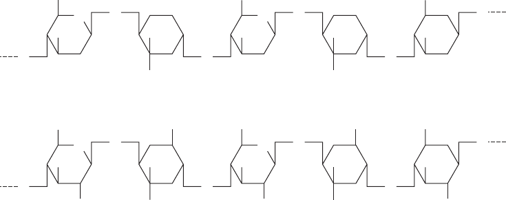
Figure 3.17 The structures of (a) cellulose and (b) chitin. Cellulose is composed of repeating
glucose units joined by β-1,4 linkages, and chitin is a polymer of N-acetylglucosamine
Cell wall
As we have just noted, not all eucaryotes possess a cell wall; among those that do are
fungi, algae and plants. Whilst the function, like that of procaryotes, is to give strength to
the cell, the chemical composition is very different, generally being a good deal simpler.
The cell walls of plants, algae and lower members of the fungi are based on cellulose
(Figure 3.17a), a repeating chain of glucose molecules joined by β-1,4 linkages, and
may also include pectin and hemicellulose, both also polymers of simple sugars. Most
fungi such as yeasts and mushrooms contain chitin, a polymer of N-acetylglucosamine
(Figure 3.17b: we have encountered N-acetylglucosamine before, as a component of
peptidoglycan in bacterial walls.) Chitin is also to be found as the major component of
insect and crustacean exoskeletons, where the function is also to provide strength and
rigidity. As in procaryotes, the cell wall plays little part in the exchange of materials
between the cell and its environment, a role fulfilled by the plasma membrane.
Some protozoans and unicellular algae are surrounded by a flexible pellicle made of
protein.
Flagella and cilia
Motility in eucaryotic cells may be achieved by means of flagella or cilia; cilia can be
thought of as, essentially, short flagella. Both are enclosed within the plasma membrane
and anchored by means of a basal body. Flagellated cells generally have a single flagel-
lum, whereas cilia are often present in very large numbers on each cell. In the microbial
world, flagella are found in protozoans and motile algal forms, whilst cilia are mostly
found in a class of protozoans called the Ciliophora. Flagella and cilia are not found
in members of the Fungi. Although they share the same thread-like gross morphology,
JWBK011-03 JWBK011-Hogg August 12, 2005 15:52 Char Count= 0
72 CELL STRUCTURE AND ORGANISATION
Figure 3.18 Eucaryotic flagella have a characteristic ‘9 + 2’ structure. Although function-
ally analogous to their procaryotic counterparts, eucaryotic flagella differ appreciably in
their fine structure. A membrane surrounds an arrangement of proteinaceous microtubules,
in which nine pairs surround a single central pair. Movement of eucaryotic flagella is by
means of an adenosine triphosphate-driven whiplike motion
eucaryotic flagella differ dramatically in their ultrastructure from those of procaryotes.
Seen in cross-section, they have a very characteristic appearance, made up of two central
microtubules, surrounded by a further nine pairs arranged in a circle (Figure 3.18). The
microtubules are made of a protein called tubulin. Flagella in eucaryotes beat in waves,
rather than rotating; cilia, present in large numbers, beat in a coordinated fashion so
that some are in forward motion while others are in the recovery stroke (rather like
a ‘Mexican wave’!). In animals, ciliary motion has been adapted to move particulate
matter across a tissue surface; ciliated cells of the respiratory tract, for example, act
as a first line of defence in the removal of inhaled particles, such as bacteria from the
airways.
Cell division in procaryotes and eucaryotes
In, unicellular procaryotes, cell division by binary fission leads to the creation of a new
individual. Growth occurs in individual cells until a maximum size is achieved and a
cross-wall forms. Before cell division takes place, the genetic material must replicate
itself (see Chapter 11), and one copy pass to each new daughter cell (Figure 3.19).
Cell division in eucaryotes also results in two identical daughter cells. In the case of
unicellular eucaryotes, this results in two individual organisms (asexual reproduction),
while in multicellular forms there is an increase in overall size. Cell division is pre-
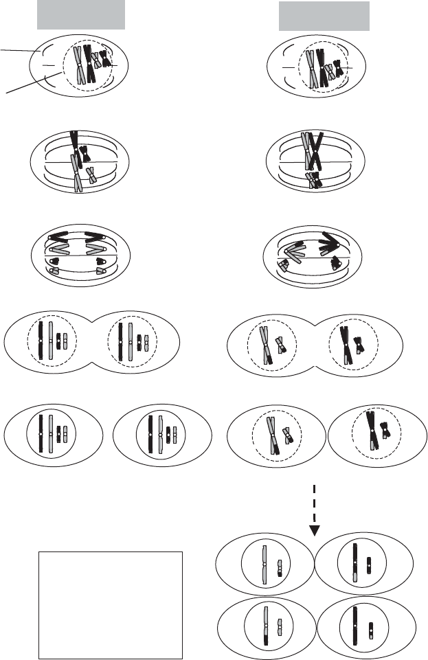
ceded by a process of nuclear division called mitosis, which ensures that both daughter
cells receive a full complement of chromosomes. The principal phases of mitosis are
summarised in Figure 3.20(a). In interphase, the chromosomes are not clearly visible
under the microscope; DNA replication takes place during this period. The duplicated
chromosomes, held together as sister chromatids by the centromere, move towards the
centre of the cell during prophase. A series of microtubules form a spindle between
JWBK011-03 JWBK011-Hogg August 12, 2005 15:52 Char Count= 0
CELL DIVISION IN PROCARYOTES AND EUCARYOTES 73
Bacterial
chromosome
DNA replicates, making a
second copy of the chromosome.
Origins of replication migrate to
ends of cell.
Cell lengthens and new
cell wall is laid down
Plasma membrane
starts to grow inwards
Septum formation is
complete and daughter
cells separate
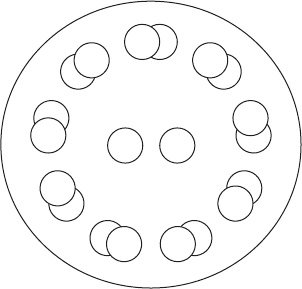
Figure 3.19 Binary fission in E. coli. Replication of the single circular chromosome is
accompanied by an increase in cell size. The plasma membrane invaginates, and a new
cross-wall is synthesised, resulting in two new daughter cells
the centrioles, and the chromosomes line up along this during metaphase. Also, during
this phase the nuclear membrane breaks down, and each centromere duplicates. One
chromosome from each pair then migrates away from the centre to opposite ends of
the spindle. This stage is called anaphase. Finally, in telophase, new nuclear membranes
surround the two sets of chromosomes, to form two nuclei. Mitosis is followed by cell
division. Overall, the process of mitosis results in two identical nuclei containing the
original (diploid) chromosome number.
At various stages of eucaryotic life cycles, a process of meiosis may occur, which
halves the total number of chromosomes, so that each nucleus only contains one copy
of each. In sexual reproduction, the haploid gametes are formed in this way, and the
diploid condition is restored when two different gametes fuse. In some eucaryotes, not
just the gametes but a substantial part of the life cycle may occur in the haploid form (see
Chapters 8 & 9). Meiosis (Figure 3.20b) comprises two nuclear divisions, the second
of which is very similar to the process of mitosis just described. In the first meiotic
division, homologous chromosomes (i.e. the two members of a pair) line up on the
spindle together and eventually migrate to opposite poles. While they are together, it
JWBK011-03 JWBK011-Hogg August 12, 2005 15:52 Char Count= 0
74 CELL STRUCTURE AND ORGANISATION
Cytokinesis: the two
new diploid nuclei
separate into two
daughter cells
Spindle
disappears,
nuclear
membrane
reforms
Telophase
Chromosomes
move towards
opposite poles
of cell
Chromosomes
line up along
spindle
Spindle
formation
begins
Nuclear
membrane
is broken
down
Prophase
Metaphase
Anaphase
Prophase II
a) MITOSIS
b
) MEIOSIS
(Intermediate
stages of
meiosis II
not shown)
Meiosis II, whose steps
are similar to those of
mitosis, results in four
haploid nuclei. Note
the recombinant
chromosomes arising
from crossing over
Figure 3.20 The main steps of (a) mitosis and (b) meiosis in an organism whose diploid
number (2n) =4. Mitosis results in two cells identical to the parent. Meiosis results in a
reduction in the chromosome number and introduces genetic variation by means of crossing
over. For details see the text
JWBK011-03 JWBK011-Hogg August 12, 2005 15:52 Char Count= 0
TEST YOURSELF 75
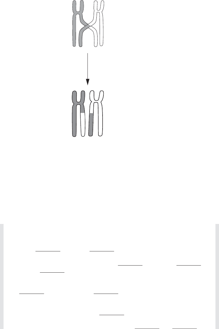
Homologous
chromosomes
Recombinant
chromosomes
Figure 3.21 Crossing over leads to recombination of genetic material. During crossing
over, portions of homologous chromosomes are exchanged. This forms the basis of genetic
recombination in eucaryotes, and ensures that offspring contain new combinations of genetic
material
is possible for crossing over to occur, a process by which the two chromosomes swap
homologous stretches of DNA (Figure 3.21). Since these may not be identical, crossing
over serves to introduce genetic variation into the daughter nuclei. In the second meiotic
division, sister chromatids separate as before, resulting in four haploid nuclei.
Test yourself
1 Procaryotic cells have a much simpler structure than eucaryotes, lacking in-
ternal
and a true .
2 Most bacterial cells are rod-shaped (
), spherical ( )or
curved (
).
3 Many bacteria commonly carry extrachromosomal pieces of DNA called
, which are able to independently of the bacterial chro-
mosome.
4 Protein synthesis takes place at
.
5 The main components of cell membranes are
and .

JWBK011-03 JWBK011-Hogg August 12, 2005 15:52 Char Count= 0
76 CELL STRUCTURE AND ORGANISATION
6 Gram-positive cell walls contain a higher percentage of than those
of Gram-negative cells.
7 Many bacteria have long, hair-like structures called
projecting from
the cell wall. These are used for
.
8 The DNA of eucaryotes is organized into chromosomes and associated with
proteins called
.
9 In eucaryotic cells, extranuclear DNA is also found in
and
.
10 Eucaryotic ribosomes may be found associated with the
or free in the cytoplasm.
11 The Golgi apparatus
and newly synthesised substances.
12
are the site of energy generation in eucaryotic cells. In procaryotic
cells, some of these reactions take place at the
.
13 The photosynthetic membranes of chloroplasts are called
.
14 The cell walls of algae are mostly made up of
.
15 The structure of eucaryotic flagella is more complex than that of procaryotes,
comprising an arrangement of
made of .
JWBK011-04 JWBK011-Hogg August 12, 2005 15:53 Char Count= 0
Part II
Microbial Nutrition, Growth
and Metabolism
77