Klipp E., Herwig R., Kowald A., Wierling C., Lehrach H. Systems Biology in Practice: Concepts, Implementation and Application
Подождите немного. Документ загружается.


simple oscillations; (3) coexistence of a stable state and a limit cycle; (4) birhythmi-
city, i.e., coexistence of two limit cycles that are reached depending on the initial
values; (5) complex periodic oscillations; (6) a strange attractor associated with
chaos; and (7) a folded limit cycle with complex but periodic oscillations. Examples
of complex periodic behavior and chaos are shown in Fig. 7.2.
7.1.3
Coupling of Oscillators
In yeast cell suspensions, the dynamics of glycolysis are usually studied on a popula-
tion level by monitoring mean concentrations. In this way, one obtains only limited
information about the behavior of single cells. A series of experiments has shown
that individual cells also must show oscillations (e.g., Richard et al. 1996). When
these oscillations are synchronized, they can be observed on the population level.
The absence of oscillations on the population level may be due to the absence of os-
cillations on the individual level or due to the fact that cells oscillate out of phase and
only the mean values are measured.
Synchronization of oscillations is mediated by a coupling compound, which is
shared by all cells of the suspension. For glycolytic oscillations, ethanol, pyruvate,
and acetaldehyde have been proposed as coupling metabolites, the latter being the
most probable.
Wolf and Heinrich (1997) employed the Higgins-Sel’kov oscillator to explain basic
features of coupled oscillators. Consider for N cells the situation shown in Fig. 7.3a.
The dynamics is described by the following equation system:
dS
i
dt
v
0
S
i
P
2
i
dP
i
dt
S
i
P
2
i
P
i
k
2
k
3
P
i
C
dC
dt
k
3
j
N
X
N
i1
P
i
N C
!
; (7-13)
where C is the extracellular concentration of the coupling metabolite, N is the num-
ber of cells, j denotes the ratio of intracellular and extracellular volume, and k
3
is
the effective rate constant for the transmembrane diffusion of the coupling com-
pound. All cells are assumed to be identical.
It is easy to see that the equation system in Eq. (7-13) for N coupled cells has a
unique steady state for all cells identical to the steady state derived for an isolated
cell (Eq. (7-5)) with
P =
C. This steady state is stable in certain regions of the para-
meter space. At the boundary of this region, Hopf bifurcations lead to synchronous
or asynchronous oscillations. Here, the solution of the characteristic equation
(Eq. (3-29)) yields a pair of purely imaginary eigenvalues. In the vicinity of the bifur-
cation points, two typical types of behavior may occur: synchronous oscillations and
regular asynchronous oscillations (see Fig. 7.3). Synchronous oscillations for cell po-
230
7 Selected Biological Processes

231
7.1 Biological Oscillations
Fig. 7.2 Decroly-Goldbeter model. The model depicted in Eqs.
(11) and (12) shows interesting patterns of temporal self-orga-
nization. The upper panels show behavior associated with
chaos, and the lower panels show trajectories for complex peri-
odic behavior. (a, d) Time courses. (b, e) Phase planes for S
and P
1
. (c, f ) Trajectories in the space spanned by all three con-
centrations. Parameters: v
0
/K
m1
= 0.5 s
–1
, s
1
= s
2
=10s
–1
,
q
1
= 50, q
2
= 0.02, , L
1
=5710
8
, L
2
= 100. (a–c): k
3
= 2.032 s
–1
,
d = 0.00001, (d–f ): k
3
= 2.5 s
–1
, d = 0.0001.
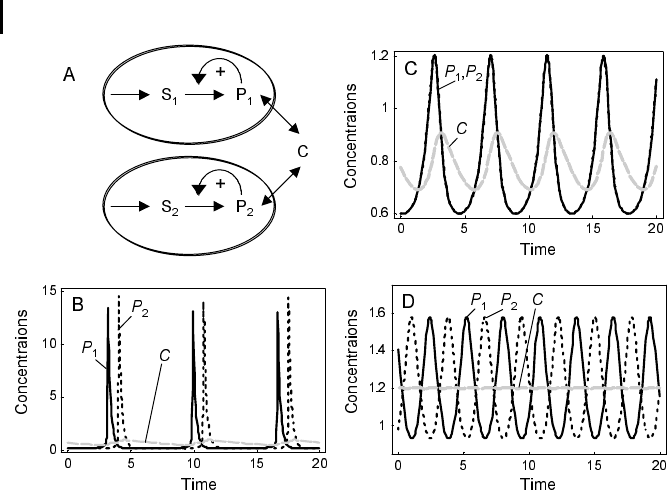
pulations can even be experimentally observed. Regular asynchronous oscillations
are hidden; upon concentration measurements for cell populations, they result in
constant concentration values at the level of the mean for all cells. Two experiments
demonstrate the character of both types of oscillations. Upon mixing of two cell po-
pulations that are synchronized internally but exhibit a phase shift of 1808 with re-
spect to each other, the oscillations are initially strongly damped but reappear after a
while (Richard et al. 1996). This means that both populations synchronize within a
time that depends on the strength of the coupling parameter k
3
. The hidden oscilla-
tions in the case of regular asynchronous oscillations can be made visible by increas-
ing or decreasing the concentration of the coupling substance in a pulse-like man-
ner. Far from the area of Hopf bifurcation, other modes of behavior may occur, e.g.,
non-regular oscillations.
232
7 Selected Biological Processes
Fig. 7.3 Dynamics of coupled oscillators ac-
cording to the model of Wolf and Heinrich
(1997). (a) Schematic representation of two os-
cillators. The metabolites S
1
and P
1
occur in a
compartment (cell) that is different from the
compartment containing the metabolites S
2
and
P
2
. The metabolite C is the coupling compound
that is present in both cells and in the extracellu-
lar space. (b–d) Time course simulations with
parameter values v
0
=3,j = 0.2. (b) Asynchro-
nous oscillations, parameter values: k
2
= 4.3,
k
3
= 1, (c) Synchronous oscillations, parameter
values: k
2
= 3.84, k
3
= 3.2. The coupling com-
pound C oscillates with the same frequency.
(d) Regular asynchronous oscillations, para-
meter values: k
2
= 2.5, k
3
= 1. Oscillations of
the coupling compound practically disappeared,
i.e., the amplitudes are very small. All para-
meters are given in arbitrary units.
7.1.4
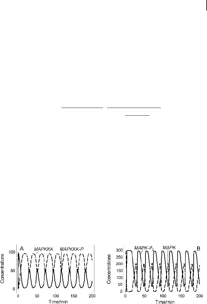
Sustained Oscillations in Signaling Cascades
Sustained oscillations are an emerging property of ultrasensitive cascades with nega-
tive feedback, as there is always a range of kinetic constants in which oscillatory be-
havior is observed. To analyze this feature, Kholodenko (2000) employed a numerical
analysis of the dynamics of the MAPK cascades as illustrated in Fig. 6.11 with the dy-
namics described in the equation systems (Eqs. (6-9)–(6-11)) with Michaelis-Menten
kinetics of the individual reaction steps (Eqs. (6-21) and (6-22)). In addition, a feed-
back from the MAPK-P
2
to the first reaction is considered that changes the rate equa-
tion of the first reaction to
v
1
V
m1
MAPKKKK
MAPKKK
K
1
MAPKKK
1
1
MAPK-P
2
K
I
n
; (7-14)
where K
1
is the Michealis constant for MAPKKK, V
m1
is the maximal reaction rate,
and K
I
is the dissociation constant for MAPK-P
2
with respect to MAPKKKK. Nega-
tive feedback can bring about oscillations in the kinase activities. Following the acti-
vation of the initial kinase and a subsequent activation of the terminal cascade ki-
nase, a strong negative feedback can in some cases be operational in turning off the
activation of a cascade. In other cases, implementation of a strong negative feedback
may have the effect that the system steady state loses its stability. Since there is no
other stable state, the phosphorylation level of cascade kinases starts to oscillate in a
sustained manner. This dramatic change in the system’s dynamic behavior is again
a Hopf bifurcation.
The time course of the active and inactive forms of the MAPK cascade kinases is
shown in Fig. 7.4 for an abrupt increase in the input stimulus (i. e., active
MAPKKKK concentration) at time point zero. At low basal stimulus, the kinases of
the cascade remained predominantly in the inactive forms, and the corresponding
233
7.1 Biological Oscillations
Fig. 7.4 Sustained oscillations in MAPK concentrations according
to the model of Kholodenko (2000). (a) Oscillations of MAPKKK
(b) Oscillations of MAPK Parameters V
m1
= 2.5,V
m2
= 0.25,
V
m3
= V
m4
= V
m7
= V
m
8
= 0.025,V
m5
= V
m6
= 0.75,V
m9
= V
m10
= 0.5,
(in s
–1
and nM s
–1
, respectively) K
m1
=1,,K
m2
=8,K
mi
=15(i =3,
…, 10) (in nM), n =1,K
I
=9.

steady state (off state) is stable. A high stimulus switches the kinases into the active
forms (on state), but this state is instable. The cascade kinases do not remain phos-
phorylated for a prolonged period of time. Due to the negative feedback from
MAPK-P
2
, the rate of activating phosphorylation of MAPKKK decreases with an in-
crease in MAPK-P
2
. As the phosphatase continues to operate, the rate of MAPKKK-P
dephosphorylation begins to exceed the phosphorylation rate and the concentration
of MAPKKK-P decreases. A decrease in the MAPKKK-P causes the kinase activity
down the cascade (MAPKK) to drop. Finally, the concentration of MAPK-P
2
de-
creases, and a new oscillation cycle begins. The amplitude of oscillation in the con-
centrations of the active biphosphorylated MAPK-P
2
and inactive MAPK can be
large. The period of oscillation is about 20 min, but it depends on the choice of ki-
netic parameters. Importantly, the oscillations appear to be stable to changes in the
initial distribution of active and inactive kinase forms. Whether oscillations can oc-
cur at all under conditions of negative feedback depends (1) on the saturation of ki-
nases and phosphatases, i.e., on the ratio between their total concentrations and
their K
m
values, and (2) on the strength of the negative feedback. Tendentially it
holds that the stronger the feedback, the more likely oscillations are.
7.2
Cell Cycle
The eukaryotic cell cycle is the repeated sequence of events accompanying the divi-
sion of a cell into daughter cells (Johnson and Walker 1999). It includes two main
sections: the doubling of the genome (DNA) and all other cell components in the S
phase (synthesis phase) and halving of the genome during the M phase (mitosis).
The periods between the M and S phases are the gap or growth phases G
1
and G
2
(Fig. 2.13). Passage through the eukaryotic cell cycle is strictly regulated by the peri-
odic synthesis and destruction of cyclins that bind and activate cyclin-dependent ki-
nases (CDKs). The notion “kinase” expresses that their function is phosphorylation
of proteins with controlling functions. Cyclin-dependent kinase inhibitors (CKI) also
play important roles in cell cycle control by coordinating internal and external sig-
nals and impeding proliferation at several key checkpoints.
The general scheme of the cell cycle is conserved from yeast to mammals. The
levels of cyclins rise and fall during the stages of the cell cycle. The levels of CDKs ap-
pear to remain constant during the cell cycle, but the individual molecules are either
unbound or bound to cyclins. In budding yeast, one CDK (Cdc28) and nine different
cyclins (Cln1 to Cln3, Clb1 to Clb6) that seem to be at least partially redundant are
found. In contrast, mammals employ a variety of different cyclins and CDKs. Cyclins
include a G1 cyclin (cyclin D), S-phase cyclins (A and E), and mitotic cyclins (A and B).
Mammals have nine different CDKs (referred to as CDK1–CDK9) that are important
in different phases of the cell cycle. The anaphase-promoting complex (APC) triggers
the events leading to destruction of the cohesions, thus allowing the sister chromatids
to separate and degrade the mitotic cyclins.
234
7 Selected Biological Processes

7.2.1
Steps in the Cycle
Let us take a course through the mammalian cell cycle starting in the G1 phase. As
the level of G
1
cyclins rises, they bind to their CDKs and signal the cell to prepare
the chromosomes for replication. When the level of S phase–promoting factor (SPF)
rises, which includes cyclin A bound to CDK2, it enters the nucleus and prepares the
cell to duplicate its DNA (and its centrosomes). As DNA replication continues, cyclin
E is destroyed, and the level of mitotic cyclins begins to increase (in G
2
). The M
phase–promoting factor (the complex of mitotic cyclins with the M-phase CDK) initi-
ates (1) assembly of the mitotic spindle, (2) breakdown of the nuclear envelope, and
(3) condensation of the chromosomes. These events take the cell to metaphase of mi-
tosis. At this point, the M phase–promoting factor activates the APC, which allows
the sister chromatids at the metaphase plate to separate and move to the poles (ana-
phase), thereby completing mitosis. APC destroys the mitotic cyclins by coupling
them to ubiquitin, which targets them for destruction by proteasomes. APC turns on
the synthesis of G
1
cyclin for the next turn of the cycle and it degrades geminin, a
protein that keeps the freshly synthesized DNA in the S phase from being re-repli-
cated before mitosis.
A number of checkpoints ensure that all processes connected with cell cycle pro-
gression and DNA doubling and separation occur correctly. At these checkpoints,
the cell cycle can be aborted or arrested. They involve checks on completion of the S
phase, on DNA damage, and on failure of spindle behavior. If the damage is irrepar-
able, apoptosis is triggered. An important checkpoint in G
1
has been identified in
both yeast and mammalian cells. Referred to as “start” in yeast and as “restriction
point” in mammalian cells, this is the point at which the cell becomes committed to
DNA replication and completing a cell cycle (Hartwell 1974; Hartwell et al. 1974;
Pardee 1974; Nurse 1975). All the checkpoints require the services of complexes of
proteins. Mutations in the genes encoding some of these proteins have been asso-
ciated with cancer. These genes are regarded as oncogenes. Failures in checkpoints
permit the cell to continue dividing despite damage to its integrity. Understanding
how the proteins interact to regulate the cell cycle became increasingly important to
researchers and clinicians when it was discovered that many of the genes that en-
code cell cycle regulatory activities are targets for alterations that underlie the devel-
opment of cancer. Several therapeutic agents, such as DNA-damaging drugs, micro-
tubule inhibitors, antimetabolites, and topoisomerase inhibitors, take advantage of
this disruption in normal cell cycle regulation to target checkpoint controls and ulti-
mately induce growth arrest or apoptosis of neoplastic cells.
For the presentation of modeling approaches, we will focus on the yeast cell cycle
since intensive experimental and computational studies have been carried out using
different types of yeast as model organisms. Mathematical models of the cell cycle
can be used to tackle, for example, the following relevant problems.
1. The cell seems to monitor the volume ratio of nucleus and cytoplasm and to trig-
ger cell division at a characteristic ratio. During oogenesis, this ratio is abnormally
235
7.2 Cell Cycle

small (the cells accumulate maternal cytoplasm), while after fertilization cells di-
vide without cell growth. How is the dependence on the ratio regulated?
2. Cancer cells represent a failure in cell cycle regulation. Which proteins or protein
complexes are essential for checkpoint examination?
3. What causes the oscillatory behavior of the compounds involved in the cell cycle?
7.2.2
Minimal Cascade Model of a Mitotic Oscillator
One of the first genes to be identified as being an important regulator of the cell cy-
cle in yeast was cdc2/cdc28 (Nurse and Bissett 1981), where cdc2 refers to fission
yeast and cdc28 to budding yeast. Activation of the cdc2/cdc28 kinase requires asso-
ciation with a regulatory subunit referred to as a cyclin.
A minimal model for the mitotic oscillator involving a cyclin and the Cdc2 kinase
has been presented by Goldbeter (1991). It covers the cascade of post-translational
modifications that modulate the activity of Cdc2 kinase during cell cycle. In the first
cycle of the bicyclic cascade model, the cyclin promotes the activation of the Cdc2 ki-
nase by reversible dephosphorylation, and in the second cycle, the Cdc2 kinase acti-
vates a cyclin protease by reversible phosphorylation. The model was used to test the
hypothesis that cell cycle oscillations may arise from a negative feedback loop, i. e.,
the cyclin activates the Cdc2 kinase while the Cdc2 kinase triggers the degradation of
the cyclin.
The minimal cascade model is represented in Fig. 7.5. It involves only two main
actors, cyclin and cyclin-dependent kinase. Cyclin is synthesized at constant rate, v
i
,
and triggers the transformation of inactive (M+) into active (M) Cdc2 kinase by en-
hancing the rate of a phosphatase, v
1
. A kinase with rate v
2
reverts this modification.
In the lower cycle, the Cdc2 kinase phosphorylates a protease (v
3
) shifting it from
the inactive (X+) to the active (X) form. The activation of the cyclin protease is re-
verted by a further phosphatase with rate v
4
. The dynamics is governed by the ODE
system
dC
dt
v
i
v
d
X C
K
md
C
k
d
C
dM
dt
V
m1
1 M
K
m1
1 M
V
m2
M
K
m2
M
dX
dt
V
m3
1 X
K
m3
1 X
V
m4
X
K
m4
X
; (7-15)
where C denotes the cyclin concentration; M and X represent the fractional concentra-
tions of active Cdc2 kinase and active cyclin protease, and (1 – M) and (1 – X) are the
fractions of inactive kinase and phosphatase, respectively. K
m
values are Michaelis
constants. V
m1
= V
1
C/(K
mc
+ C) and V
m3
= V
3
7M are effective maximal rates (com-
pare Section 5.1.3). Note that the differential equations for the changes of M and X are
modeled with the so-called Goldbeter-Koshland switch (compare Section 6.4).
236
7 Selected Biological Processes
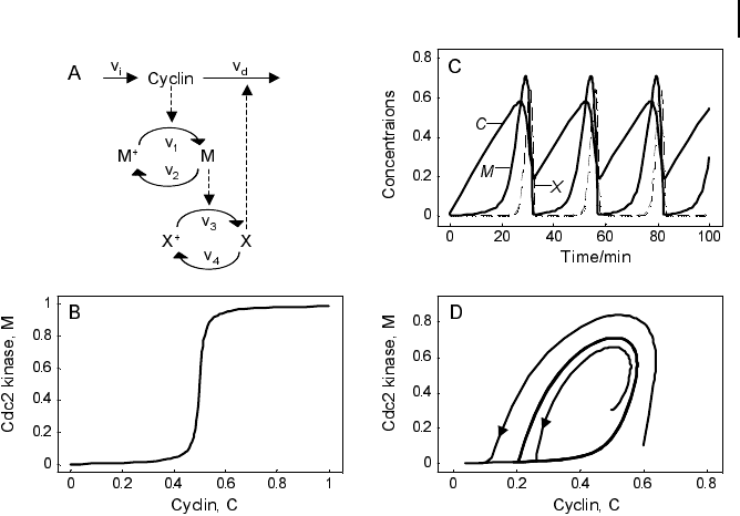
This model involves only Michaelis-Menten type kinetics, but no form of positive
cooperativity. It can be used to test whether oscillations can arise solely as a result of
the negative feedback provided by the Cdc2-induced cyclin degradation and of the
threshold and time delay involved in the cascade. The time delay is implemented by
considering post-translational modifications (phosphorylation/dephosphorylation
cycles v
1
/v
2
and v
3
/v
4
). For certain parameters they lead to a threshold in the depen-
dence of steady-state values for M on C and for X on M (Fig. 7.5b). Provided that this
threshold exists, the evolution of the bicyclic cascade proceeds in a periodic manner
(Fig. 7.5c). Starting from low initial cyclin concentration, this value accumulates at a
constant rate, while M and X stay low. As soon as C crosses the activation threshold,
M rises. If M crosses the threshold, X starts to increase sharply. X in turn accelerates
cyclin degradation and consequently, C, M, and X drop rapidly. The resulting oscilla-
tions are of the limit cycle type. The respective limit cycle is shown in phase plane re-
presentation in Fig. 7.5d.
237
7.2 Cell Cycle
Fig. 7.5 Goldbeter’s minimal model of the mi-
totic oscillator. (a) Illustration of the model com-
prising cyclin production and degradation, phos-
phorylation and dephosphorylation of Cdc2
kinase, and phosphorylation and dephosphoryla-
tion of the cyclin protease (see text). (b)Thresh-
old-type dependence of the fractional concentra-
tion of active Cdc2 kinase on the cyclin concen-
tration. (c) Time courses of cyclin (C), active
Cdc2 kinase (M), and active cyclin protease (X)
exhibiting oscillations according to the equation
system in Eq. (7-15). (d) Limit cycle behavior, re-
presented for the variables C and M. Parameter
values: K
mi
= 0.05 (i = 1, …, 4), K
mc
= 0.5,
k
d
= 0.01, v
i
= 0.025, v
d
= 0.25,V
m1
=3,V
m2
= 1.5,
V
m3
=1,V
m4
= 0.5. Initial conditions in (b):
C(0) = M(0) = X (0) = 0.01, and in (c):
X(0) = 0.01. Units: µM and min
–1
.
7.2.3
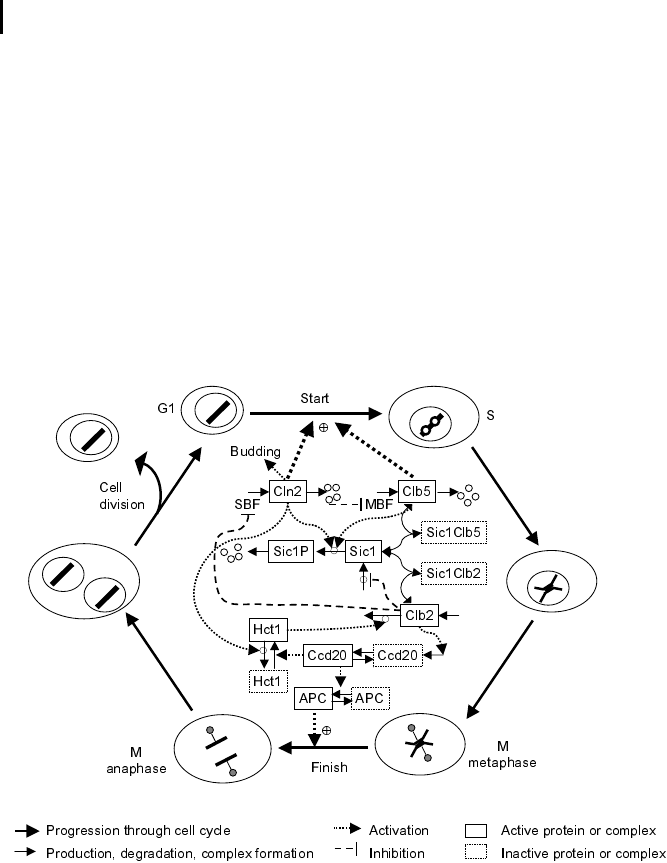
Models of Budding Yeast Cell Cycle
Tyson, Novak, and colleagues have developed a series of models describing the cell
cycle of budding yeast in detail (Tyson et al. 1996; Novak et al. 1999; Chen et al.
2000, 2004). These comprehensive models employ a set of assumptions that are
summarized in the following.
The cell cycle is an alternating sequence of the transition from the G
1
phase to the
S/M phase, called “Start”, and the transition from S/M to G
1
, called “Finish”. An
overview is given in Fig. 7.6.
238
7 Selected Biological Processes
Yeast Cell Cycle
Fig. 7.6 Schematic representation of the yeast
cell cycle (inspired by Fall et al. [2002]). The outer
ring represents the cellular events. Beginning
with cell division, the G
1
phase follows. The cells
possess a single set of chromosomes (shown as
one black line). At Start, the cell goes into the S
phase and replicates the DNA (two black lines).
The sister chromatids are initially kept together
by proteins. During the M phase they are
aligned, attached to the spindle body, and segre-
gated to different parts of the cell. The cycle
closes with formation of two new daughter cells.
The inner part represents the main molecular
events driving the cell cycle, comprising (1) pro-
tein production and degradation, (2) phosphory-
lation and dephosphorylation, and (3) complex
formation and disintegration. For sake of clarity,
CDK Cdc28 is not shown. The Start is initiated
by activation of CDK by cyclins Cln2 and Clb5.
The CDK activity is responsible for progression
through the S and M phases. At Finish, the pro-
teolytic activity coordinated by APC destroys the
cyclins and thereby renders the CDK inactive.
The CDK (Cdc28) forms complexes with the cyclins Cln1 to Cln3 and Clb1 to
Clb6, and these complexes control the major cell cycle events in budding yeast cells.
The complexes Cln1-2/Cdc28 control budding, the complex Cln3/Cdc28 governs the
executing of the checkpoint Start, Clb5–6/Cdc28 ensures timely DNA replication,
Clb3-4/Cdc28 assists DNA replication and spindle formation, and Clb1-2/Cdc28 is
necessary for completion of mitosis.
The cyclin-CDK complexes are in turn regulated by synthesis and degradation
of cyclins and by the Clb-dependent kinase inhibitor (CKI) Sic1. The expression of
the gene for Cln2 is controlled by the transcription factor SBF, and the expression
of the gene for Clb5 is controlled by the transcription factor MBF. Both transcrip-
tion factors are regulated by CDKs. All cyclins are degraded by proteasomes fol-
lowing ubiquitination. APC is one of the complexes triggering ubiquitination of
cyclins.
For the implementation of these processes in a mathematical model, the following
points are important. Activation of cyclins and cyclin-dependent kinases occurs in
principle by the negative feedback loop presented in Goldbeter’s minimal model (see
Section 7.2.1). Furthermore, the cells exhibit exponential growth. For the dynamics
of the cell mass M, it holds that dM/dt = mM. At the instance of cell division, M is re-
placed by M/2. In some cases uneven division is considered. Cell growth implies
adaptation of the negative feedback model to growing cells.
The transitions Start and Finish characterize the wild-type cell cycle. At Start, the
transcription factor SBF is turned on and the levels of the cyclins Cln2 and Clb5 in-
crease. They form complexes with Cdc28. The boost in Cln2/Cdc28 has three main
consequences: it initiates bud formation, it phosphorylates the CKI Sic1 promoting
its disappearance, and it inactivates Hct1, which in conjunction with APC is respon-
sible for Clb2 degradation in the G
1
phase. Hence, DNA synthesis takes place and
the bud emerges. Subsequently, the level of Clb2 increases and the spindle starts to
form. Clb2/Cdc28 inactivates SBF and Cln2 decreases. Inactivation of MBF causes
Clb5 to decrease. Clb2/Cdc28 induces progression through mitosis. Cdc20 and Hct1,
which target proteins to APC for ubiquitination, regulate the metaphase-anaphase
transition. Cdc20 has several tasks in the anaphase. Furthermore, it activates Hct1,
promoting degradation of Clb2, and it activates the transcription factor of Sic1.
Thus, at Finish, Clb2 is destroyed and Sic1 reappears.
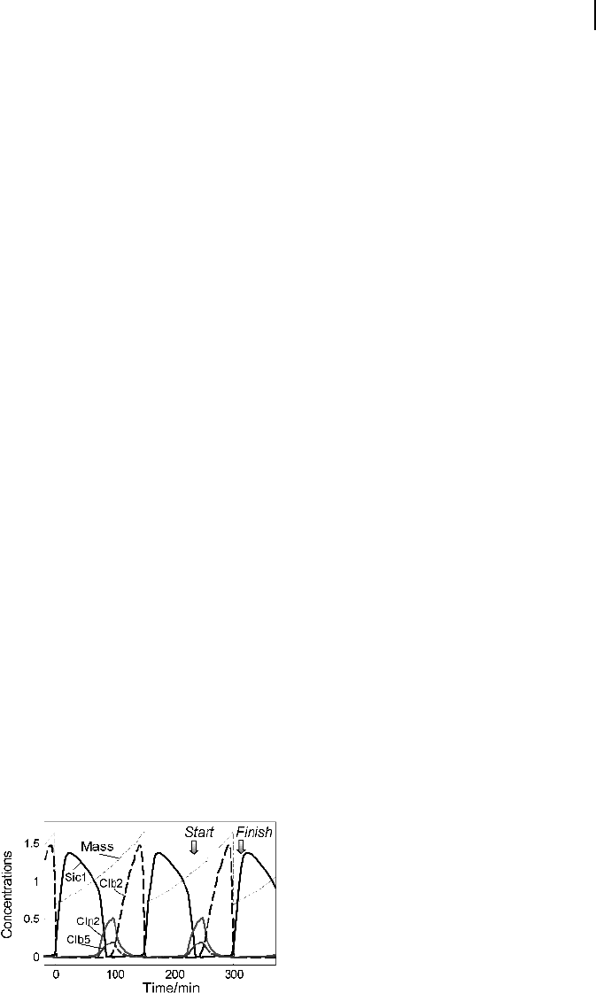
The dynamics of some key players in the cell cycle according to the model given
in Chen et al. (2000) is shown in Fig. 7.7 for two successive cycles. At Start, Cln2 and
239
7.2 Cell Cycle
Fig. 7.7 Temporal behavior of some key players
during two successive rounds of the yeast cell
cycle. The dotted line indicates the cell mass
that halves after every cell division. The levels of
Cln2, Clb2
total
, Clb5
total
, and Sic1
total
are simu-
lated according to the model presented by Chen
et al. (2000).