Lopes H.S., Cruz L.M. (eds.) Computational Biology and Applied Bioinformatics
Подождите немного. Документ загружается.

21
Analysis of Transcriptomic and Proteomic Data
in Immune-Mediated Diseases
Sergey Bruskin
1
, Alex Ishkin
2
, Yuri Nikolsky
2
,
Tatiana Nikolskaya
2
and Eleonora Piruzian
1
1
Vavilov Institute of General Genetics, Russian Academy of Sciences, Moscow
2
Thomson Reuters, Professional, Saint Joseph, MI
1
Russia
2
USA
1. Introduction
Psoriasis (a skin disease) and Crohn’s disease (a disease of the intestinal epithelium) are
multifactorial diseases caused by abnormalities in genetic machinery regulation. Both
pathologies disturb the immune system, and the pathological processes are triggered by
environmental factors. In the case of psoriasis, these factors are psychoemotional stresses,
infections (group A streptococci and Staphylococcus aureus), drugs (lithium-containing,
antimalarial, and antituberculous agents and Novocain), smoking, and mechanical damages
(the so-called Koebner phenomenon) [Bowcock A et al., 2004]. Psoriasis vulgaris is one of
the most prevalent chronic inflammatory skin diseases affecting approximately 2% of
individuals in Western societies, and found worldwide in all populations. Psoriasis is a
complex disease affecting cellular, gene and protein levels and presented as skin lesions. The
skin lesions are characterized by abnormal keratinocyte differentiation, hyperproliferation
of keratinocytes, and infiltration of inflammatory cells [Boehncke WH et al. 1996; Ortonne
JP, 1996]. The factors triggering Crohn’s disease include psychoemotional stresses, infections
(Mycobacterium avium ssp. paratuberculosis and invasive Escherichia coli variants), drugs
(antibiotics and nonsteroid antiimflammatory agents), smoking, and nutritional regimen
[Sartor R., 2006]. Crohn’s disease known only since the 1920s [Crohn B et al., 1932] and now
affecting up to 0.15% of the northwest European and North American population [Binder
V., 2005].
Both psoriasis and Crohn’s disease are now regarded as incurable, and the goal of their
therapy is to extend the remission periods and decrease the severity of the disease. These
two diseases are tightly related at the genetic level, as over five genetic loci are involved in
the development of both psoriasis and Crohn’s disease.
The mechanisms of both psoriasis and Crohn’s disease are complex and involve genetic and
environmental factors. As we gain more knowledge about molecular pathways implicated
in diseases, novel therapies emerge (such as etanercept and infliximab that target TNF-α or
CD11a- mediated pathways [Pastore S et al., 2008; Gisondi P et al., 2007]).
We have studied earlier the components of AP-1 transcription factor as psoriasis candidate
genes. This study was performed by bioinformatics analysis of the transcription data using
the GEO DataSets database (http://www.ncbi.nlm.nih.gov/geo/) [Piruzian ES et al., 2007].
Computational Biology and Applied Bioinformatics
398
The same approach was used by other researchers to detect potential therapeutic targets for
psoriasis [Yao Y., et al., 2008]. In next step, we performed a comparative analysis of the
molecular processes involved in the pathogenesis of two diseases, psoriasis and Crohn’s
disease [Piruzian ES et al., 2009].
Despite the fact that psoriasis and Crohn`s disease affect completely different body systems
(skin and intestine), they are much more similar that it may seem at first glance. Both skin
and intestinal epithelium are barrier organs, that are the first to resist the environmental
factors, including microorganisms. Both pathologies are immune-mediated inflammatory
diseases, that is also marked by the same drug therapies. Finally, they have a lot of common
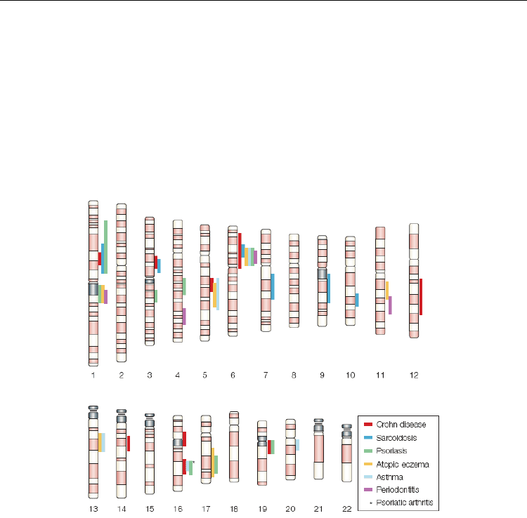
susceptibility loci (Fig. 1).
Fig. 1. Localization of various linkage regions for barrier diseases on human chromosomes
map [Schreiber S et al., 2005])
In recent years, microarray mRNA expression profiling [Oestreicher JL et al., 2001; Bowcock
AM et al., 2001; Zhou X et al., 2003; Quekenborn-Trinquet V et al., 2005] of lesional psoriatic
skin revealed over 1,300 differentially expressed genes. Enrichment analysis (EA) showed
that these genes encode proteins involved in regeneration, hyperkeratosis, metabolic
function, immune response, and inflammation and revealed a number of modulating
signaling pathways. These efforts may help to develop new-generation drugs. However,
enrichment analysis limits our understanding of altered molecular interactions in psoriasis
as it provides a relative ranking based on ontology terms resulting in the representation of
fragmented and disconnected perturbed pathways. Furthermore, analysis of gene
expression alone is not sufficient for understanding the whole variety of pathological
changes at different levels of cellular organization. Indeed, new methodologies have been
applied to the analysis of OMICs data in complex diseases that include algorithm-based

Analysis of Transcriptomic and Proteomic Data in Immune-Mediated Diseases
399
biological network analysis [Nikolskaya T, et al., 2009; Nikolsky Y et al., 2005; Bhavnani SK
et al., 2009; Ideker T et al., 2008; Chuang HY et al., 2007] and meta-analysis of multiple
datasets of different types [Cox B et al., 2005; Wise LH et al., 1999; Ghosh D et al., 2003;
Warnat P et al., 2005; Hack CJ, 2004; Menezes R et al., 2009]. Here, we applied several
techniques of network and meta-analysis to reveal the similarities and differences between
transcriptomics- and proteomics-level perturbations in psoriasis lesions. We particularly
focused on revealing novel regulatory pathways playing a role in psoriasis development
and progression.
2. Transcriptomic and proteomic data, network analysis
Data preparation. The data deposited with the public database of microarray experiments,
GEO (http://www.ncbi.nlm.nih.gov/geo/), were analyzed. The expression data on
psoriasis were contained in entry GDS1391, and on Crohn’s disease, in entry GDS1330. Since
these data were obtained using different microarrays and experimental schemes, analysis
was individually performed for each disease with subsequent comparison of the lists of
genes with altered expression for each case.
Two sets were selected from the overall data on psoriasis, namely, four experiments with
gene expression in psoriatic skin lesions, and four, with gene expression in the healthy skin
of the same patients. The selected data for Crohn’s disease were also represented by two
sets: 10 experiments on expression in intestinal epithelial lesions, and 11, on expression in
the intestinal tissue of healthy individuals. The data were prepared for analysis using the
GeneSpring GX (http://www.chem.agilent.com/scripts/pds.asp?lpage=27881) software
package. This processing comprised discarding of the genes with poorly detectable
expression and normalization of the remaining data. In addition to the values of expression,
the so-called absent call flags were added for psoriasis cases; these flags characterize the
significance of the difference in expression of a particular gene from the background noise.
The genes displaying the flag value of A (i.e., absent, which means that the expression of a
particular gene in experiment is undetectable) in over 50% of experiments were discarded
from further analysis. This information was unavailable for Crohn’s disease; therefore, this
step was omitted. The results were normalized by the median gene expression in the
corresponding experiment to make them comparable with one another.
Detection of the genes with altered expression. Differentially expressed genes were sought
using Welch’s t-test [Welch B.L., 1947]. This test does not require that the distribution
variances for the compared samples be equal; therefore, it is more convenient for analyzing
expression data than a simple t-test. FDR algorithm [Benjamini Y et al., 1995] with a
significance threshold of 0.1 was used to control the type I errors in finding differentially
expressed genes; in this case, the threshold determined the expected rate of false positive
predictions in the final set of genes after statistical control.
Detection of common biological processes. The resulting gene lists were compared, and the
molecular processes mediated by the genes displaying altered expressions in both diseases
were sought using the MetaCore (GeneGo Inc., www.genego.com) program. The
significance of the biological processes where the genes displaying altered expressions in
both diseases was assessed according to the degree to which overlapping between the list of
differentially expressed genes and the list of genes ascribed to the process exceeded random
overlapping. Hypergeometric distribution [Draghici S et al., 2007] was used as a model of

Computational Biology and Applied Bioinformatics
400
random overlapping between the gene lists. The measure of signifi- cance for the input gene
list, the p value, in this distribution is calculated as
() ()
()
(
)
()()
()()( )
()
()
min ,
max ,
min ,
max ,
,, , ,, ,
!! ! !
!
1
!!! !'
nR
irRnN
nR
irRnN
pVal r n R N P i n R N
Rn N R N n
N
iR i n i N R n i
=+−
=+−
=
−−
=
×
−− −−+
∑
∑
where N is the number of genes in the MetaCore database; R, the number of genes ascribed
to a particular process; n, the size of the input gene list; and r, the number of genes from the
input list related to this process.
Three ontologies of biological processes were used in this work: GO (www.gene-
ontology.org) and two ontologies included in the MetaCore, Canonical pathways and
GeneGo process networks. The processes contained in the MetaCore ontologies are gene
networks, which reflect the interaction of proteins involved in a particular biological
regulatory or metabolic pathway. The processes for all three ontologies were prioritized by
the negative logarithm of p value.
The common molecular biological pathways were determined based on the analysis of
significant biological processes and expressions of the genes involved in these processes.
The MetaCore contains the algorithms providing for detection in the total network of gene
interactions the particular regulatory pathways and subnetworks saturated with the objects
of research interest, in this case, the genes with altered expression. The resulting
assumptions on the pattern of common biological pathways were visualized as a gene
network using the MetaCore.
Skin biopsies. Acquisition of the human tissue was approved by the Vavilov Institute of
General Genetics of Russian Academy of Sciences review board and the study was
conducted after patient's consent and according to the Declaration of Helsinki Principles. A
total of 6 paired nonlesional and lesional (all were plaque-type) skin biopsies from 3
psoriatic patients were profiled using 2D electrophoresis. All the donors who gave biopsy
tissue (both healthy controls and individuals with psoriasis) provided a written informed
consent for the tissue to be taken and used in this study. Clinical data for all patients are
listed in Table 3. Full-thickness punch biopsies were taken from uninvolved skin (at least 2
cm distant from any psoriatic lesion; 6 mm diameter) and from the involved margin of a
psoriatic plaque (6 mm diameter) from every patient.
Sample preparation, two-dimensional electrophoresis, gel image analysis and
massspectrometry was carried out using the standard procedure [Gravel P & Golaz O, 1996;
Mortz E, et al., 2001].
Microarray data analysis. We used recently published data set [Yao Y, et al., 2008] from
GEO data base (http://www.ncbi.nlm.nih.gov/geo/; accession number GSE14095). We
compared 28 pairs of samples (in each pair there was a sample of lesional skin and a sample
of healthy skin from the same patient). Values for each sample were normalized by sample
median value in order to unify distributions of expression signals. For assessment of
differential expression we used paired Welch ttest with FDR correction [Benjamini Y et al.,

Analysis of Transcriptomic and Proteomic Data in Immune-Mediated Diseases
401
1995]. Probe set was considered as differentially expressed if its average fold change
exceeded 2.5 and FDR corrected p-value was less than 0.01.
Overconnection analysis. All network-based analyses were conducted with MetaCore
software suite http://www.genego.com. This software employs a dense and manually
curated database of interactions between biological objects and variety of tools for
functional analysis of high-throughput data. We defined a gene as overconnected with the
gene set of interest if the corresponding node had more direct interactions with the nodes of
interest than it would be expected by chance. Significance of overconnection was estimated
using hypergeometric distribution with parameters r - number of interactions between
examined node and the list of interest; R - degree of examined node, n - sum of interactions
involving genes of interest and N - total number of interactions in the database:
() ()
()
()
min ,
max ,
,, , ,, ,
nR
irRnN
p
Val r n R N P i n R N
=+−
=
∑
Hidden nodes analysis. In addition to direct interacting objects, we also used objects that may
not interact directly with objects of interest but are important upstream regulators of those
[Dezso Z et al., 2009]. The approach is generally the same as described above, but the shortest
paths instead of direct links are taken into account. As we were interested in transcriptional
regulation, we defined a transcriptional activation shortest path as the preferred shortest path
from any object in the MetaCore database to the transcription factor target object from the data
set. We added an additional condition to include the uneven number of inhibiting interactions
in the path (that's required for the path to have activating effect). If the number of such paths
containing examined gene and leading to one of objects of interest were higher than expected
by chance, this gene was considered as significant hidden regulator. The significance of a
node's importance was estimated using hypergeometric distribution with parameters r -
number of shortest paths between containing currently examined gene; R - total number of
shortest paths leading to a gene of interest through transcriptional factor, n - total number of
transcription activation shortest paths containing examined gene and N - total number of
transcription activation shortest paths in the database.
Rank aggregation. Both topology significance approaches produced lists of genes
significantly linked to a gene or protein set of interest, ranked by corresponding p-values.
To combine results of these two approaches, we used a weighted rank aggregation method
described in [Pihur V et al., 2009]. Weighted Spearman distance was used as distance
measure and the genetic algorithm was employed to select the optimal aggregated list of
size 20. This part of work was accomplished in R 2.8.1 http://www.r-project.org.
Network analysis. In addition to topology analysis, we examined overexpressed genes and
proteins using various algorithms for selecting connected biologically meaningful
subnetworks enriched with objects of interest. Significance of enrichment is estimated using
hypergeometric distribution. We first used an algorithm intended to find regulatory pathways
that are presumably activated under pathological conditions. It defines a set of transcription
factors that are directly regulating genes of interest and a set of receptors whose ligands are in
the list of interest and then constructs series of networks; one for each receptor. Each network
contains all shortest paths from a receptor to the selected transcriptional factors and their
targets. This approach allows us to reveal the most important areas of regulatory machinery
affected under the investigated pathological condition. Networks are sorted by enrichment p-

Computational Biology and Applied Bioinformatics
402
value. The second applied algorithm used was aimed to define the most influential
transcription factors. It considers a transcriptional factor from the data base and gradually
expands the subnetwork around it until it reaches a predefined threshold size (we used
networks of 50 nodes). Networks are sorted by enrichment p-value.
3. A comparative analysis of the molecular genetic processes in the
pathogenesis of psoriasis and Crohn’s disease
Constructing List of the Genes with Altered Expression in Both Pathologies We detected the
lists of differentially expressed genes separately in each dataset and compared these lists at
the system level. This approach to analysis was dictated by the properties of expression data
in general (a high noise level and a large volume of analyzed data) and individual
properties of datasets selected for analysis, which were obtained using different microarrays
with different numbers of probes. That is why the datasets were incomparable in a direct
fashion. The dataset on psoriasis initially contained information on the expression levels of
12626 probes from eight experiments (four specimens of skin lesions, and four of the healthy
skin from the same patients). After discarding the probes with poorly detectable expression
(see Materials and Methods), the set was reduced to 5076 probes. The list of the probes with
statistically significant differences in expression between the lesion and healthy tissue
contained 410 items at a significance level of 0.1.
The dataset on Crohn’s disease contained information on the expression level of 24016
probes from 21 experiments (11 specimens of epithelial lesions and 10 specimens of healthy
epithelium). The list of probes displaying statistically significant differences in expression
between the lesion and healthy tissue contained 3850 probes at a significance level of 0.1.
This pronounced difference in the sizes of gene lists result from the fact that the algorithm
used for controlling type I errors (FDR) depends on the input set. The larger the initial gene
list, the larger number of genes will pass the FDR control at a similar p-value distribution; in
our case, the number of analyzed probes in the dataset for Crohn’s disease is five times
larger than that in the dataset for psoriasis.
The lists of differentially expressed genes were input into the MetaCore program. Because
microarrays contained not only gene probes, but also a large number of ESTs with
unidentified functions, the size of gene lists at this stage changed because not all the probes
had the corresponding gene in the MetaCore database and because some probes
corresponded to more than one gene. The lists of recognized genes comprised 425 and 2033
items for psoriasis and Crohn’s disease, respectively.
The common part for the compared lists comprised 49 genes, which is a significant
overlapping (p value = 4.94 × 10 –2 ). The significance was estimated using Fisher’s test. The
complete set contained 9017 genes present in both studied datasets (this set was identified
by comparing the complete lists of genes for both microarrays in MetaCore). The lists of
genes with altered expression were reduced to the subset of genes present in both datasets.
Thus, these particular 49 genes were selected for further analysis (Table 1).
It was of interest to determine the molecular processes with which the genes common to
psoriasis and Crohn’s disease are associated. Table 2 consolidates the most probable cell
processes with involvement of the genes listed in Table 1, as determined by the MetaCore
software tools. These processes (Table 2) fall into two main groups—related to inflammation
and cell cycle. Indeed, the pathological lesions in both psoriasis and Crohn’s disease
are inflammatory foci. The cell cycle is also considerably affected in both pathologies.

Analysis of Transcriptomic and Proteomic Data in Immune-Mediated Diseases
403
An increased proliferation of keratinocytes is observed in the psoriatic skin, an
inflammatory focus.
GNA15 SFPQ IFI35 IER2 OAS2 RFK UBE2L6
CBX3 CG018 CSNK1D SYNCRIP PSME2 CTSC CASP4
GPM6B UGT1A4 STAT3 S100A8 FOXC1 SOSTDC1 ETS2
UGT1A6 VKORC1 TRIM22 RARG TRAK2 SERPINB5 MECP2
IFI44 H2AFY TXNDC1 ARMET ZNF207 KIAA1033 QPCT
DEGS1 MIB1 IRF9 DDOST DNAJC7 RBPMS JUNB
LONRF1 HMGN1 MRPL9 FGFR2 CDC42EP1 S100A9 PHGDH
Table 1. Genes displaying altered expression in both psoriasis and Crohn's disease.
Process
p value
Inflammation: interferon signaling pathways 2.19E-03
Signal transduction: Wnt signaling pathways 1.20E-02
Regulation of translation initiation 5.66E-02
Morphogenesis of blood vessels 9.76E-02
DNA repair 1.17E-01
Inflammation: amphoterin signaling pathways 1.19E-01
Proteolysis determined by the cell cycle and apoptosis 1.29E-01
Interleukin regulation of the cell cycle in G1-S phase 1.29E-01
Signal transduction: androgen receptor signaling pathways 1.34E-01
Table 2. Cell processes common to psoriasis and Crohn's disease.
For a more detailed description of the inflammatory response and cell cycle in the parts of
them most tightly related to the genes listed in Table 1, we constructed gene networks,
which are fragments of the larger gene networks describing the inflammatory response (Fig.
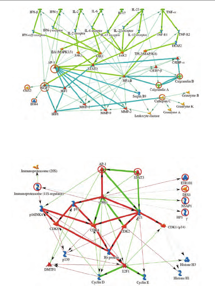
2) and cell cycle control (Fig. 3). Figure 2 shows that the inflammatory response is initiated
by such well-known cytokines as TNF-α, IFN-γ, IL-2, IL-6, IL-17, and IL-23. Then protein
kinases activate the transcription factors AP-1, STAT3, C/EBP, NF-κB, ISGF3, and others.
Figure 3 shows that the key cell cycle regulators that changed gene expression are the
transcription factors AP-1, c-Myc, and STAT3. It is also evident that the genes encoding AP-
1 transcription factor components are involved in both the inflammatory response and cell
cycle control. It is known that the genes depending on AP-1 play an important role in
regulation of proliferation, morphogenesis, apoptosis, and cell differentiation. Induction of
cell differentiation activates transcription of the genes encoding the components of AP-1
complex [Turpaev K.T., 2006]. We assume that the genes of AP-1 transcription factor are the
candidate genes involved in the pathogenesis of both psoriasis and Crohn’s disease;
moreover, this hypothesis is particularly based on the bioinformatics analysis of microarray
data. Therefore, it was interesting to compare our data with the available information about
the chromosome localization of the loci associated with psoriasis and Crohn’s disease.
Computational Biology and Applied Bioinformatics
404
Fig. 2. Detail of the gene network describing inflammatory response. Green arrows indicate
activation of the corresponding network elements, from the level of cytokines to
transcription factors; light blue arrows, the activation of effector genes by transcription
factors; and red circles, genes from the list.
Fig. 3. Detail of the gene network describing cell cycle control. Green arrows indicate
activation of the corresponding elements; red arrows, inhibition; and red circles, genes from
the list.
Analysis of Transcriptomic and Proteomic Data in Immune-Mediated Diseases
405
4. Integrated network analysis of transcriptomic and proteomic data in
psoriasis
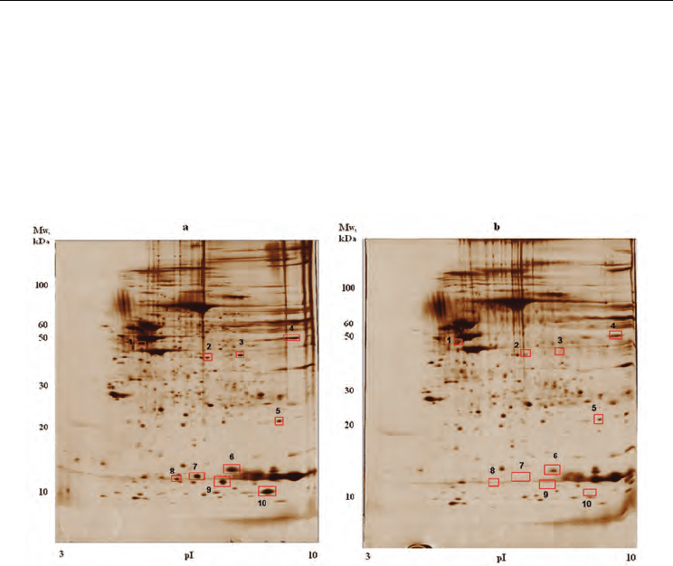
Differentially abundant proteins. Protein abundance was determined by densitometric
quantification of the protein spots on 2D-electophoresis gel (Figure 4) followed by MALDI-
TOF mass spectrometry. Total of 10 proteins were over-abundant at least 2-fold in lesional
skin compared with uninvolved skin: Keratin 14, Keratin 16, Keratin 17, Squamous cell
carcinoma antigen, Squamous cell carcinoma antigen-2, Enolase 1, Superoxide dismutase
[Mn], Galectin-7, S100 calcium-binding protein A9 and S100 calcium-binding protein A7.
Fig. 4. Representative silver-stained 2DE gel images of lesional and uninvolved skin biopsy
lysates. a) - gel image of lesional skin biopsy lysate; b) - gel image of uninvolved skin biopsy
lysate. Spots corresponding to proteins overexpressed in lesions are marked with red
rectangles and numbered. Spot 1 correspond to 3 proteins of keratin family, spot 2 - SCCA2,
spot 3 - SCCA1, spot 4 - enolase 1, spot 5 - SOD2, spot 6 - galectin-7. S100A7 is found in
spots 7 and 8 and S100A9 corresponds to 9 th and 10 th spots.
Several of these proteins were previously reported to be over-abundant in psoriatic plaques
[Leigh IM et al., 1995; Madsen P et al., 1991; Vorum H et al., 1996; Takeda A et al.., 2002]. The
proteins belonged to a diverse set of pathways and processes.
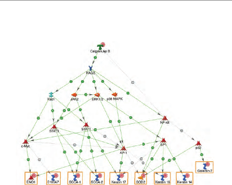
We attempted to connect the proteins into a network using a collection of over 300,000
manually curated protein interactions and several variants of "shortest path" algorithms
applied in MetaCore suite [Nikolsky Y et al, 2009] (Figure 5). The genes encoding
overabundant proteins were found to be regulated by several common transcription factors
(TFs) including members of the NFkB and AP-1 complexes, STAT1, STAT3, c-Myc and SP1.
Moreover, the upstream pathways activating these TFs were initiated by the overabundant
S100A9 through its receptor RAGE [Ghavami S et al., 2008] and signal transduction kinases
(JAK2, ERK, p38 MAPK). This network also included a positive feedback loop as S100A9
expression was determined to be controlled by NF-kB [Schreiber J et al., 2006]. The topology
of this proteomics-derived network was confirmed by several transcriptomics studies
Computational Biology and Applied Bioinformatics
406
[Tsuruta D, 2009; Sano S et al., 2008; Ghoreschi K et al., 2003; Piruzian ES et al., 2009;
Gandarillas A & Watt FM, 1997; Arnold I & Watt FM, 2001] which showed overexpression
of these TFs in psoriasis lesions. Transiently expressed TFs normally have low protein level
and, therefore, usually fail to be detected by proteomics methods.
Fig. 5. Network illustrating regulatory pathways leading to transcription activation of
proteomics markers. Red circles denote upregulated proteins.
RAGE receptor is clearly the key regulator on this network and plays the major role in
orchestrating observed changes of protein abundance. This protein is abundant in both
keratinocytes and leukocytes, though normally its expression is low [Lohwasser C et al.,
2006]. RAGE participates in a range of processes in these cell types, including inflammation.
It is being investigated as a drug target for treatment of various inflammatory disorders
[Santilli F et al., 2009]. Thus, we may propose that RAGE can also play significant role in
psoriasis.
We used Affymetrix gene expression data set from the recent study [Yao Y et al., 2008]
involving 33 psoriasis patients. Originally, more than 1300 probe sets were found to be
upregulated in lesions as compared with unlesional skin of the same people. We identified
451 genes overexpressed in lesional skin under more stringent statistical criteria (28 samples
of lesional skin were matched with their nonlesional counterparts from the same patients in
order to exclude individual expression variations, genes with fold change >2.5 and FDR-
adjusted p-value < 0.01 were considered as upregulated). The genes encoding 7 out of 10
proteomic markers were overexpressed, well consistent with proteomics data. Expression of
Enolase 1, Keratin 14 and Galectin 7 was not altered.
Despite good consistency between the proteomics and expression datasets, the two orders of
magnitude difference in list size make direct correlation analysis difficult. Therefore, we