Yao N. Focused Ion Beam Systems: Basics and Applications
Подождите немного. Документ загружается.

in SEMs have also been used for positioning multi-wall carbon nanotubes
onto cantilever tips [ 54] and pre-patterned electrodes [55].
From the mid 1990s until recently the main technique used for the pre-
paration of TEM lamellae was the ex-situ lift-out technique, and many
groups do not possess an in-situ micromanipulator as such. Unfortunately, as
these micromanipulators are relatively expensive it is difficult for many
groups, especially in academia, to find sufficient funding to purchase one.
However, De Veirman [56] has shown that it is possible to attach a metal
needle to a modified gas injection (GIS) needle, which is a cost effective
solution if the system has a spare GIS.
The first step in this technique involves positioning the needle’s tip at the
stage eucentric position, which for an FIB/SEM system is also the beam
coincident point. Once this is done the needle is not moved in the x or y
directions. Instead the stage is used to move and position the sample and
TEM grid relative to the needle tip. Having positioned the needle it is then
retracted and a slice of material approximately 500–1000 nm wide is prepared
using the same steps as in the ex-situ lift-out technique. The sample is then
tilted by 45
and the base and side edges of the slice are cut through (one
completely and the other only 80% along its length) (Figure 8.5(b)). Next the
sample is lowered by 100 mm (to prevent accidentally crashing the needle tip
into the sample) and the manipulator needle and gas insertion needle for the
Pt ‘‘welding’’ are inserted. The sample is then raised, and if using an FIB/SEM
system the cut end of the slice is positioned 100–200 nm beneath the tip of the
needle. Owing to the two imaging directions this is relatively straightforward
to do in an FIB/SEM system. Electron beam imaging may be used to position
the edge of the slice beneath the tip of the needle while ion beam imaging can
be used for the z-positioning. However, if using a single beam system and the
sample is perpendicular to the ion imaging direction it is difficult, due to the
large depth of field, to position the slice’s edge a few hundred nanometers
beneath the needle. If the needle and slice are too far apart (>300 nm) the
deposited Pt will not be able to bridge/‘‘weld’’ them together. Therefore, in a
single beam system it is better to push the slice against the tip of the needle.
Contact between the two is indicated by a change in the image contrast,
movement of the needle tip, or the touch alarm being triggered.
The tip of the needle is then ‘‘welded’’ to the slice using in-situ Pt
deposition (Figure 8.5(c)). The size of the Pt square deposited is 1 mm
2
and
the deposition time is about 30 seconds when using a beam current of 50 pA.
Care has to be taken not to use too large a beam current when depositing the
Pt (i.e., >100 pA) in order not to locally deplete the Pt precursor gas. In
this technique the two most critical steps are that the base of the slice is
Focused ion beam systems228
completely cut through and the needle is Pt welded to the slice’s top edge. To
ensure that the base is cut through the points highlighted in the ex-situ lift-
out technique should be followed. To make sure that the needle is being
‘‘welded’’/attached to the slice during the Pt deposition the grounding current
can be monitored. As the two become ‘‘welded’’ (i.e., electrically connected)
there will be a sharp change in the magnitude of the grounding current. After
the Pt has been deposited another method to check that the two are ‘‘welded’’
is to gently tap the plinth of the system. If the two are fixed together the
needle tip will not be able to vibrate.
On ce t he n ee dle a nd s l ic e are we lde d to ge the r the ma ter ial h ol din g th e
slicetothesampleiscut. Theplinthisthenagaingentlytappedtocheckif
the s lice is complet ely free from the s ample; i f it is t he needl e and slice
should vibrat e together. If the sl ice is free the stage is then lowered and
re pla ce d b y a T EM slo t g rid (whi ch ha s b ee n c ut in ha lf) an d t he sli ce is Pt
welded to the top edge of the s lot gri d (Figure 8.5 (d)) in essentiall y the
reverse process of the lift-out pr oc edure . Once the sli ce is fixed to the gri d
th e n ee dl e i s c ut fro m it an d ret rac te d. Th e fin al ste p i nv olv es milli ng th e
sli ce to e lectron trans parency u sing the tilt ing and li ne m ill ing procedur e
whil e b eing careful not to mill away the Pt fixing ‘‘wel d.’ ’ In addition to slot
grids, TEM gri ds with tabs and V -shaped g rooves (Figure 8.5 (e) ) can also
be use d. A n a dv an tag e o f usi ng a gri d wi th t abs is th at a s the ed ges o f th e
sli ce a re str aight ( as a gains t the b ase w hich has b een cut at 45
)itiseasierto
Pt weld thi s to the si de of a tab a nd there is n o m aterial b eneath the sli ce t o
be sputtered and rede posite d onto the lamella as it is being thinne d. Figure
8. 5(f) s hows SE images of thinne d s lices Pt welded to a V-shaped groove
and tab (insert).
8.2.4 Comparison of the different techniques
The choice of technique to use is dictated by numerous factors such as the
secondary equipment available, the type of information required, the geo-
metry of the sample, the amount of material, how unique a defect or sample
is, the turn around time, and also to some extent the familiarity of a user with
a given technique. Probably, the most decisive factors affecting the choice
are the yield, how unique a defect is, and the turn around time. In the
semiconductor industry the down time of a production line is very costly,
while within academia the large numbers of users on a system may limit the
available access time. In the following section the relative advantages of the
different techniques are discussed and these should ideally form the basis for
selecting which technique to use.
Preparation for physico-chemical analysis 229
The success yields of the H-bar and in-situ slice techniques are similar, with
both approaching 100%. Unfortunately, for reasons discussed in the pre-
vious sections the yield of the ex-situ lift-out technique can be as low as 60%
depending on the experience of the user. Thus if a defect or sample is unique
the H-bar or in-situ slice techniques should be used.
Although the in-situ slice technique is currently in vogue because of its high
success yield, its main drawback is that the FIB or FIB/SEM system is used for
the lift-out step, which typically takes 30–60 minutes per sample. Also, after
the lift-out step the slice still has to be thinned to electron transparency. Thus
to prepare three TEM lamellae with this technique typically takes 3–6 hours in
addition to the time spent (typically overnight) milling the slices. In contrast,
the FIB milling in the H-bar and ex-situ lift-out techniques can be automated
to produce up to ten specimens overnight that are approximately 150–200 nm
thick and as such require only 5–10 minutes per sample to thin them to 100 nm.
If there are many users on a system making lamellae, another important
consideration is how many sessions are required before a user becomes pro-
ficient at a technique. Typically it takes three or four 4-hour sessions to
become competent at the H-bar technique and seven or eight 4-hour sessions
to become experienced with the other two techniques.
In the ex-situ lift-out and in-situ slice techniques, the pre-FIB sample
preparation time is minimal (the samples can generally be placed directly into
the system), whilst in the H-bar technique the preparation of the slice can
take 2–3 hours if it is being prepared by hand polishing. In contrast, after
FIB milling the H-bar’s samples can be placed immediately into the TEM
whilst the lifting out of the lamella and placing it onto a TEM support
membrane in the ex-situ technique can take between 10 and 30 minutes each.
The primary drawback of the H-bar samples is that their geometry limits
the angles of tilt or rotation during TEM analysis; the base or sidewalls of the
trenches may block the path of the electron beam. For example, if a trench is
50 mm long and 10 mm deep, the maximum angle of tilt, if the electron beam is
to pass through the center of the lamella, is only 6
. For ex-situ lift-out
lamellae and slice samples the maximum angle of tilt is determined by the
TEM holder. The trenches in the H-bar samples may also affect energy
dispersive X-ray (EDS) analysis [57,58]. Electrons scattered from the lamella
can irradiate the sidewalls and generate additional X-rays. The intensity of
these additional X-ray signals can be reduced by 90% by preparing the
lamella near the side edge of the slice [59], i.e., U-shaped samples.
Another advantage of the ex-situ lift-out and in-situ slice techniques over
the H-bar technique is that the surrounding sample remains intact, which
enables numerous lamellae to be prepared in close proximity to one another.
Focused ion beam systems230
Another advantage of the in-situ slice and H-bar samples is that they can be
returned to the system for further thinning if they have been prepared too thick.
In a single-beam system this has also been used to prepare site-specific TEM
lamellae of subsurface features in DRAM samples [ 60 ] and to cross section
through the center of 1 mm diameter stratified Fe particles [61 ]embeddedina
resin. For the subsurface features in the DRAM samples, the TEM lamellae
were prepared 500 nm thick and the position of the defect within the lamellae
was determined using HV SEM and energy filtered TEM, while for the Fe
particles, the lamellae were initially prepared several micrometers thick and the
diameter of the particles was determined using STEM. If an FIB/SEM system is
being used then electron beam imaging can be used to end the FIB milling.
As will be discussed in Section 8.6.3, using high-energy FIB beams (30 keV)
can result in damage being generated at the sidewalls of a lamella. To reduce
the amount of damage post-FIB BIB milling using a low energy (100–500 eV)
argon beam can be used. The geometries of the ex-situ and in-situ samples are
both suitable for BIB milling whereas the geometry of the H-bar samples is
such that they are not suitable. If the lamella is at a shallow angle to the BIB
direction then the base and sidewalls of the trenches will be at a large angle
(resulting in a higher sputter rate), and thus material can be sputtered from
these and onto the lamella.
Ideally, if there is access to all the required secondary equipment (i.e., high-
precision saws, micromanipulators) then the technique used should be chosen
based on the required compositional and physical information, the geometry
of the sample, and how unique the sample is. For example, for semiconductor
devices on a silicon wafer from which only critical dimension measurements
are required, if a high-precision diamond saw is available then the H-bar
technique is the most appropriate to use. However, if the samples to be
analyzed are unique and access time to an instrument is not limited, then
because of its high yield the in-situ slice technique is the best method to use. If
access time is limited and the samples are not unique then the ex-situ method
should be used as it can prepare numerous specimens overnight, and if in the
worse case scenario only 50% of the lamellae are successfully lifted out this is
still around 25% more than can be prepared in the equivalent time using the
in-situ lift-out technique.
8.3 Preparation of plan-view lamellae and SEM specimens
In addition to preparing cross section lamellae, FIBs are also used to prepare
plan-view lamellae. One of the first methods was reported by Young et al.[62]
who milled from the underside of the sample. Shortcomings with this
Preparation for physico-chemical analysis 231
approach include making site-specific lamellae and stopping at the layers of
interest.
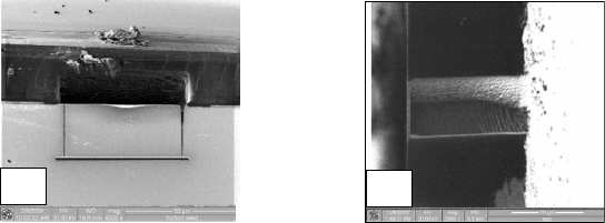
Methods based on both the trench and ex-situ lift-out techniques have been
used to prepare plan-view lamellae: the sample is cleaved or polished and
mounted end-on in the FIB system. Figures 8.6(a) and (b) show SE images of
a plan-view lamellae prepared using the ex-situ lift-out and H-bar techniques,
respectively. In the approach based on the in-situ lift-out technique, it has
been reported that the cut free lamella invariably falls into the FIB cut box
and that during the lift-out step the needle should be swung towards the
staircase cut so that the lamella is knocked into the FIB cuts [63]. These
methods have been used to prepare plan-view lamellae of tungsten plugs [ 64],
CeO
2
/Gd
2
Zr
2
O
7
multi-layers [65], silicon nanowires [66], and to prepare
numerous plan-view specimens at different heights in an yttria stabilized
zirconia layer to study the change in texture through the layers [67].
Unfortunately, it is difficult to make site-specific plan-view lamellae using
methods based on the H-bar and ex-situ lift-out techniques. The fracture
position of the cleave cannot be controlled to better than 1 mm and polishing
close to the ROI to reduce the amount of FIB milling can be time consuming.
Site-specific plan-view lamellae can be prepared using the in-situ slice tech-
nique [ 29] by mounting the TEM grid at 90
to the sample and thus the slice.
Other advantages of using the in-situ lift-out method are that the sur-
rounding material is conserved, numerous specimens can be prepared in close
proximity to one another, and the success yield can be near to 100%.
For some defects and materials it is advantageous to image them in both
plan view and cross section. Methods to do this based on both the ex-situ and
in-situ lift-out techniques have been reported by Langford et al.[68] and
Minowa et al.[69]. Langford et al. used TEM grids without any support
membrane (see Figure 8.4(c)) which enabled the lamella to be lifted off after
imaging in the TEM and to be placed onto a piece of silicon where it could
Cleaved
edge
(a)
(b)
Polished
edge
Figure 8.6 SE images of plan-view lamellae prepared using (a) the lift-out
and (b) the H-bar techniques.
Focused ion beam systems232
then be cross-sectioned. Minowa et al.[69] Pt welded the needle to the plan-
view lamella and then cut this out and placed it onto the top edge of another
slice sample where it was then cross-sectioned.
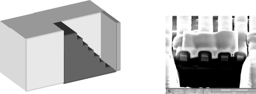
The steps outlined previously for preparing the lift-out lamella can also be
used to prepare cross sections, i.e., a Pt strap is deposited, a staircase-shaped
cut is milled, and then the line milling procedure is used to mill up to the ROI.
Figure 8.7 (a) shows a schematic of an FIB cross section and Figure 8.7(b)
shows an SE image of a cross section through a series of metal interconnects.
Cross sections have been used to investigate diffractive optical elements [ 70],
grain sizes [71], and the vertical structure of nanoporous aluminum oxide
membranes [ 72], and to prepare samples for other analytical techniques such
as electron backscatter diffraction analysis (EBSD) and Auger [12]. For
example, Prasad et al. [73] prepared cross sections through shallow wear scars
in electroformed Ni and used EBSD to study wear induced microstructural
changes.
8.4 FIB sample preparation for other analytical techniques
8.4.1 Electron holography
FIBs have also been used to prepare site-specific lamellae for electron holo-
graphy. In electron holography the region for analysis has to be adjacent to
the vacuum therefore it is not possible to deposit a Pt protective layer over
the ROI. To prevent ion milling the sample and the curtain effect occurring
(which affects the interpretation of the data), both Dunin-Borkowski et al.
[74] and Scharz et al.[75] prepared lamellae for electron beam holography by
using the silicon beneath the ROI as a planarizing protective strap. Newcomb
et al. milled from the back of the silicon wafer to create a free-standing
cantilever and milled the lamella into this, while Mcarthy et al. used the
in-situ slice technique to mount a slice upside down on the TEM grid.
(b)(a)
Figure 8.7 (a) A schema tic of an FIB cross section for SEM and (b) an SE
image of a cross section through some metal interconnects.
Preparation for physico-chemical analysis 233
8.4.2 Modification of scanning probe microscopy tips
and the fabrication of atom probe specimens
FIBs have been used to modify the shape of scanning probe microscopy
(SPM) tips for measuring structures with high aspect ratios and to deposit
metal layers for electric force probe microscopy [76– 79]. The AFM tips were
modified by milling a series of annular rings with decreasing inner diameters
down to a few hundred nanometers. The sharpening of the AFM tips to a
radius of curvature of 5–20 nm is due to the milling by the tails of the ion
beam distribution.
FIB based techniques have also been used to prepare specimens for AP
analysis [80,81 ] from planar samples. AP field-ion microscopy (APFIM) is an
atomic resolution technique, which requires a needle-shaped specimen, with an
apex radius of curvature of 10–100 nm. Prior to using FIB milling and
micromanipulation, the metal layers had to be deposited onto pre-sharpened
tips thus preventing direct correlation with macroscopic properties. AP speci-
mens have been prepared using methods based on both the ex-situ [82]andin-
situ lift-out [83] techniques. In the approach based on the ex-situ technique the
metal layers were grown onto Si wafers that had been patterned into pillars
using reactive ion etching. The base of the pillars was then mechanically broken
and the ex-situ micromanipulator was used to pick up a pillar and to mount it
onto an electro-polished needle. The pillars were then shaped and sharpened by
milling a series of annular rings, with the tails of the ion beam distribution again
being used to sharpen the tips to 10–100 nm. The trimming and shaping of the
pillar is considerably easier in an FIB/SEM system where real-time imaging can
be used. In a method based on the in-situ technique a 2 m m
2
pillar has been FIB
milled from the substrate and this has been lifted out and Pt welded to the
electro-polished needle. If numerous pillars are being prepared the repeated
milling and lifting out of the pillars from the substrate is a time consuming step.
The total time to prepare many pillars can be reduced by milling a 20–30 mm
slice, fixing its end onto the electro-polished needle, and cutting the pillar from
this slice and then repeating this step. Figure 8. 8(a) shows an SE image of an
FIB prepared slice that contains numerous pillars along its length as one of the
pillars is being Pt welded to a needle, and Figure 8. 8(b) after a pillar has been
welded and the electro-polished needle lowered.
8.4.3 Three-dimensional reconstruction
The capability to make site-specific cross sections is also exploited for 3D
reconstruction [84]. In this process, sequential slices (like cutting a loaf of
Focused ion beam systems234
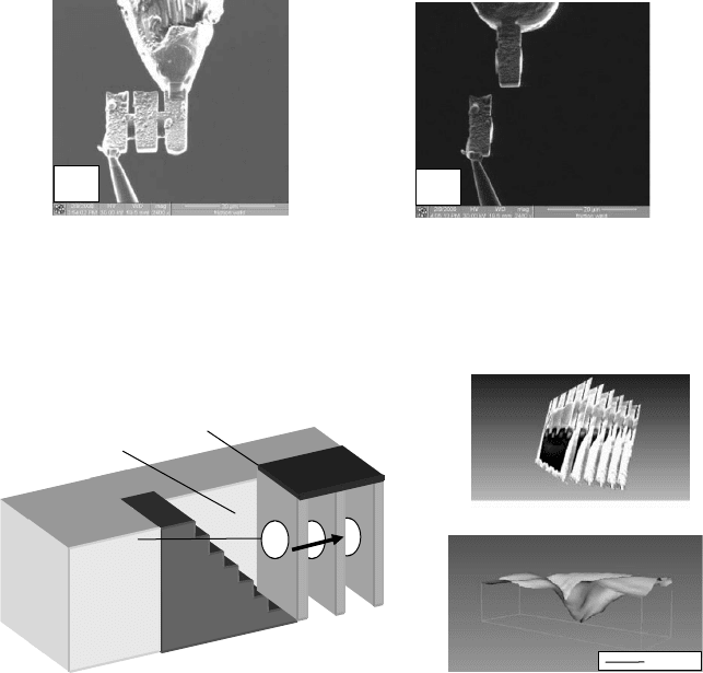
bread) are milled through a feature or ROI, and the information from each
cross section is then used for the 3D reconstruction. Figure 8.9(a) shows a
schematic of the serial sectioning and a set of slices taken through the metal
interconnects shown in Figure 8.7(b). Figure 8.9(b) shows a reconstruction of
the surface of the nanoindent into the 4 m m thick PVAC layer shown in
Figure 8.1 (a).
FIB sequential slicing and 3D reconstruction have been used to investigate
the shape of FIB milled vias in photonic structures [85], to examine Cu-Al
multi-layers which had been deformed by nanoindentation [86], and have
been combined with both Auger and SIMS for chemical analysis of bonding
wires in an integrated circuit [87]. (3D chemical analysis can also be per-
formed using image depth profiling in SIMS in which the beam is rastered
(a)
(b)
Figu re 8.8 SE imag es of (a) a slice, into which num erous pillars ha ve be en
FIB mil led, being mou nted onto an electro-pol ished ne edle, and (b) having
cut a pill ar from the slice and lowered the ne edle.
Staircase-shaped
cut
Pt protective strap
Face extend
leader of class
section
(b)(a)
5 µm
Figure 8.9 (a) Schematic of sequential FIB slicing for 3D reconstruction and
a set of sli ces taken through the sampl e shown in Figure 8.7 (b), and (b) the
reconstructed surface of a nanoindented PVAC film.
Preparation for physico-chemical analysis 235
perpendicular to the surface of the sample. However, the roughening of the
base of the crater due to preferential milling, redeposition, and different
sputter rates limits the z (depth) resolution [ 88].)
8.5 Advantages of using an FIB/SEM system for
the preparation of TEM lamellae
There are numerous advantages of using FIB/SEM systems for preparing
TEM lamellae relative to single beam FIB systems [89,90] and these include:
1. Navig ation to a ROI can be perfor med using the elect ron be am, thus pre venting
any ion da mage to the surroun ding regions of the sample.
2. The Pt pro tective strap can be deposite d using electron beam assi sted de position
thereby pr eventing ion beam da mage to the sampl e unde rneath.
3. As the tw o co lumns are inclined at a large angle relat ive to ea ch other (52
), this
enables the face of a TEM specimen to be imag ed with the electron beam whilst it
is being milled to electron transparen cy. This reduces the probabil ity of mil ling
through a feat ure of interest .
4. The higher imag e resol ution pos sible when using the electron beam (the spo t size
of the elect ron beam can be more than five tim es smaller than that of the ion
beam) , coup led with the different image contras t mechani sms, can enable a
feature to be imag ed that can not otherwis e be resolved when using ion induced
SE imag ing.
5. The change in the brightness of the SE imag e of a lamella as it is thinned can be
used to monitor its thickne ss and therefore prepare sampl es of optim al thickne ss
for TEM analys is.
6. The two imaging direct ions enab le the relative height s of the needle to the sample
and the TEM grid to be readil y determined, whi ch can be difficul t to do in a single
beam system, owi ng to the large depth of field.
7. Analy sis techn iques such as EDS or EBSD may also be pe rformed.
8.6 Artefacts resulting from the use of FIBs
Although FIBs are invaluable tools to prepare TEM lamellae, there are
several detrimental effects associated with their use. Typically, the incident
energies of the ion beams range between 30 and 50 keV. Therefore, during
milling and ion imaging, Ga will be implanted into the top and sidewalls of
the specimens. The implanted Ga may, through collision cascades, create
vacancies and interstitials which, if of sufficient density, can result in the
formation of dislocations or an amorphous layer. Figure 8.10(a) shows
schematically the damage to a TEM lamella and Figure 8.10(b) is a BF TEM
Focused ion beam systems236
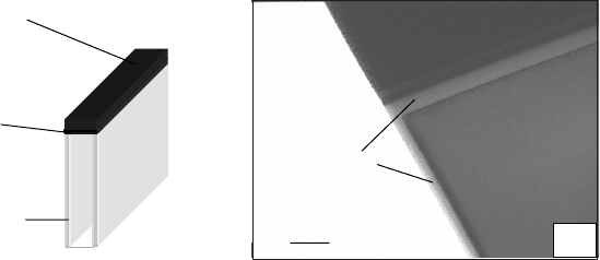
image in which the amorphous layer at the side of a lamella, created when it
was cut free from the sample, is visible as a bright band. The increase in point
defect density and the rise in local temperature may also result in intermixing
at interfaces and diffusion giving rise to changes in the local composition and
structure of a sample. Furthermore, preferential sputtering, redeposition, and
segregation effects may also occur [91], which, depending on the material,
may result in a phase transformation [92].
Numerous studies of the artefacts associated with using FIBs to prepare
TEM lamellae have been made and these can be divided into two categories:
those in which a comparison is made between lamellae prepared by FIB
milling and other techniques and those that measured the depth of damage at
the sidewalls and investigated methods to decrease it. Generally, in the FIB
literature, the depth of the damage refers to the thickness of the amorphous
layer, i.e., the region where the depth of the damage exceeds the critical point
defect density [93]. However, it should be noted that the damage and its effect
on the properties of a material may penetrate considerably further than this
into the samples.
8.6.1 Comparisons of lamellae prepared by FIBs and other techniques
Ma [94] compared the microstructure of Inconel 783 lamellae prepared by jet
polishing, BIB, and FIB milling. The precipitation of spherical Ni
3
Al was only
clearly observed in the lamella prepared using jet polishing. In the BIB pre-
pared TEM lamella only part of the microstructure seen in the electro-polished
lamella was visible and preferential sputtering of some of the spherical pre-
cipitates had occurred. For the FIB prepared lamella the morphology of the
different phases and even the grain boundaries were not visible; however,
50 nm
Amorphous Si
Si
Pt
Pt
Pt damage
Sidewall
damage
(a)
(b)
Figure 8.10 (a) Schematic of FIB damage to a lamella and (b) a BF TEM
image of the FIB induced damage at the sidewalls of an FIB prepared
lamella.
Preparation for physico-chemical analysis 237