Yao N. Focused Ion Beam Systems: Basics and Applications
Подождите немного. Документ загружается.


An electron passing through this field will feel a Lorentz force as described by
the Lorentz force equation:
F ¼e E þ v · BðÞ: ð1:6Þ
According to the equation, in a uniform magnetic field B, a moving charged
particle will follow a curved path. In the case of a magnetic lens, there is no
electric field and the E term is dropped, leaving F ¼eðv · BÞ. As an electron
travels down the column, it first encounters the horizontal component (B
r
)of
the magnetic field. This causes the electron to begin a rotation about the axis
along a helical path. With a nonzero angular velocity about the axis, the
electron begins to feel an inward force from the vertical field component (B
z
)
that draws it toward the axis. Finally, as the electron emerges through the
bottom horizontal field component, it receives a reverse angular impulse that
cancels its rotation about the axis. The constricted beam then continues to
narrow toward the focal point.
The lens equation (relevant here since the fields are relatively weak com-
pared to the velocities of the electrons) is
1
f
¼
1
p
þ
1
q
; ð1:7Þ
where f is the focal length of the lens, and p and q the distances between the
original source and the lens and the distance from lens to image, respectively.
The diameter of the source at p is demagnified by a factor of M ¼q/p through
the lens (i.e., the beam diameter is reduced by this factor with no loss
in intensity). Integrating the Lorentz force through the lens gives a focal
length of
1
f
¼
e
8m
e
V
Z
1
1
B
2
z
dz; ð1:8Þ
which depends only on the field strength along the axis.
The electrostatic lenses found in ion beam systems operate in a simpler
manner than magnetic lenses, although the underlying principle is analogous.
Figure 1.7 depicts a three-electrode design. Positively charged particles enter
the lens from the left and encounter an electrical field formed by the large
voltage difference between the first two electrodes. The ions follow the field
lines, receiving an impulse toward the optical axis and a boost in velocity by
the increasingly negative field. As the ions pass the second electrode, they are
pulled outward, but since they are now closer to the axis and have more
momentum, the change in direction is less than from the first impulse. Upon
Focused ion beam systems18
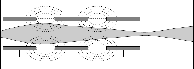
passing the second field, the beam is once again constricted, but this time the
ions are decelerated by the increasingly positive field to near their original
velocity [14]. Just as in the magnetic lens, the charged particle beam exits and
continues to narrow toward its focal point downstream in the column. It is
worth noting that, if the potential difference between the center electrode and
end electrodes were reversed, it would create not a converging but a diverging
lens system (commonly used in the transmission electron microscope, but not
actually used in any SEM or FIB optics).
1.4.2 Aberrations
Most electron columns have more than one lens, usually with apertures along
the beam zone axis to reduce aberrations. A small imperfection or a funda-
mental limit in one lens will be multiplied by the number of lenses and cause a
significant loss in resolution. It is known that such optical aberrations in the
electrostatic lenses of ion columns are also quite severe. As a result, charged
particle lenses are incapable of completely faithful imaging, and to optimize
their performance, it is critical to operate them in the paraxial mode. (This
means that, for high-resolution images, the angle of the trajectory of particles
with respect to the lens axis must be kept extremely small: less than 10 mrad.)
The aberrations can be categorized into three basic groups, as follows:
spherical aberration, chromatic aberration, and astigmatism.
Spherical aberration is the effect of a nonlinear dependence of beam
deflection on radius within a lens. In an ideal lens, the larger the radius from
the axis at which a particle is found, the sharper the angle of deflection: a
linear relationship that holds true from the center of the lens out to the edges.
In a real lens, however, the fields necessarily experience a degree of fringing
V
1
V
2
V
3
Figure 1.7 Schematic of a three-electrode electrostatic lens in an ion beam
column. Field lines generated by the voltages on the electrodes are shown as
dotted lines, and the ion trajectories traveling from left to right are in gray.
Introduction to the focu sed ion beam system 19

and edge effects closer to the substance of the lens, causing the beam to
deflect at an angle that may not match that needed to reach the desired focus.
Thus, parallel vectors traveling through the lens may be focused at different
points as a result of intrinsic lens properties, and the focal length varies with
radius instead of remaining constant. In the case of a positive spherical
aberration, a ray at distance r
1
from the axis will be focused less strongly than
a ray at distance r
2
> r
1
from the axis; a negative aberration is obtained if the
inequality is reversed. In magnetic lenses the mutual dependence of B
r
and B
z
means that the variation in focal length as a particle travels through the lens
further from the center is great enough to cause significant spherical aber-
ration. The phenomenon can be reduced by careful lens design, or with an
aperture in place to reduce the aberration; the diameter of the beam resulting
from a spherical aberration becomes
d
s
¼ 0:5C
s
fi
3
; ð1:9Þ
where C
s
is the spherical aberration coefficient, and fi is the aperture angle, a
function of the focal length and the diameter of the lens field.
Chromatic aberration is the chief limitation on the focusing ability of elec-
tron and ion optics. This problem occurs when a spread of energies 1E is
present in the beam. Since the lenses rely on the interaction between fields,
charges, and velocities, according to the Lorentz force law, a slight difference
in velocity owing to a difference in initial energy will result in a different focal
length for a particle. The spread in energies thus translates to a spread in focus.
Particles with higher-than-expected energies will be focused beyond the surface
of the sample, while particles with lower-than-expected energies will pass
through focus above the sample and spread out again, producing a larger spot
size than desired. A point object under this condition will be imaged as a disc,
the radius of which is proportional to the aperture angle and to the magnitude
of the relative energy spread 1E/E. The chromatic aberration (so called
because a similar focal length spread is observed for different energies, and
thus different colors, of light) is a fundamental property of the source, and
represents the most serious practical limitation on the performance of current
ion beam designs. In the approximation implicit in the above equations, it is
assumed that all electrons or ions have the same kinetic energy due to having
gone through the same accelerating voltage. With an energy spread of 1E,
however, the new focused beam diameter becomes
d
c
¼ C
c
1E
E
fi; ð1:10Þ
where C
c
is the chromatic aberration coefficient of the lens.
Focused ion beam systems20
The last issue, astigmatism, is an effect of asymmetry in the focusing field,
whereby the cross section of the beam in a given plane is not circular, but
rather ellipsoid, causing the defocused beam to appear elliptical rather than
circular. The astigmatism increases the beam diameter to
d
A
¼ 1f
A
fi; ð1:11Þ
and the image of a point is mapped onto two perpendicular lines lying in
front of and behind the image plane, known as the tangential and sagittal
planes, respectively. The distance of these planes from the image plane varies
as the square of the distance of the point object from the lens axis and as the
aperture angle. This problem can be corrected by using a set of alignment
coils near the beam’s exit to reshape the beam.
It should be noted that while chromatic and spherical aberrations are
properties of the lenses and electron source and can only be corrected by
apertures and the addition of other lenses with opposing aberrations, astig-
matism can be dynamically corrected by the use of stigmators, a set of
magnetic coils with deliberately asymmetric fields that can be used to move
the two focal lines until the distortion is corrected. The total beam diameter
resulting from the combination of these aberrations is given by
d
2
p
¼ d
2
0
M
2
þ d
2
A
þ d
2
s
þ d
2
c
; ð1:12Þ
where d
0
is the initial source beam diameter. This calculation is known as the
quadrature method.
The first major difference between electron and ion beam columns is in the
emission source. As discussed previously, instead of a heated tungsten fila-
ment or a field-emission region above a tip, the ion emitter is a liquid surface
drawn by electrostatic fields into a sharpened cone about 5 nm wide at the
apex. Understanding the balance of forces needed to achieve this shape is
somewhat involved, so we will take it as given and assume a beam diameter
d
0
as in the electron source with similar parameters for kinetic energy (we will
assume that the ions have one electron charge’s worth of ionization charge,
and are thus given a kinetic energy equivalent to that of the electrons). It is
important to note that metal ions used in a focused ion beam system have
much higher mass than electrons, meaning that they travel slower at the same
kinetic energy and require more force to divert. As discussed above, this
means that their lens apparatus needs to use much larger fields than in the
electron column. Thus the fact that, in general, electrostatic fields are used for
ion lenses, rather than magnetic fields. This comes from the practical pro-
blems of generating a large enough magnetic field in the confined space of the
Introduction to the focu sed ion beam system 21
lens to focus the ions and the issue of the radius of the spiral path that the
ions will follow; the radius of this path will be much larger than that of
electrons in a comparable field due to the ions’ higher inertia. In order to
produce the necessary forces, carefully shaped electrodes with precisely
controlled potentials are used, generating electric fields that focus the slower-
moving and heavier ions, as seen in Figure 1.7. The ions undergo a slight
acceleration in the process, which must be accounted for when considering
the impact energy of the beam on the sample.
The high potentials used to generate the electric fields take advantage of the
Lorentz force’s e-field term; this term is not a cross-product and so the ions do
not travel in a spiral path, making them somewhat easier to control. High
electrostatic fields are easier to create than magnetic fields, and in general make
for a more stable lens. Other than replacing the magnetic term in the focal
length equation with the radial component of the electric field (the only
component that exists, if edge effects are disregarded, which is a reasonable
approximation for such strong fields and high energies), there is functionally
no difference between ion optics and electron optics, as far as the physics of
operation goes. Since the aberrations depend on the same factors, it should be
noted that many ion systems contain stigmator coils even though astigmatism
is not as much a concern in ion beams. In fact, as the half-angle fi of the beam
arriving at the final focal spot diminishes to < 1mrad, the d
0
M virtual source
diameter term dominates in the quadrature equation above. Thus the funda-
mental limit for present FIB technology, in the minimum beam diameter and
the energy spread resulting from mutual repulsion and space charge limita-
tions, overshadows other aberration effects to the point that improvement in
any of them will not improve ion beam resolution significantly.
One more point must be made about ion beam systems. In an electron
optics column, the apertures serve by and large to correct aberrations. High
electron beam intensities and smaller beam size are desirable as they lead to
better imaging; the amount of damage an electron beam does to a conductive
sample is negligible for most applications. Ion beams, however, must have
their beam current carefully controlled, as they constantly damage and
change the surface of a sample. As such, apertures serve as current-limiting
devices, reducing the ion current to whatever level the user deems appro-
priate, rather than simply screening out errant ions from the focusing system.
1.5 Detection of electron and ion signals
The differences between ions and electrons can be extended to their respective
machines, the FIB system and the scanning electron microscope (SEM).
Focused ion beam systems22
Though they are designed and work very similarly, the FIB’s use of gallium
ions from a liquid-metal ion source rather than electrons provides func-
tionality and applicability different from that of the SEM. It uses the focused
beam of gallium ions and rasters the surface of the material of interest. The
small amount of material sputtered from the surface during this process may
form secondary ions and electrons which are then collected and analyzed as
signals to form an image on a screen. This allows high-magnification
microscopy with the FIB system.
In both machines a source emits charged particles that are focused into a
beam and rastered over small areas of the sample using deflection plates or
scan coils. The SEM uses magnetic lenses to focus its beam of electrons;
however, since ions are much heavier and, therefore, much slower with a
lower corresponding Lorenz force, magnetic lenses are less effective. The FIB
system is instead equipped with electrostatic lenses (shown schematically in
Figure 1.8), which have proven to be much more effective.
Both the SEM and the FIB form high resolution images by collecting the
secondary electrons (SE) that are emitted from the interactions between the
beam and the surface atoms, although images may also be formed from
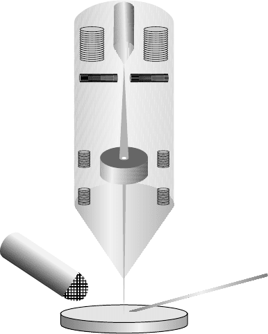
Liquid gallium ion source
Suppressor
Extractor
Lens 1
Aperture
Objective lens
Secondary electron detector
Sample
Gas injection system
Gallium ion beam
Figure 1.8 A schematic diagram illustrates the major components in an FIB
system.
Introduction to the focu sed ion beam system 23
backscattered electrons (BSE) or secondary ions (SI). SE detection is the chief
method, however, and two main detector types exist: multi-channel plate and
electron multiplier. A multi-channel plate is generally mounted directly above
the sample, and, as a result, offers negligible topographical information. The
Everhart–Thornley electron multiplier detector is the most common design
used today for secondary electron detection. Also known as a scintillator-
PMT (photomultiplier tube) detector, it consists of three main parts. The first
part is the collector grid and screen, which is located to the side of the sample
stage, usually at an angle of 45
to the beam. Secondaries are attracted
toward the wire mesh screen by a potential of several hundred volts, and
most of them continue to be accelerated into the scintillator. Captured
electrons cause the scintillator to ‘‘scintillate,’’ that is, to emit visible-light
photons by virtue of its cathodoluminescence, and the number of photons
generated per electron is on the order of 100 for a typical scintillator voltage
of 10 kV. Not unlike a fiber optic cable, a light pipe extends from the scin-
tillator and internally reflects (due to its high index of refraction) the photons
to the photomultiplier tube (PMT). The PMT is a highly sensitive visible
photon detector that consists of a sealed glass tube containing a high
vacuum. At the entrance to the PMT, the incoming photons strike a low-
work-function material that comprises the photocathode, liberating valence
electrons that are subsequently accelerated as photoelectrons toward the
first of a series of (usually) eight dynode electrodes. Each dynode is biased
positively with respect to the photocathode, and each of them is also biased
100–200 V positively with respect to the preceding one. The photoelectrons
generate secondary electrons at the first dynode, and these secondaries are
then amplified by a factor of about 10
6
after they have completed striking
the remaining dynodes. Recalling that each SE originally produced at the
specimen surface generated about 100 photoelectrons, the overall magnifi-
cation of the scintillator-PMT detector can be as high as 10
8
, depending on
the applied dynode voltage. Thus, although it may seem unnecessarily
complicated to convert SEM secondary electrons into photons, then into
photoelectrons, and finally back into secondaries, the high amplification and
low electronic noise – versus a simple metal plate to absorb the electrons – in
fact fully justifies the system. Backscattered electrons can also be detected by
a scintillator-PMT if the bias on the first grid is made negative instead of
positive, therefore repelling lower-energy secondaries but not BSE, which
retain most of their kinetic energy after impact.
Raised areas of the sample (hills) produce more collectable secondary
electrons, while depressed areas (valleys) produce less, thereby creating
a contrast that is interpreted by the machine and at the same time intuitively
Focused ion beam systems24
understood by the operator as light and shadow. Also, to increase the sec-
ondary electron yield, the whole sample is often tilted away from the hor-
izontal plane and toward the detector in order to increase the SE signal
without interfering with the contrast-based topography. A viewing monitor
synched to the scan coils controls the beam so that as it scans across the
sample surface, the image of the sample is reproduced on the screen, with a
magnification inversely proportional to the area of scan.
Images obtained from secondary ions can be below 10 nm resolution and
show topographic and materials information complementary to that
obtained from an SEM image. Although material contrast arising from
differences in specimen chemistry can be significant in FIB secondary elec-
tron images, it is most readily observed in secondary ion images, where it is
often the dominant effect. While SE images provide uniformly good depth of
field, SI images reflect more selective depths that depend on different mate-
rials and sample structure. Information about the grain size and crystal
orientation can also be obtained using an FIB because of the dependency of
the ion–atom interaction upon the crystal grain orientation; this is known as
channeling contrast. Thus, from a materials science standpoint, secondary
ion imaging is an invaluable capability. SI imaging is also superior for
insulating materials when used in conjunction with a charge neutralizer
(electron flood gun). Although ions move slower than electrons, they still
move faster than the image can be collected. It is important to note that, since
the sample is continually sputtering during the FIB imaging process, small
beam currents (< 100 pA) are advisable to minimize sample erosion.
Detection methods for secondary ions fall into one of two categories:
microprobe mode and microscope (or direct) mode. The microprobe mode is
essentially analogous to the process used in SEMs [15]: the primary ion beam
is rastered while the SI signal is synchronously detected. The key difference is
that the particle detector grid is biased to a highly negative voltage to repel
both secondary and backscattered electrons and to attract positive ions,
which are subsequently amplified. Typically, SI microprobe images are of the
‘‘total detected positive ion’’ type, in which virtually all positive ions are
collected and amplified regardless of mass. A more sophisticated recent
development is mass-resolved ion imaging, which is present on FIB systems
that are equipped with secondary ion mass spectrometers (SIMS); this allows
elemental analysis of the sample combined with imaging [16]. In the alter-
native microscope or direct mode, ion image formation is nonraster based
and relies on electrostatic lenses in the secondary ion column. Several types of
position sensitive detectors can currently be used with this process. The most
common of these is a microchannel plate connected to a phosphor screen,
Introduction to the focu sed ion beam system 25
and the resulting image is captured by a highly sensitive CCD. Alternatively,
a direct ion-imaging detector such as the resistive anode encoder (RAE) can
be used to capture the ion images. The resolution in microscope mode
imaging is limited by the electric field strengths of the electrostatic lenses to
about 1 mm.
1.6 The two-beam system
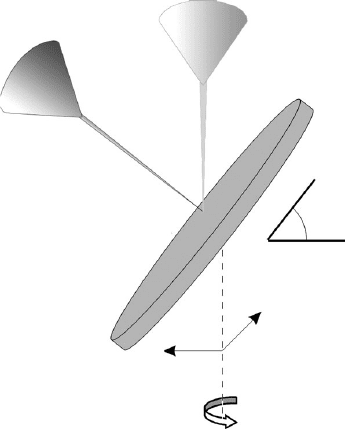
When we combine both SEM and FIB into one system, called a two-beam
system, the ion beam and electron beam are placed in fixed positions, with
an angle of 45–52
between two beams for the best performance as illu-
strated in Figure 1.9. The two beams are co-focused at what is called the
‘‘coincidence point,’’ an optimized position for the majority of operations
taking place within the machine, with a working distance of typically a few
millimeters. The two-beam system allows SEM imaging and FIB sample
modification without having to move the sample. In addition, the stage can
be tilted, allowing changes in the sample-beam orientation. Similarly to the
FIB system, integrated software with a single user interface controls the
two-beam.
Electron beam
Ion beam
Sample
Translation axes
Rotation axis
X
Y
52 degrees til
t
Figure 1.9 Schematic of the two-beam system, in which both electron and
ion beams are co-focused at the coincidence point on the sample surface.
Focused ion beam systems26
The two-beam system provides new advantages that simplify as well as
improve nanoscale imaging, analysis, and fabrication as detailed in the fol-
lowing chapters. One such advantage is in imaging. Whereas FIB imaging
has high contrast abilities but can cause damage to the sample, SEM images
have relatively lower contrast, but provide a higher resolution and do not
damage the sample. The result is a more complete set of data. A study done
at Portland State University showed that using this combination of beams
could provide a more comprehensive imaging and characterization of carbon
nanotubes [ 17]. Also the reconstruction of three-dimensional structure and
chemistry of a sample can be simplified using the two-beam system to
interpolate two-dimensional SEM and FIB images and ionassisted SIMS
chemical maps of layers that have been exposed using the milling feature of
the ion beam [18].
The charging effects of ion and electron beams are worth consideration.
A typical two-beam system setu p will have the ion beam work ing in
tandem with a scanning electron micro scop e, placing both types of beams
at the oper ator’s disposal. One of the great challenges in the use of elec-
tron beam for i magi ng i s that if the sample is not fairly conductive, the
electrons from the beam will build up charge in the material. This charge
will then distort both t he inco ming b eam an d the out going s econd ar y and
backscattered electrons, producing elec tron artefacts and distortions in the
data. While the i on beam does to some degree incre ase local c harge at the
pointofimpact,thenatureoftheionsthemselves,generallymetals,causes
the imbalance to be quickly rectified. This can be exploited to assist in
electron-bea m imaging of t he sample, by using th e ion beam at a low
setting over a large area to reduce l ocal charging effe cts and increase
surface conductivity [19].
The complementary nature of the negatively charged electrons and the
positively charged ions also eliminates the charging problem found in the
single beam FIB. The accumulation of charge would hurt the resolution of
the image, but because of the availability of both charges, this is no longer a
problem with the two-beam system.
Not only does the combined system produce a better and more extensive
collection of data, but it also allows for precise monitoring of FIB operation
through the SEM. By using the slice-and-view technique to observe the
progress of the ion beam cross section, the operator can stop the milling
process at a precise point in order to obtain local information. Also, the two-
beam system allows for the use of both the ion beam and the electron beam
simultaneously without interference, doing away with the necessity to switch
back and forth. The sample can be imaged in real time with the SEM while
Introduction to the focu sed ion beam system 27