Graef M. Introduction to conventional transmission electron microscopy
Подождите немного. Документ загружается.

198 The transmission electron microscope
S
A
W
F
u
v
C
1
C
1
a
(a)
S
A
W
F
u
v
C
1
C
1
a
(b)
C
2
a
C
2
θ
c
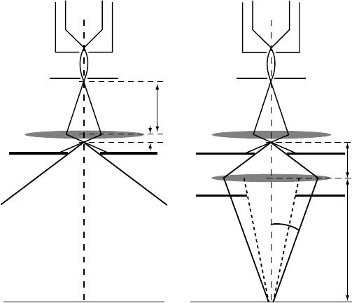
Fig. 3.27. (a) Ray diagram for a strongly excited C
1
lens. Electrons leave the C
a
1
aperture
at large angles with respect to the optical axis. (b) Ray diagram with a second condensor
lens C
2
. The lens has the first condensor cross-over as the object plane, and the specimen
as the image plane. The diameter of the C
2
cross-over on the specimen is known as the spot
size. The variable diameter aperture C
a
2
limits the angular range of electron trajectories at
the expense of beam current.
under-focused; if the image cross-over falls before the sample, then the lens is said
to be over-focused. The lens is in-focus when the smallest possible spot diameter
is observed on the sample. Although over- and under-focused conditions may give
rise to a spot with as identical diameter on the sample, one must be aware of the fact
that the beam divergence angle θ
c
is different in the two cases. Observation modes
that require a nearly parallel incident beam (such as high-resolution observations,
see Chapter 10) must hence be carried out with as underf-ocused C
2
lens (unless
additional condensor lenses are present). On microscopes with rotary lens control
knobs, the under-focus condition usually corresponds to a counterclockwise rotation
(i.e. less current through the lens).
Using the two-lens condensor system, we can routinely obtain 100 nm spot
sizes. The first condensor lens control is often known on the microscope console
as the spot size; the spot size can only be varied in discrete steps. The C
2
current
is often labeled as intensity or brightness on the microscope console; it can be
varied continuously. Since the C
1
cross-over changes location whenever the spot
size is changed it is necessary to change the C
2
current to obtain the smallest beam
diameter on the sample. The second condensor aperture C
a
2
can be set to one of
3.9 The specimen stage 199
three or four different diameters. It can be seen from Fig. 3.27(b) that a change in
C
a
2
diameter does not change the size of the illuminated area on the specimen, only
the beam intensity changes (less beam current for a smaller aperture diameter). A
smaller aperture will also change the maximum beam divergence angle by limiting
the solid angle subtended by the C
1
cross-over at the sample. This improves the
coherence of the incident beam at the expense of lower image intensity. Modern
microscopes often have a third so-called mini-condensor lens, located right above
the objective lens upper pole piece. Since this lens is an integral part of the objective
lens stage, we will discuss it in the next section.
3.9 The specimen stage
3.9.1 Types of objective lenses
We have seen that the objective lens of a TEM is a type of immersion lens, since the
sample is located well within the magnetic field of the lens. There are two major
types of objective lenses: the high-contrast lenses, also known as asymmetric
lenses,
and the twin or symmetric lenses. In an asymmetric objective lens the sample is
located at one-third of the distance between the upper and lower pole pieces, as
shown in Fig. 3.28. The objective or diffraction aperture is located two-thirds of
the way down and coincides with the lens back focal plane. The upper pole piece
usually has a narrower bore diameter than the lower pole piece, hence the name
“asymmetric”. In a symmetric lens design, the sample is located in the center
between the two pole pieces, as indicated in Fig. 3.29. Both bore diameters are the
same in the symmetric lens.
1/3
1/3
1/3
B(z)
z
Back focal plane
Specimen
Objective aperture
Fig. 3.28. Schematic diagram of a high-contrast objective lens. The specimen and back
focal plane are located at one-third and two-thirds the distance along the axis of the lens.
The angles between the rays and the optical axis are highly exaggerated and are actually
only a few tens of milliradians. The lens forms a first intermediate image.
200 The transmission electron microscope
B(z)
z
Back focal plane
Specimen
Objective aperture
Objective
condensor lens
Objective
imaging lens
Condensor
mini lens
Microprobe mode
B(z)
z
Back focal plane
Specimen
Objective aperture
Objective
condensor lens
Objective
imaging lens
Condensor
mini lens (off )
Nanoprobe mode
(a)
(b)
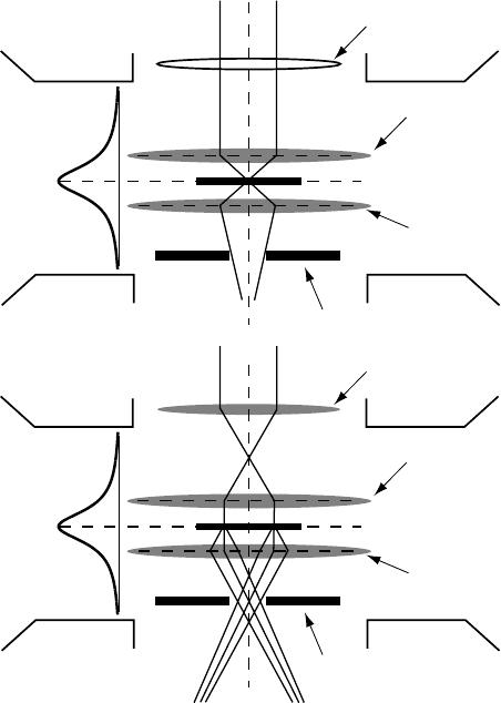
Fig. 3.29. Schematic diagram of a symmetric objective lens. With the condensor mini-lens
switched off (a) a fine probe is obtained (nanoprobe mode). When the condensor mini-lens
is switched on (b) a parallel beam is obtained (microprobe mode).
Asymmetric lenses have a much longer focal length than symmetric lenses (often
in the range 5–10 mm), and hence a smaller lateral magnification M or, equivalently,
a larger angular magnification M
α
, which makes them well suited for large camera
length diffraction observations. Since the incident electrons experience only a frac-
tion of the objective lens magnetic field before reaching the sample (as indicated
by the B(z) profile in Fig. 3.28), this prefield acts only as a weak third condensor
lens. Recalling that the primary task of the condensor lenses is to demagnify the gun
cross-over, it is clear that the asymmetric objective lens cannot be used to obtain
nanometer probe sizes.
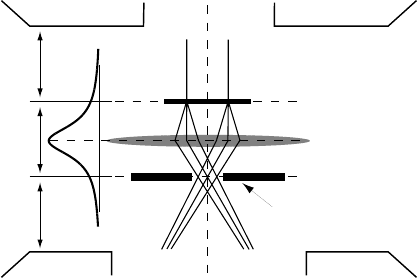
In the symmetric lens design (the so-called Riecke–Ruska lens) the electron
experiences nearly half of the objective lens magnetic field before reaching the
sample, and this upper half of the field is known as the objective condensor lens. It
can be used to further demagnify the incident probe, down to the single nanometer
3.9 The specimen stage 201
size range. This observation mode is thus known as nanoprobe mode (Fig. 3.29a).
When an additional mini-condensor lens is present, often as part of the objective
lens circuit, then this lens can be used to create another cross-over, this time right
above the objective condensor lens (Fig. 3.29b). The electrons leaving this cross-
over are then focused in a parallel but very narrow beam and after passing through
the sample and the objective imaging lens, they go through a symmetric cross-
over. This mode is known as microprobe mode. In microprobe mode, the electrons
travel in a parallel beam through the sample, whereas in nanoprobe mode they are
focused into a small probe. Microprobe mode is therefore useful as a diffraction
technique, since the parallel beam can be focused readily into diffraction spots,
while nanoprobe mode is more useful for analytical observation techniques. The
focal length of the symmetric lens is much smaller than that of the asymmetric
lens, and is usually in the range 1–3 mm. This means that the objective aperture is
located very close to the sample, which imposes limitations on the tilt range of the
specimen holder. The lens gap of a symmetric lens (the distance between the upper
and the lower pole pieces) is of the order of a few millimetres; the asymmetric lens
has a much larger lens gap, in the range of 10–20 mm.
While the primary function of the illumination stage is to create an electron
beam in a well-defined reference state, the primary function of the objective lens
is to bring the various diffracted electron beams to a cross-over while introducing
minimal lens aberrations (see also Section 3.5). When we discussed the Fraunhofer
diffraction conditions we saw that we do not need to have a lens in order to have
a diffraction pattern; indeed, simply projecting a laser beam through a fine optical
grating onto a wall on the other side of a room will produce a diffraction pattern
without using a lens. The main purpose of the objective lens is to reduce the infinite
“focal length” of the lensless system to a finite length, so that both diffraction
pattern and image can then be magnified by other lenses. It is, in principle, possible
to create microscopes without lenses, since all of the relevant physics occurs in the
interaction of the electron beam with the specimen. One such microscope is the
point projection microscope [SQM93], which uses no lenses at all, and hence does
not suffer from lens aberrations.
3.9.2 Side-entry, top-entry and special purpose stages
There are two different types of specimen stages: those where the sample enters
the objective lens from the side (side-entry) and those where the sample is lowered
into the lens from the top (top-entry).
The side-entry stage is by far the most common stage and is used for both
conventional and analytical electron microscopy and for most in situ experiments.
As shown in the schematic drawing in Fig. 3.30, the sample is mounted at the end
202 The transmission electron microscope
primary
tilt axis
secondary
tilt axis
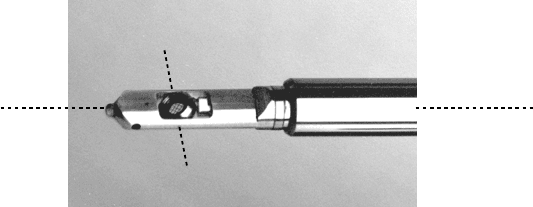
Fig. 3.30. Side-entry double-tilt holder; the primary and secondary tilt axes are indicated.
(Photograph reproduced with permission of B. Armbruster, Gatan, Inc.)
of a rod-like specimen holder. The sample fits into a 3.05 mm diameter cup, which
can either pivot around one axis in the plane of the sample, or rotate around an
axis perpendicular to the sample. The rod-axis represents the primary rotation axis.
These holders are referred to as double-tilt and rotation-tilt, respectively. Depending
on the objective lens gap the maximum tilt angle may be as high as 60
◦
or as low
as 15
◦
. Note that most specimen holders are made out of a soft aluminum alloy
(or pure Be for some analytical holders), so utmost care is necessary whenever a
holder is manipulated. Depending on the diameter of the microscope column, the
specimen rod is anywhere from about 25–60 cm long. This rod is entered into the
column through a vacuum airlock. Refer to the microscope manuals for the correct
procedure to insert a sample holder into the column.
For most microscopes the sample has five degrees of freedom as illustrated in
Fig. 3.31: two lateral movements in the horizontal plane (x and y, controlled by the
specimen translation wheels or a joystick/trackball system), one vertical movement
(z, specimen height) and two rotation angles α and β (either double-tilt or rotation-
tilt). The five numbers {x, y, z,α,β} describe the specimen attitude. The rotation
angles are defined as positive for counterclockwise rotations, when looking from
the positive side of the x, y,orz axes towards the specimen. The reader should
carefully study the geometry of the holder(s) he/she will be using, in particular
how the holder is oriented inside the microscope column; for several microscopes,
the holder is actually placed upside down in the column.
†
For top-entry stages, the
reader should consult the microscope manual to determine the relative orientation
of the specimen holder and rotation axes with respect to the microscope column.
In modern microscopes, some or all of the five coordinates may be computer
controlled, while in older instruments x, y, and z motion are manually controlled.
The primary tilt angle α can be changed manually or via a motor, and the secondary
tilt/rotation angle β is always controlled by a motor. Both motors can be actuated
†
Upside down with respect to the orientation of the specimen cup during loading.
3.9 The specimen stage 203
+y
+x
-z
+z
-y
-x
+β
+y
+α
+x
+β
+z
(a)
(b)
(c) (d)
Fig. 3.31. Degrees of freedom for a typical specimen holder: (a) translation axes (positive
x points along
the length axis of the holder, positive
y points to
wards the front of the
microscope column); (b) primary tilt angle α; (c) secondary tilt angle β for a double-tilt
holder; (d) secondary angle β for a rotation-tilt holder.
by floor-mounted pedals, or, in some microscope models, by rotary or push but-
tons on the microscope console near the column. More recent computer-controlled
microscopes are equipped with a joystick-like mechanism to control the specimen
attitude.
We have seen in Section 3.5 that lens aberrations increase rapidly for electrons
traveling at large angles with respect to the optical axis of the microscope. This
axis is defined as the line connecting the filament tip to the center of the viewing
screen, and for a well-aligned microscope column the center line of each lens
should coincide with the optical axis. The size of the electron transparent region in
a thin foil can range from a few to several hundred micrometers, and the specimen
translation controls x and y are used to bring a region of interest in the sample onto
the optical axis of the column and therefore in the electron beam.
For both top- and side-entry stages, the specimen is located inside the objective
lens magnetic field. Surrounding the specimen holder is the so-called cold-finger or
anti-contamination device (ACD). It is a piece of metal, typically copper, linked to
a liquid nitrogen dewar. By keeping the cold finger at liquid nitrogen temperature,
residual gases in the microscope vacuum (or petroleum derivatives from an oil
diffusion pump) are trapped onto this cold surface instead of on the sample. The
ACD keeps the sample clean so that prolonged observations are possible. On some
microscope models, one can insert an ACD-heater into the dewar at the end of a
microscope session so that the remaining nitrogen is boiled off.
204 The transmission electron microscope
A transmission electron microscope is an ideal instrument for in situ experiments.
A variety of special purpose stages have been designed:
r
heating and cooling;
r
magnetic field (one must use special low-field pole pieces in order to apply a controlled
magnetic field on the sample);
r
electric field, e.g. to study ferroelectric domain walls;
r
straining stages, mostly tensile, some torsion or mode I deformation.
Combinations of two or more externally applied fields (e.g. electric field and defor-
mation) are also possible. We refer to [Val79] for an extensive overview of different
stages. Each stage has its own peculiarities and one should always be aware of the
fact that performing an experiment on a thin-foil sample may not give rise to the
same sample response as that expected for a bulk sample. The recent advances in
spherical aberration correctors will undoubtedly rekindle interest in in situ obser-
vations, since the lens gap was kept small to reduce the spherical aberration. If C
s
can be varied at will, then there is no longer a need for a small lens gap.
3.9.3 The objective lens and electron diffraction geometry
The objective lens is used with a high excitation current which means that a high
lateral magnification is obtained. From our discussion on electron optics we know
that the angular magnification M
α
is the inverse of the lateral magnification M,
which means that rays leaving the objective lens will travel at very small an-
gles to the optical axis in all subsequent lenses – this is why most aberrations
of the subsequent lenses are unimportant and can be ignored. The aberrations
of the objective lens cannot be ignored since in this lens electrons travel at the
largest angles with respect to the optical axis. The spherical aberration C
s
and
the chromatic aberration C
c
determine the final image quality for conventional
TEM work (assuming correctable aberrations, such as astigmatism, have been taken
care of).
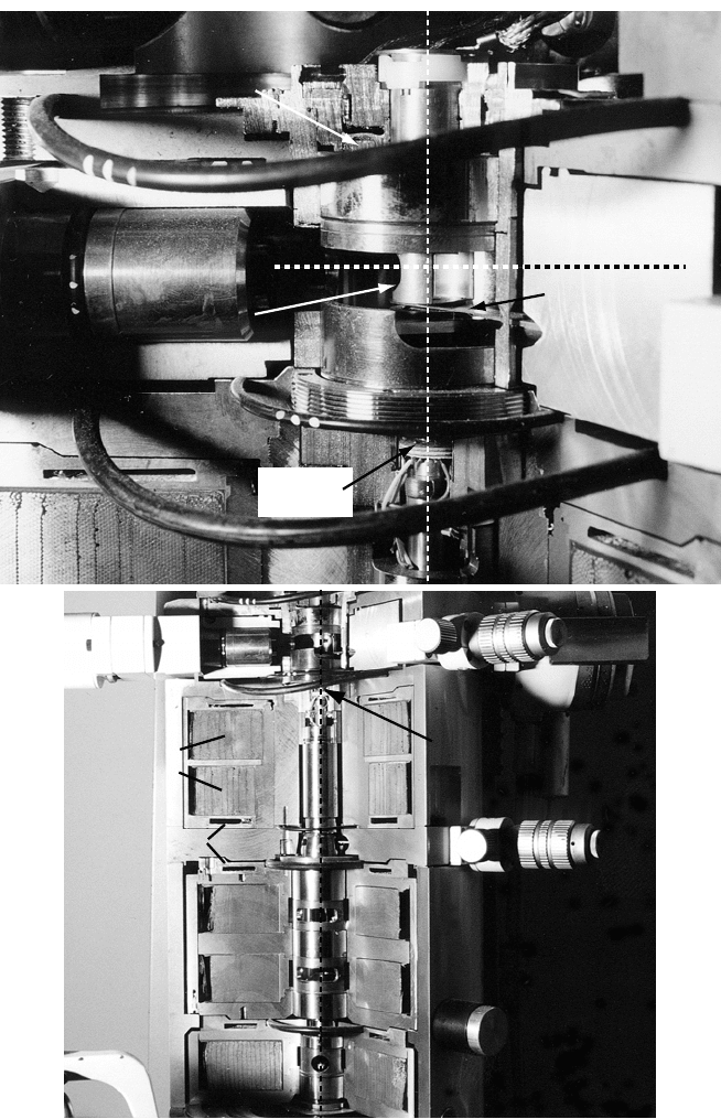
A cross-sectional view of the goniometer area of the JEOL 120CX microscope
is shown in Fig. 3.32(a). The specimen rod enters from the right and is firmly
seated in a metal cup which is linked to one of the specimen translation controls.
The specimen holder sits right above the bottom plate of the anti-contamination
device (cold finger) which, in turn, sits above the objective aperture. The objective
stigmator follows a few millimeters below the lower objective pole piece. The OL
pole pieces are located above the lens coil which is visible in the lower left-hand
corner. Figure 3.32(b) shows the entire imaging stage of the microscope, from
the objective lens area down to the projector lens; the bottom three lenses will be
discussed in more detail in the following section.
3.9 The specimen stage 205
diffraction
aperture plate
ACD
scan coil
sample holder axis
"cold finger"
objective lens coil
specimen rod
support
objective
stigmator
optical axis
diffraction
aperture
selected area
aperture
objective
lens coils
cooling water
diffraction
lens
intermediate
lens
projector lens
objective stigmator
(a)
(b)
Fig. 3.32. (a) Cross-sectional view of the goniometer area of the JEOL 120CX microscope;
(b) cross-sectional view of the objective lens and magnifying lenses.
206 The transmission electron microscope
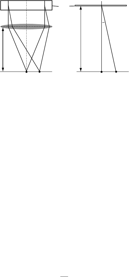
f
2θ
λfg
2θ
r ~ 2θL
L
(a)
(b)
thin
foil
bfp
screen
Fig. 3.33. (a) Schematic representation of the diffraction geometry; (b) definition of the
diffraction camera length L.
The OL is characterized by two different planes: the back focal plane, in which
the diffraction pattern is formed, and the image or selected area plane. In the
back focal plane we can introduce an aperture, typically known as the diffraction
aperture. Similarly, in the image plane we can introduce the so-called selected area
aperture (see Fig. 3.32b). Since electrons leaving the OL form an image at a large
distance compared to the sample dimensions, Fraunhofer diffraction conditions are
satisfied and hence the back focal plane contains the Fourier transform of the object
function. The reciprocal lattice points in the back focal plane are at a distance λ f |g|
from the origin of reciprocal space, as shown in Fig. 3.33(a) (this follows from
equation 3.41). The BFP hence contains an “image” of reciprocal space, magnified
by a factor of λ f . Only those reciprocal lattice points that lie near the Ewald sphere
and satisfy the Bragg condition will give rise to a diffracted beam.
Figure 3.33(b) defines the camera length L of the microscope in diffraction mode.
The camera length is a measure for the magnification of the diffraction pattern. The
relation between the camera length L, the electron wavelength λ, the lattice spacing
d and the measured spacing r on an electron micrograph can be derived easily as
follows: from the Bragg equation we know that 2d sin θ = λ. Since Bragg angles
for high-energy electrons are small, it is a good approximation to replace sin θ
by θ, and we have 2dθ = λ. From Fig. 3.33(b) we have r = L tan(2θ) ≈ 2Lθ .
Combining the two relations we find
r =
λL
d
= λLg, (3.71)
where g = 1/d. The constant L is known as the camera length, and the product
λL as the camera constant; it has dimensions of [nm mm]. For a given lattice
spacing d, an increase in the acceleration voltage will cause a corresponding de-
crease in the electron wavelength and therefore a decrease of the distance r in the
3.9 The specimen stage 207
diffraction pattern. The camera length L converts the diffraction angle 2θ into the
distance r .
For the aluminum example in Table 2.3 on page 102, 2θ = 12.38 mrad for the
(200) planes (at 200 kV), and for a typical camera length (the distance between
the sample and the observation screen) of L = 1000 mm we find r = 12.38 mm.
Rather than changing the magnitude of L by physically changing the location of
the observation screen (a bit impractical), we can use the imaging lenses to change
the effective value of L, while keeping the object and image planes constant. All
microscopes have a predefined set of camera lengths, and the microscope operator
can select the value of L best suited for the observation at hand. This value would
typically be shown on the microscope console and on the diffraction micrograph.
Care must be taken to accurately calibrate the camera length; this will be discussed
in Section 4.5 in the following chapter.
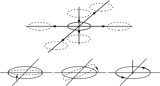
From the preceding paragraphs we can deduce the following geometrical in-
terpretation of electron diffraction (see Fig. 3.34a): the incident electron beam is
represented by the wave vector k, which is parallel to the optical axis of the ob-
jective lens. According to the Ewald sphere construction, this wave vector ends in
the origin O of reciprocal space, which is located at the intersection of the optical
Ewald
sphere
Incident
beam
Laue
circle
05
1015
(a)
(b)
(c)
(d)
Fig. 3.34. Perspective representation of the spatial relation between the Ewald sphere and
a plane in reciprocal space. In (a) the plane is tangential to the sphere, and the relrods cor-
responding to individual reciprocal lattice points intersect the sphere. When the reciprocal
lattice plane is tilted around an axis in the plane, the intersection of this plane and the Ewald
sphere becomes a circle through the origin, the Laue circle, indicated by a white arc. With
increasing tilt angle (b, c, and d) the circle becomes larger.