Vaccari D.A., Strom P.F., Alleman J.E. Environmental Biology for Engineers and Scientists
Подождите немного. Документ загружается.

helps provide muscle tone, which is the contraction of some muscle fibers even when resting.
Muscle tone helps keeps bones and joints in position and protects against sudden shocks.
9.4 NERVOUS SYSTEM
The nervous system is one of the two main control mechanisms of the body, the other
being the endocrine system. Nervous tissue is among the most sensitive in the body to
toxins. Because nervous tissue has a very high metabolic rate, its role is so critical that
even small amounts of damage can have significant effects, and some toxins (i.e., the
insecticides) are actually designed to be neurotoxic in order to be effective against insects
at low dosages.
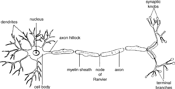
The primary functional components of the nervous system are the nerve cel ls, or neu-
rons. They can have many shapes; Figure 9.2 shows a common type. The main parts are
the cell body, or soma, the highly branched dendrites, and the axon. The soma contains
the nucleus and cytoplasm, with its typical cell machinery, such as mitochondria and ribo-
somes. The neuron lacks a centriole, which makes it impossible for it to divide. Thus,
neurons cannot regenerate once damaged. This also means that neurons cannot become
cancerous. Brain cancer in adults occurs in other nervous system cells, called glial cells.
The dendrites provide most of the sites for the reception of nerve signals. The axon is a
long extension of the cell that serves to transmit the nerve signal over a great distanc e. It
may have branches, and it is often sheathed in a lipid coating called myelin that serves as
an insulator for the signal transmission. Neurons transmit signals from one cell to another
at a specialized point of contact called a synapse.Anerve is a bundle of neuron axons.
9.4.1 Nerve Signal Transmission
Nerve signals are transmitted along the axon as a wave of electrochemical charges caused
by movement of potassium and sodium ions across its membrane. A membrane would be
Figure 9.2 Typical motor neuron. (From Fried, 1990. # The McGraw-Hill Companies, Inc. Used
with permission.)
178
THE HUMAN ANIMAL
fairly impermeable to ions except for the presence of special pores called membrane
channels that provide a path for the ions. There are special membrane channels for potas-
sium and others for sodium. One type of membrane channel allows a continuous but slow
leak of ions across the membrane. Another type opens and closes in response to voltage
across the membrane. In addition, there is a sodium–potassium exchange pump that
uses ATP to exchange three intracellular sodium ions with two extracellular potassium
ions. In other words, it pumps two potassium ions into the cell for every three sodium
ions pumped out. When the nerve cell is resting, the exchange pump maintains an elec-
trostatic potential, or voltage, of 70 mV across the membrane. That is, the inside of the
cell is negatively charged, or has a deficiency of positive anions. This voltage is called
the resting potential of the cell. The pump also ensures that under resting conditions,
the inside of the cell has far more potassium than sodium, and the reverse is true
outside the cell.
Nerve signal propogation along the axon begins when a sodium channel is opened at
some point, such as by a chemical signal from a synapse. This causes the voltage to
increase toward zero, a process called depola rization. Depolarization triggers a sequence
of sodium and potassium pore openings and closings. First one, then the other ion floods
across the membrane, causing the voltage to increase and then decrease back to the resting
potential. This sequence moves along the axon in a wave, transmitting the signal. The
entire sequence at one point may occur in 1 ms.
The largest axon fibers, from 4 to 20 mm in diameter, are well insulated by myelin
and can propagate signals at speed up to 140 m/s. Others are unmyelinated and are
less than 2 mm in diameter; their transmission speed is only about 1 m/s. The faster
fibers are used to transmit the senses of balance, body position, and delicate touch.
Slower neurons take l ess space and are used to transmit information on te mperature
and pain as well as information for organs and glands. About one-third of an adult’s
neurons are my elinated. Children lack myelina tion until early adolescence, which
part ly accounts for their reduced coordinati on ability. Mul tiple sclerosis is a disease
involving the loss o f mye lination of axons, which results in muscle paralys is and loss
of sensations.
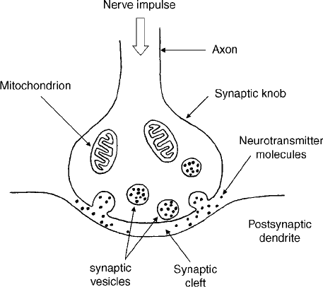
9.4.2 Synaptic Transmission
At the end of the axon, the n euron must transmit the signal to another neuron or to an
effector. This is done through the synapse. Most synapses involve the use of a chemical,
called a neurotransmitter, to commun icate the signal across a gap from one cell to
another. One of the most widespread neurotransmitters is acetylcholine (ACh). This neu-
rotransmitter is a target of many insecticides, in particular the organophosphorus and
carbamate pesticides.
The axon ends in a synaptic knob (Figure 9.3), which contains as many as 1 million
vesicles containing ACh. The membrane of the synaptic knob is separ ated from the
membrane of a target neuron by a gap of about 20 nm width. When the depolarization
wave from an axon reaches the synaptic knob, ACh is released and diffuses across the
gap to the target neuron. There it stimulates pores to open, depolarizing the target neuron
membrane and initiating a nerve signal transmission. An enzyme, acetylcholinesterase,
rapidly decomposes the ACh into choline and acetate. The choline is reabsorbed by the
synaptic knob and recycled into more ACh. It is the enzyme acetylcholinesterase that is
affected by organophosphorus and carbamate pesticides (see Section 17.4.7).
NERVOUS SYSTEM 179
There are many other neurotransmitters. Many hormones serve this function, including
epinephrin ( adrenaline), ADH , oxytocin, insulin, and glucagon. The amino acids glycine,
glutamine, and aspartic acid are neurotransmitters, as are the gases carbon monoxide and
nitric oxide. A group of compounds called endorphins modifies the effect of neurotrans-
mitters and may be involved in mood and pain reduction. They are similar in structure to
morphine. It is thought that exercise produces a natural release of endorphins. Dopamine
is a central nervous system neurotransmitter that can be inhibitory or excitatory, depend-
ing on the receptor. A decline in dopamine production produces Pa rkinson’s disease,in
which the inhibitory action of dopamine is missing. As a result, the neurons that control
muscle tone become overstimulated. All movement requires overcoming the tension of the
opposing muscle. Dopamine cannot cross the blood–brain barrier, but the drug
L-dopa
can, and it is converted to dopamine in the brain, providing relief from symptoms.
9.4.3 Nervous System Organization
The nervous system may be the best example of ‘‘the whole is more than the sum of its
parts.’’ Even accounting for the fact that the behavior of individual neurons is much more
complex than described above, it is difficult to explain our higher behaviors, such as lan-
guage, abstract reasoning, and self-consciousness, in terms of them. That is a far greater
task than explaining the functioning of a computer in terms of the action of individual
transistors. Those higher behaviors depend on neuronal activity, but in ways far from
well understood and beyond our scope here, in any case. Here we can only summarize
the basic organization of the nervous system.
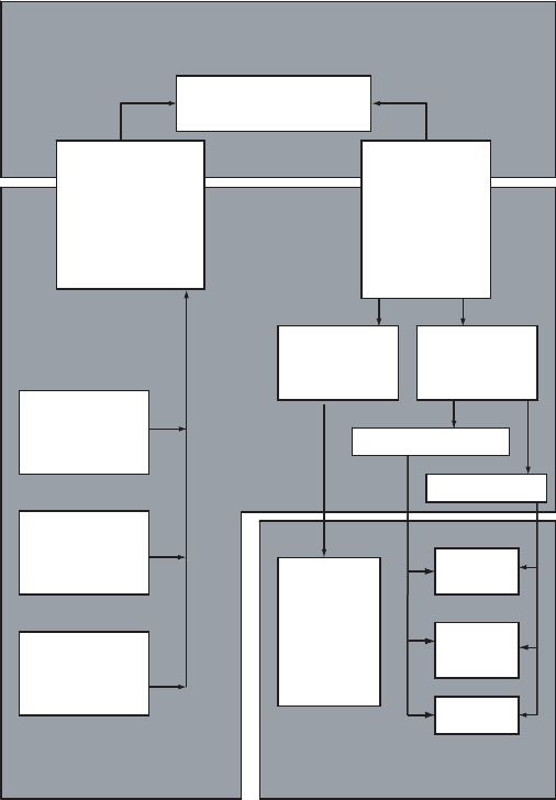
The nervous system can be divided into a central nervous system (CNS) and a per-
ipheral nervous system (PNS) (Figure 9.4). The CNS consists of the brain and spinal
cord. The CNS performs integration of information and coordination of actions. The
actual source of information and distributor of commands to the body is the PNS,
which includes all of the other neurons in the body.
Figure 9.3 Neural synapse. (Based on Fried, 1990.)
180
THE HUMAN ANIMAL
The brain is where all the higher-level activity occurs. The human brain weighs about
1.4 kg and has a volume averaging 1200 cm
3
. Brain size in humans correlates with body
size and not with intelligence. It contains about 10
12
neurons, each with an average of
about 1000 connections. The brain has three main parts: the cerebrum, the cerebellum,
and the brainstem. The brainstem is the connection between the brain and the spinal
cord. It regulates some of the functions most basic to our survival. Within the brainstem,
the medulla oblongata controls basic functions such as breathing, heartbeat, and blood
pressure. Above the medulla are the pons and midbrain, which act as relay centers for
sensory information. The hypothalamus is above this. It regulates the pituitary gland,
forming a connection between the nervous system and the endocrine system. It also reg-
ulates many functions of homeostasis: body temperat ure, salt and water balance, hunger,
and the digestive system. It also contains areas associated with pleasure and ecstasy. The
thalamus is above the hypothalamus (as the name implies) and also serves as a relay sta-
tion. From the thalamus down to the spinal cord runs the reticular formation, which is
Sensory
Neurons
Motor
Neurons
Somatic
Nervous
System
Autonomic
Nervous
System
Skeletal
Muscle
Smooth
muscle
Cardiac
muscle
Glands
Sympathetic
Parasympathetic
Special
Sensory
Receptors
Somatic
Sensory
Receptors
Visceral
Sensory
Receptors
Effectors
Information
Processing
Receptors
Sensory
Neurons
Motor
Neurons
Somatic
Nervous
System
Autonomic
Nervous
System
Skeletal
Muscle
Smooth
muscle
Cardiac
muscle
Glands
Sympathetic
Parasympathetic
Special
Sensory
Receptors
Somatic
Sensory
Receptors
Visceral
Sensory
Receptors
Effectors
Information
Processing
Receptors
Figure 9.4 Organization of the nervous system. (Based on Martini, 1998.)
NERVOUS SYSTEM 181
connected with sleeping and awareness and the ability of a student to concentrate during
long, boring lectures.
The cerebellum is located dorsal to the medulla and controls fine motor skills.
Although the decision to make some motion is made in the cerebrum, the cerebellum pro-
vides feedback control to accomplish the task with precision. It is most developed in birds
and mammals, since they have much higher dexterity than that of the other animals.
The cerebrum is the part of the brain responsible for voluntary control of skeletal mus-
cles, speech, vision, memory, thoughts, and consciousness. Most of its activity relates to
sensory and motor functions. It is the largest part of the brain, forming the familiar highly
folded structure divided into two parts, a right and a left hemisphere. The outer layer of
the cerebrum is the cerebral cortex. It is only 3 mm thick but accounts for 90% of the
brain’s cell bodies. Many specific capabilities can be localized to very small areas of
the cerebral cortex. For example, the ability to recognize your mother’s face can be
eliminated by the destruction of a very small area of the cortex, such as by a stroke.
Another area controls learned eye movements. People with damage to this area can under-
stand writing but cannot read because they cannot follow lines of type on a page with their
eyes.
The endothelial cells that line the blood vessels in the brain are joined to each other
very tightly. Thus, for any chemical to pass from the blood to the brain, it must pass into
and out of the epithelial cells rather than through spaces between them as is possible else-
where in the body. This is called the blood–brain barrier, and it limits the passage of
hydrophilic (water-soluble) compounds, thus protecting the brain against many types of
toxins and drugs. The brain uses glucose for energy almost exclusively and does not store
glycogen; thus, it is dependent on a continuous supply in the blood. It also uses oxygen at
a high rate. If the oxygen is cut off for more than 4 or 5 minutes, or if glucose is cut off
for more than about 15 minutes, brain damage will occur.
The peripheral nervous system has two parts, corresponding to the two functions of
obtaining information and distributing commands. Information is provided by the nerves
associated with the senses through the neurons of the sensory system. Action is stimu-
lated by the motor system, which connect to the muscles and initiate their contractions.
Both of these systems branch out from the brain and spinal cord. Some sensory neurons,
such as some of those connected with pain sensors in the skin, are connected to motor
neurons in the spinal column. This forms a reflex arc, which enables rapid resp onse to
danger without taking the time to transmit signals all the way to the brain for additional
CNS signal processing. For example, the sudden pulling of a finger away from a hot
object is accomplished by the reflex arc.
The motor system is itself divided into two parts: the somatic nervous syst em and the
autonomic nervous system. The somatic nervous system transmits signals for voluntary
control of the skeletal muscles. Their neurons, called motor neurons, have large-diameter
myelinated axons and tend to pass directly from the brain or spinal cord to the target mus-
cle without intermediate synaps es. All the motor neurons stimulate muscle contractions;
none are inhibitory. The only neurotransmitter used is ACh.
The autonomic nervous system controls involuntary activities, the body functions
that occur without our conscious awareness. The effectors (the target organs that act
upon receiving a nerve signal) include smooth muscle, the heart, and glands. Most of
the neurons of the autonom ic nervous system do not pass directly from the CNS to the
effector. Instead, each path consists of two neurons, joined by a synapse in a structure
called a ganglion, most of which are located alongside the spinal column. Autonomic
182 THE HUMAN ANIMAL
system neurons may be stimulatory or inhibitory. Most autonomic neurons use ACh, but
other neurotransmitters are also involved. One ‘‘ganglion’’ is actually a gland, the adrenal
medulla, in which the neurotransmitters epinephrine and norepinephrine are released to
the bloodstream instead of to another neuron. Thus they act as hormones in this situation.
This activity is described in Section 9.5.
The autonomic nervous system is divided into the sympathetic and parasympathetic
divisions. These are both anatomically and functionally distinct. The parasympathetic
division neurons originate in the brainstem and in the sacral region at the base of the
spine. The parasympathetic division stimulates activity of the visceral organs, which
occurs while in a relaxed state. Its effects can be summarized as follows: (1) metabolic
rate is decreased; (2) heart rate and blood pressure are decreased; (3) secretion by salivary
and digestive glands increases; (4) muscle contractions in the digestive tract increase; and
(5) urination and defecation are stimulated.
The sympathetic division branches out from the spinal column in the thoracic and
lumbar regions. Most sympathet ic paths release norepinephrine at the effector, although
some release ACh or nitric oxide. It is the sympathetic division that stim ulates the release
of epinephrine (adrenaline) and norepinephrine by the adrenal gland. In fact, the overall
effect of sympathetic division stimulation can be summarized as producing the fight or
flight response, which prepares the body for an emergency that might require intense
physical activity. It is stimulated by emotions such as fear or stress. In summary, the
effects of the sympathetic division are (1) increased mental alertness, (2) increased meta-
bolism, (3) inhibited digestive and urinary function, (4) activation of energy reserves, (5)
increased respiration, (6) increased heart rate and blood pressure, and (7) stimulation of
sweat glands.
Both the sympathetic and parasympathetic divisions innervate most of the same organs
and other effectors, but they usually have opposite effects: One will stimulate while the
other inhibits. This gives finer control over activity, like having both a brake and an accel-
erator on a car. The heart, for example, is stimulated to decr ease its output by ACh from
the parasympathetic division and to increase output by norepinephrine from the sym-
pathetic division. Some of the neurotransmittor receptors in the autonomic system are
stimulated by nicotine, the active ingredient in tobacco. As a result, nicotine poisoning
produces increased heart rate and blood pressure, plus vomiting and diarrhea.
Information is provided by sensory receptors, which are neurons that are specialized
to produce signals in response to physical or chemical stimulus . The receptors can be
divided into three divisions. The special sensory receptors are associated with complex
sensory organs; the special senses are vision, hearing, taste, smell, and balance. The
somatic sensory receptors include the senses of touch, pain, temperature, pressure,
vibration, and proprioception. Proprioception is the sense of position of the skeletal
muscles and joints. The visceral sensory receptors monitor the internal organ syst ems,
including the cardiac, digestive, respiratory, urinary, and reproductive systems. Since
an action potential is always of the same strength, a receptor signals the strength of a
sensation by varying the frequency of action potentials. That is, a stronger sensation pro-
duces a more rapidly repeated action potential. Ultimately, sensory signals are interpreted
by the central nervous system in what is called perception. A receptor can be as simple as
an ordinary dendrite of a neuron. Pain receptors may be of this form. They respond to
many different types of stimulus. Other receptors are enclosed in complex structures
that admit only a highly selective type of stimulus. The special sensory receptors are of
this type.
NERVOUS SYSTEM 183
9.5 ENDOCRINE SYSTEM AND HOMEOSTASIS
The nervous system is not the only way the body controls bodily functions. It also uses
chemical messengers. Some cells communicate directly with their contact neighbors
through special junctions. This is usually to coordinate local activity such as ciliary move-
ment or muscle contractions. Others release chemicals into the intercellular spaces that
primarily affect cells in the same tissue. An example is the prostaglandins, a powerful
fatty acid with many functions. Prostaglandins are released by damaged tissues and
stimulate inflammation and the sensation of pain. Aspirin and other analgesics act by
inhibiting the formation of prostaglandins and similar compounds. Many tissues issue
chemicals that inhibit cell division locally. This prevents uncontrolled growth such as
occurs in cancer tumors. Compounds such as these are called local hormones or
paracrine factors. Some specialized cells produce chemicals that are excreted through
ducts onto epitheli al surfaces, such as inside the intestines or onto the skin. These cells are
in structures called exocrine glands. Saliva and sweat are secretions of exocrine glands.
The focus in this section is on chemicals that are secreted by glands into the blood
supply for the regulation of bodily function. These glands form the endocrine system,
which consists of endocrine organs that produce hormones . The chemical messengers
produced by the endocrine system are called hormones, which are chemical messengers
that influence the response of cells and tissues at locations remote from the hormone-
producing cells. The endocrine system is just as vital for proper functioning of the body
as the nervous system is. Although its response time is not as fast, its effects can be
long lasting. Of interest from an environmental point of view is the idea that some pollu-
tants, including 2,3,7,8-TCDD (dioxin), mimic the female sex hormone estrogen. Such
toxins are called xenoestrogens or endocrine disrupters .
Figure 9.5 shows some of the glands of the endocrine system. The pituitary gland has
two main parts. The anterior pituitary produces a number of important hormones, the
release of all of which is controlled by other hormones produced by the hypothalamus.
The posterior pituitary does not produce its own hormones. However, it stores several
hormones produced by the hypothalamus and releases them upon receiving a neural com-
mand from the hypothalamus.
The adrenal glands are located atop each kidney and also have two main parts. The
outer part, or cortex, produces steroid hormones; the inner part, called the medulla,
produces epinephrine and norepinephrine. Ninety-nine percent of the pancreas serves
an exocrine function, producing digestive enzymes . The other 1% performs a critical
endocrine function: controlling blood glucose.
9.5.1 Homeostasis
The nervous system and the endocrine system share major responsibility for maintaining
homeostasis, a stable internal environment, in the face of external changes. Homeostasis
requires that a number of vital factors be controlled within a proper range. Some of these
vital factors include body temperature, blood glucose concentration, the concentration of indi-
vidual electrolytes (sodium, potassium, calcium) in the blood, and blood pressure and v olume.
Homeostatic control is accomplished mostly by the use of negative feedback, in which
the movement of a condition outside the vital range stimulates an effect that tends to
move it in the opposite direction. For example, most adults have a resting body tempera-
ture between 36.7 and 37.2
C (98.1 to 99.0
F). The ‘‘normal’’ value for a person’s vital
factor is termed the set point. If the temperature rises about 0.2
C above the set-point
184 THE HUMAN ANIMAL
temperature, the hypothalamus stimulates blood vessels in the skin to dilate and sweat
glands to increase their secretion. Both of these tend to increase the loss of heat from
the body, limiting or reversing the temperature increase. A body temperature decrease
causes the reverse effects, plus other activities, such as shivering.
Body temperature also involves feedforward control, in which events that would alter
a vital factor are dete cted and the body responds before the alteration actually occurs. The
hypothalamus also receives signals from temperature sensors in the skin. These stimulate
the temperature control effects before the sensors within the hypothalamus actually detect
an internal change.
A few mechanisms involve positive feedback, in which a stimulus produces an effect
that increases the stimulus. This usually involves processes that need to be com pleted
quickly, such as blood clotting. When childbirth begins, stretch receptors in the wall of
the uterus stimulate the brain to cause the pituitary gland to release stored oxytocin,a
hormone that stimulates uterine contractions. This increases the rate of contractions,
expelling the baby faster. Once the baby has left the birth canal, the uterine receptors
relax, leading to a drop in oxytocin levels, breaking the positive feedback loop.
Figure 9.5 Some of the most important endocrine glands. (From Van de Graaff and Rhees, 1997.
# The McGraw-Hill Companies, Inc. Used with permission.)
ENDOCRINE SYSTEM AND H OMEOSTASIS 185

Factors that cause deviation from homeostasis are termed stresses. Stress can be phy-
sical, emotional, environmental, or metabolic. When stress initiates, the body enters what
is called the alar m phase and responds with the fight or flight response described in
Section 9.4.3. If the stress continues for the long term, the body switches to the resistance
phase, in which glucocorticoid hormones are released, especially cortisol, plus lesser
amounts of epinephrine, growth hormones, and thyroid hormones. These maintain the
rate of energy supply at elevated levels by mobilizing lipid and protein reserves and
saving glucose for nervous tissue. If the stress is starvation, the resistance phase ends
when reserves run out. Otherwise, it may end due to the side effects of the hormones,
such as the slowing of wound healing due to the anti-inflammatory effects of gluc ocorti-
coids, elevated blood pressure and volume, and altered mineral balance (especially loss of
potassium in the blood) due to aldosterone and ADH, or exhaustion of the ability o f the
adrenal cortex to continue glucocorticoid hormone production, destroying the ability of
the body to maintain blood glucose levels. If any of these occur, the result is the exhaus-
tion phase, in which homeostasis breaks down. One or more organs may malfunction. For
example, excessive potassium loss can cause heart failure.
9.5.2 Hormones
Hormones are classified into three groups based on chemical structure:
Amino acid derivatives: derivatives of either tyrosine (e.g., catecholamines and
thyroid hormones) or tryptophan (e.g., melatonin).
Peptide hormones: glycoproteins, short peptides, or small proteins (under 200 amino
acids). Examples include growth hormone and insulin.
Lipid derivatives: either eicosanoids (derivatives of arachidonic acid), such as
prostaglandins, or steroids (derivatives of cholesterol), such as hydrocortisone.
Tables 9.1, 9.2, and 9.3 summarize most of the hormones and their functions. Although
others are known, some are still poorly understood.
TABLE 9.1 Peptide Hormones
Gland Hormone Target and Function
Hypothalamus Antidiuretic hormone,
or vasopressin (ADH)
Stored in posterior pituitary; when released, it
increases water resorption by the kidneys; it is
inhibited by alcohol.
Oxytocin Stored in posterior pituitary; when released,
stimulates milk ejection and uterine contractions
in females, ductus deferens and prostate in males;
secreted by uterus and fetus; released in orgasm in
both sexes.
Releasing and
inhibiting hormones
Controls the release of hormones of the anterior
pituitary, a different one for each.
Anterior
pituitary
Thyroid-stimulating
hormone (TSH)
Stimulates synthesis and secretion of thyroid
hormones; stimulated by hypothalamic
thyrotropin-releasing hormone (TRH).
Follicle-stimulating
hormone (FSH)
Females: stimulates follicle maturation and estrogen
synthesis; males: stimulates production of sperm;
stimulated by hypothalamic gonadotropin-
releasing hormone (GnRH).
186
THE HUMAN ANIMAL

TABLE 9.1 (Continued )
Gland Hormone Target and Function
Leuteinizing
hormone (LH)
Females: stimulates ovulation, formation of corpus
luteum, and synthesis of estrogen and progester-
one; males: stimulates synthesis of testosterone;
stimulated by hypothalamic GnRH.
Prolactin (PL) Stimulates growth of mammary gland and synthesis
of milk.
Growth hormone
(somatotropin, GH)
Stimulates growth of all cells, but the cells of the
bone and cartilage are especially sensitive.
Adrenocorticotropic
hormone (ACTH)
Stimulates production of steroids in adrenal cortex,
part of the response to stress.
Intermediate
pituitary
Melanocyte-stimulating
hormone (MSH)
Increases melanin synthesis in epidermis (not active
in adults except pregnant women).
Parathyroid Parathyroid hormone
(PTH)
Increases calcium in circulation by stimulating bones
to release it, enhances digestive uptake, and
inhibits kidney removal.
C cells of the
thyroid
Calcitonin (CT) Opposite effect of PTH, decreases calcium in blood.
Heart Atrial natriuretic
peptide (ANP)
Release stimulated by stretch receptors in cardiac
muscle; it promotes loss of sodium and water in
kidney, supresses thirst and water-conserving
hormones.
Thymus Thymosins A blend of hormones that induce T-cell differentia-
tion in the immune system.
Pancreas Insulin Stimulates the uptake and use of glucose by cells
throughout the body, the production of glycogen
by the liver and skeletal muscles, and the
formation of fats by adipose tissue. The effect of
insulin on glucose uptake by cells is indirect; it
stimulates an increase in the production of
membrane proteins that transport glucose through
the membrane by facilitated diffusion.
Glucagon Released when blood glucose is low: stimulates cells
throughout the body to release glucose, the liver to
break down glycogen into glucose, and the
breakdown of fats by adipose tissue.
Digestive tract Gastrin Stimulates hydrochloric acid secretion by the
stomach.
Cholecystokinin Stimulates the exocrine cells of the pancreas to
secrete digestive enzymes.
Kidneys Erythropoietin (EPO) Produced in response to low oxygen in the kidney;
stimulates red blood cell production by bone
marrow.
Renin (actually
an enzyme)
Converts angiotensinogen to angiotensin I in the
blood.
Liver Angiotensinogen
(a plasma protein)
After conversion by renin in the blood and other
enzymes in the lungs, it becomes angiotensin II,
which stimulates thirst and production of
aldosterone and ADH, causing sodium and water
retention. This is part of the response to low blood
volume.
ENDOCRINE SYSTEM AND H OMEOSTASIS 187